PLOS ONE logoLink to PLOS ONE
. 2020 Dec 3;15(12):e0243421. doi: 10.1371/journal.pone.0243421

Pregnancy outcomes after hysteroscopic surgery in women with cesarean scar syndrome

Shunichiro Tsuji 1,*,#, Akimasa Takahashi 1,#, Asuka Higuchi 1, Akiyoshi Yamanaka 1, Tsukuru Amano 1, Fuminori Kimura 1, Ayumi Seko-Nitta 2, Takashi Murakami 1
Editor: Antonio Simone Laganà3
PMCID: PMC7714235  PMID: 33270754

Abstract

Cesarean scar defect often causes postmenstrual abnormal uterine bleeding, dysmenorrhea, chronic pelvic pain, and infertility, which are collectively known as cesarean scar syndrome (CSS). Several studies have reported that hysteroscopic surgery can restore fertility in women with CSS. The study aimed to identify factors that influence subsequent pregnancy following hysteroscopic surgery. Therefore, we studied 38 women with secondary infertility due to CSS who underwent hysteroscopic surgery at Shiga University of Medical Hospital between July 2014 and July 2019. Our hysteroscopic procedure included inferior edge resection and superficial cauterization of the cesarean scar defect under laparoscopic guidance. Patients were followed up for 3 to 40 months after surgery. Surgery was successful in all cases and no complications were observed. Twenty-seven patients (71%) became pregnant (pregnant group), while 11 (29%) did not (non-pregnant group). Baseline characteristics of age, body mass index, gravidity, parity, previous cesarean section, presence of endometriosis, retroflex uterus, and preoperative residual myometrial thickness were not significantly different between the groups. However, the median residual myometrium thickness was significantly higher after surgery than before surgery in the pregnant group (1.9 [1.1–3.6] vs 4.9 [3.4–6.6] mm, P<0.0001), whereas this difference was not significant in the non-pregnant group. Of those who became pregnant, 85% conceived within 2 years of surgery. Although three pregnancies resulted in abortion and one is ongoing at the time of writing, 23 pregnancies resulted in healthy babies at 35–38 gestational weeks by scheduled cesarean sections with no obstetrical complications due to hysteroscopic surgery. The average birth weight was 3,076 g. Our findings support that hysteroscopic surgery is a safe and effective treatment for secondary infertility due to CSS. The thickness of the residual myometrium may be a key factor that influences subsequent pregnancy in women with CSS.

Introduction

The rate of cesarean section is increasing worldwide. In Japan, it reached 18.5% in 2013, which is nearly a two-fold increase in the past two decades [1]. With the increase in cesarean sections, the incidence of cesarean section scar-related complications has also risen. Cesarean section often results in a cesarean scar defect (CSD), also known as isthmocele, which reportedly occurs in 24–84% of women after cesarean section [2,3]. Although there is still no clear universal definition of this term, it is commonly used in the literature to indicate a myometrial discontinuity or a hypoechoic region in the lower anterior uterine wall via transvaginal ultrasound detection [3,4]. CSD can cause secondary infertility with postmenstrual abnormal uterine bleeding, dysmenorrhea, and chronic pelvic pain [5]. These symptoms are collectively known as cesarean scar syndrome (CSS) [6]. We have previously investigated the management of secondary infertility for patients with CSS in Japan and found that surgical treatment, including laparotomy, laparoscopy, and hysteroscopy, was effective for restoring fertility in such patients [5]. Although there is an ongoing debate regarding the best surgical approach, hysteroscopic treatment is considered less invasive than other approaches and may be an effective treatment option for restoring fertility in women with CSS [79]. However, the predictors of subsequent reproductive outcomes following hysteroscopic treatment are still not clear.

Therefore, we aimed to investigate the reproductive and obstetric outcomes and the interval between treatment and conception following hysteroscopic procedures.

Materials and methods

Study population and recruitment

The participants of this retrospective study were patients who underwent hysteroscopic surgery for CSS between July 2014 and July 2019 at the Shiga University of Medical Science. The inclusion criteria were women diagnosed with CSS. CSS was diagnosed by detecting both CSD and the presence of abnormal uterine bleeding or liquid pooling on transvaginal ultrasonography. Exclusion criteria were women who were not intending to conceive. All patients underwent cancer screenings to exclude abnormal cervical cytology. Subsequent pregnancy was confirmed by the presence of a gestational sac in the uterus. Participants were categorized into two groups, pregnant or non-pregnant, according to the outcome of conception after surgery. Written informed consent was obtained from all patients prior to surgery. All data were fully anonymized before we accessed them, and patients' medical records were accessed between June 2020 and July 2020. This study was approved by the Ethics Committee at Shiga University of Medical Science (approval number; R2020-039) and performed at the Shiga University of Medical Science.

Surgical procedures

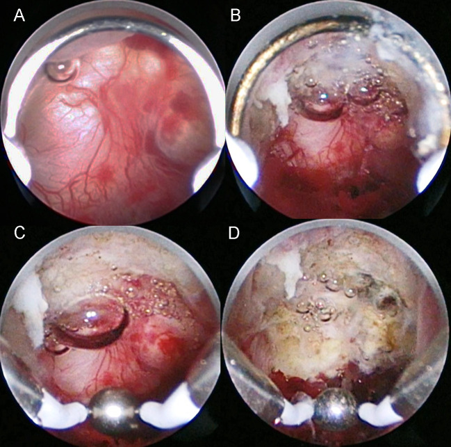
We performed hysteroscopic surgery as previously described [10]. Briefly, hysteroscopic surgery was performed using a rigid 30° hysteroscope (4 mm telescope) and working elements (#27050, KARL STORZ, Germany) connected to a video camera and monitor (Olympus, Tokyo, Japan). Diagnostic laparoscopy was performed simultaneously to monitor accidental perforation at the site of the CSD and to treat other causes of infertility, such as endometriosis, because the aim of this operation was to restore fertility. Cervical dilation was carried out the day before surgery. First, hysteroscopic resection of the CSD inferior edge was performed using a cutting loop electrode to enable visualization of the diverticulum. Next, the entire CSD was cauterized using a ball electrode (Fig 1 and S1 File). Patients were discharged 2 to 3 days after surgery.

Fig 1. Intraoperative images of the hysteroscopic surgery procedure.

Fig 1

(A) Abnormal hypervascularity is observed in the cesarean scar defect. (B) Cutting of the inferior edge of the cesarean scar defect using a cutting loop electrode. (C) Cauterization of all areas including the abnormal vasculature in the cesarean scar defect. (D) Appearance after cauterization using a ball electrode.

Data collection

Baseline characteristics included age, body mass index, gravidity, parity, previous cesarean section(s), frequency of endometriosis, and retroflex uterus. Residual myometrial thickness (RMT) was measured by magnetic resonance imaging (MRI) before and 2 months after surgery using a 1.5-T instrument (SIGNA HDxt; GE Healthcare Waukesha, WI, USA) with a cardiac coil. The settings for MRI were applied as described previously [10]. All measurements were conducted by one senior radiologist using a high-resolution monitor. The interval from operation to conception was evaluated based on patient medical records. For patients who did not continue to attend our hospital, we confirmed their current situation via a medical information provision form from their referral hospital or by telephone.

Statistical analysis

All data were analyzed using GraphPad Prism ver.7 (GraphPad Software, Inc., San Diego, CA, USA). The D’Agostino-Pearson test was used to evaluate data distribution. Normally distributed data are expressed as mean ± standard deviation. Data with a non-normal distribution are presented as median (interquartile range). Categorical data were compared by Fisher’s exact test. Comparisons between the pregnant and non-pregnant participants were carried out using an unpaired two-tailed t-test or the Mann-Whitney U test for parametric and non-parametric data, respectively. The cumulative pregnancy rate was evaluated using the Kaplan-Meier method. Statistical significance was defined as P < 0.05 in all cases.

Results

Thirty-eight patients who met the inclusion criteria during the study period were included in the analysis. No complications were experienced by any patients. Twenty-seven patients (71%) became pregnant (pregnant group) and 11 patients (29%) did not become pregnant (non-pregnant group) (S1 Table). Baseline characteristics did not significantly differ between the two groups (Table 1). Two cases did not undergo laparoscopy for individual reasons; therefore, information on endometriosis was not available at the time of hysteroscopic surgery. Endometriosis was detected in 19 patients (52.8%) during hysteroscopic surgery. A blueberry spot was detected in one case on the surface of the CSD (S1 Fig).

Table 1. Comparison of patients and clinical data.

Pregnancy (n = 27) Non-pregnancy (n = 11) P
Age, yrs 35.6±3.4 37.0±4.2 n.s.
BMI 22.2±3.7 21.5±3.1 n.s.
Gravidity 1 (1–2) 2 (1–2) n.s.
Parity 1 (1–1) 1 (1–2) n.s.
Previous CS 1 (1–1) 1 (1–2) n.s.
Endometriosis (%) 14 (52)* 5 (45) n.s.
Retroflexion(%) 14(52) 3(27) n.s.
RMT preoperatively (mm) 2.3(1.3–3.8) 2.1 (0.8–3.9) n.s.

BMI: Body mass index, CS: cesarean section, RMT: Residual myometrium thickness, Data are median (quartiles)

*Two patients did not undergo laparoscopy.

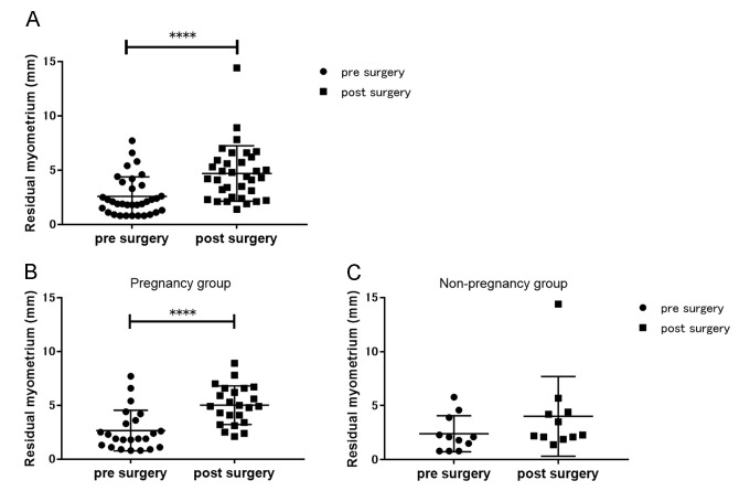
Pre- and postoperative RMTs were measured in 34 patients (postoperative measurement was not carried out in four cases due to individual reasons). The median pre- and postoperative RMT measurements were 2.0 (1.1–3.7) mm and 4.4 (2.5–6.0) mm, respectively (P < 0.0001) (Fig 2A). Considering the pregnant group alone, the postoperative RMT was significantly higher than the preoperative RMT (1.9 [1.1–3.6] mm vs 4.9 [3.4–6.6] mm, P < 0.0001); however, the difference was not significant in the non-pregnant group (2.1 [0.8–3.9] mm vs 2.3 [2.1–4.4] mm) (Fig 2B and 2C). A significant difference was observed in postoperative RMT between the pregnant and non-pregnant groups (4.9 [3.4–6.6] mm vs 2.3 [2.1–4.4] mm, respectively; P = 0.02).

Fig 2. Graphical representations of the residual myometrial thickness before and after hysteroscopic surgery.

Fig 2

The pre- and postoperative residual myometrial thicknesses of (A) the entire study population, (B) pregnant women, and (C) non-pregnant women. Significant differences between pre- and postoperative measurements were detected among the entire cohort and pregnant women. ****P < 0.0001.

After hysteroscopy, 17 patients became pregnant within 1 year and six became pregnant during the following year. The cumulative pregnancy rate is illustrated in Fig 3. Pregnancy is ongoing in one case and three cases resulted in spontaneous abortions. The mean birth weight among all the patients who gave birth was 3,076 ± 435 g. One patient underwent a scheduled cesarean section at 35 gestational weeks due to placenta previa. All other deliveries were scheduled cesarean sections following the individual policies of the obstetric hospitals; four delivered at 36 gestational weeks, eight delivered at 37 gestational weeks, and 10 delivered at 38 gestational weeks. No severe obstetrical complications, such as uterine rupture, occurred up to the day of cesarean section in any case.

Fig 3. Cumulative pregnancy rate after hysteroscopic surgery in women with infertility due to cesarean scar syndrome (n = 38).
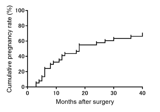
Fig 3

Discussion

To the best of our knowledge, this is the first report to evaluate the changes in residual myometrium thickness after surgery in relation to subsequent reproductive outcomes. The present retrospective observational study demonstrates the safety and efficacy of hysteroscopic surgery for treating secondary infertility caused by CSS. Our results found that the pregnancy rate was high after hysteroscopic surgery and the postoperative thickening of the residual myometrium was associated with successful reproductive outcomes.

Hysteroscopic surgery in the context of CSS is mainly performed to treat abnormal uterine bleeding [1113]; however, many recent reports have demonstrated the effectiveness of this technique for restoring fertility [8,10,1417]. In women with CSS, infertility may arise due to abnormal uterine bleeding originating from a small hemorrhage in the CSD that interferes with implantation [8,14]. Previous reports performed hysteroscopic resection of both the superior and inferior edges of the CSD, whereas the surgical procedure used in this study resected only the inferior edge of the CSD; therefore, the present study is valuable to the field as we provide further validation of the effectiveness of hysteroscopic surgery for the treatment of infertility. Furthermore, we demonstrated the safety of our hysteroscopic surgery method, as no surgery-related complications, such as perforation, occurred, even in cases with a thin residual myometrium, and no obstetrical complications, such as uterine rupture, occurred during pregnancy. The first prospective study on the effectiveness of hysteroscopic surgery was carried out by Gubbini et al. [16] who reported that among 41 patients, no complications were noted during the perinatal period after hysteroscopic surgery. Thus, the minimally invasive technique of hysteroscopy is a safe treatment, both intraoperatively and postoperatively, in regard to subsequent pregnancy.

Regarding the main reason of infertility in participants, 68% of women became pregnant spontaneously in their prior pregnancy (S1 Table). However, they became infertile after cesarean section regardless of whether various treatments, including IVF, were conducted. On the other hand, 32% of women became pregnant by assisted reproductive technology (ART) in their prior pregnancy. Therefore, ART was performed after cesarean section during the long period; however, these patients could not become pregnant. Furthermore, abnormal uterine bleeding or liquid pooling in the CSD or uterine cavity was an obvious abnormal finding associated with infertility. Taken together, we speculate that the infertility observed in these participants was caused by CSS. However, we considered that CSS may not have been the sole cause of infertility in these patients, because around half of the patients also had endometriosis.

Although the study population had CSS-related infertility, we treated endometriosis and the area of the CSD because endometriosis is a well-known cause of infertility [18,19]. Interestingly, laparoscopic investigation revealed that around half of the patients in the present study had endometriosis in the peritoneal cavity. In the general population, the frequency of endometriosis is around 10%; however, there was a higher rate of endometriosis in present study [18]. Several reports on the presence of endometriosis in patients with CSD [17,20] are in agreement with our detection of the blueberry spot in one patient with CSD, supporting the potential association between CSS and endometriosis. In addition, the interval between surgery and pregnancy was within 2 years for most patients in the present study, and endometriosis usually recurs within 2 years of surgery [18,19]. Therefore, we suggest that this surgery may provide a sufficient “endometriosis-free” window to enable conception.

A laparoscopic retractor was sometimes useful during cauterization of the CSD with a ball electrode because it can be difficult to access the inner wall of the diverticulum due to large defects, especially when they are located on the lateral side. In the present study, the uterus was moved to the left side using laparoscopic forceps when defects were located on the right lateral side, and the side of the CSD was gently pressed from the outside of the uterus using Endo Peanut (Medtronic, MN, USA). Therefore, hysteroscopic treatment with laparoscopy is a safe option for the treatment of infertility; specifically, laparoscopy can be beneficial during hysteroscopic surgery, especially in cases with large defects.

We have previously reported that RMT increases following our procedure of hysteroscopy [10], which is supported by the present study and a previous study [21]. Both reports used a roller ball electrode to electrocauterize the bottom of the diverticulum, with no resection of the bottom in the diverticulum. In contrast, another study that resected CSD scar tissue did not identify changes in RMT after hysteroscopic resection [17]. Therefore, different methods of hysteroscopy may lead to varying results. Although a consensus statement from the global congress on hysteroscopy scientific committee appealed that the laparoscopic approach should be favored if the myometrial thickness is less than 3 mm, Gubbini et al. proposed that it could still be disputable to consider the indication of hysteroscopic surgery [22,23]. The mode of delivery following resectoscopic surgery in this study was planned cesarean section in all cases. Although cesarean section can result in several complications for the mother and baby, we considered the mode of delivery was better than trial of labor after cesarean section because there was no evidence of the risk of uterine rupture following hysteroscopic surgery for isthmocele [24,25].

This study has several limitations that should be considered. First, endometriosis treatment affected subsequent conception. However, the presence of endometriosis before surgery did not influence the rate of conception after surgery. We also suspect that endometriosis treatment did not affect conception following hysteroscopic surgery. Second, although there was a significant change in postoperative RMT in the pregnant group, this study could not reveal the exact mechanism of thickening. We consider that the treatment of inflammatory tissue in the CSD might promote the regeneration of fibrotic tissue or elimination of pressure as liquid pools in the defect, resulting in the thickening of the residual myometrium. Liquid pools in the CSD might affect RMT thinning via pressure. We consider that improving the environment in the uterine cavity might contribute to successful conception. In the non-pregnant group, myometrial regeneration in the CSD might have been inhibited due to insufficient treatment. It is evident that there is room for further improvement in our procedure to increase the pregnancy rate after hysteroscopic surgery. Third, this study included a small patient cohort and was a short-term study. Therefore, further investigation of a larger patient population is needed to verify the safety and efficacy of this procedure for infertile women with CSS. Fourth, due to our study’s non-randomized design, the contributions of hysteroscopic surgery to subsequent pregnancy were controversial in women who became pregnant after the long period following hysteroscopic surgery.

In conclusion, hysteroscopic surgery is a safe, minimally invasive treatment for the restoration of fertility in women with CSS. Our findings suggest that the thickening of the residual myometrium following hysteroscopic surgery may influence subsequent reproductive outcomes.

Supporting information

S1 Fig. Intraoperative image showing the blueberry spot on the surface of the cesarean scar defect.

(B) Enlargement of the area indicated with the square in (A).

(TIF)

S1 Table. Patients included in this study.

(XLSX)

S1 File. Video of the procedure details.

(MP4)

Acknowledgments

We would like to thank Editage (www.editage.com) for English language editing.

Data Availability

All relevant data are within the paper and its Supporting Information files.

Funding Statement

This study was supported by the Grants-in-Aid for Scientific Research (KAKENHI; 20K09616). The grant provided financial support for preparation of the article, such as English language editing services and Open Access Publication Fee. https://www.jsps.go.jp/english/index.html.

References

  • 1.Maeda E, Ishihara O, Tomio J, Sato A, Terada Y, Kobayashi Y, et al. Cesarean section rates and local resources for perinatal care in Japan: A nationwide ecological study using the national database of health insurance claims. J Obstet Gynaecol Res. 2018;44: 208–216. 10.1111/jog.13518 . [DOI] [PubMed] [Google Scholar]
  • 2.van der Voet LF, Bij de Vaate AM, Veersema S, Brolmann HA, Huirne JA. Long-term complications of caesarean section. The niche in the scar: a prospective cohort study on niche prevalence and its relation to abnormal uterine bleeding. BJOG. 2014;121: 236–244. 10.1111/1471-0528.12542 . [DOI] [PubMed] [Google Scholar]
  • 3.Kremer TG, Ghiorzi IB, Dibi RP. Isthmocele: an overview of diagnosis and treatment. Rev Assoc Med Bras (1992). 2019;65: 714–721. 10.1590/1806-9282.65.5.714 . [DOI] [PubMed] [Google Scholar]
  • 4.Donnez O. Cesarean scar defects: management of an iatrogenic pathology whose prevalence has dramatically increased. Fertil Steril. 2020;113: 704–716. 10.1016/j.fertnstert.2020.01.037 . [DOI] [PubMed] [Google Scholar]
  • 5.Tsuji S, Murakami T, Kimura F, Tanimura S, Kudo M, Shozu M, et al. Management of secondary infertility following cesarean section: Report from the Subcommittee of the Reproductive Endocrinology Committee of the Japan Society of Obstetrics and Gynecology. J Obstet Gynaecol Res. 2015;41: 1305–1312. 10.1111/jog.12750 . [DOI] [PubMed] [Google Scholar]
  • 6.Morris H. Surgical pathology of the lower uterine segment caesarean section scar: is the scar a source of clinical symptoms? Int J Gynecol Pathol. 1995;14: 16–20. 10.1097/00004347-199501000-00004 [DOI] [PubMed] [Google Scholar]
  • 7.Abacjew-Chmylko A, Wydra DG, Olszewska H. Hysteroscopy in the treatment of uterine cesarean section scar diverticulum: A systematic review. Adv Med Sci. 2017;62: 230–239. 10.1016/j.advms.2017.01.004 . [DOI] [PubMed] [Google Scholar]
  • 8.Florio P, Filippeschi M, Moncini I, Marra E, Franchini M, Gubbini G. Hysteroscopic treatment of the cesarean-induced isthmocele in restoring infertility. Curr Opin Obstet Gynecol. 2012;24: 180–186. 10.1097/GCO.0b013e3283521202 . [DOI] [PubMed] [Google Scholar]
  • 9.Bhagavath B, Lindheim SR. Optimal management of symptomatic cesarean scar defects. Fertil Steril. 2018;110: 417–418. 10.1016/j.fertnstert.2018.06.035 . [DOI] [PubMed] [Google Scholar]
  • 10.Tsuji S, Kimura F, Yamanaka A, Hanada T, Hirata K, Takebayashi A, et al. Impact of hysteroscopic surgery for isthmocele associated with cesarean scar syndrome. J Obstet Gynaecol Res. 2018;44: 43–48. 10.1111/jog.13464 . [DOI] [PubMed] [Google Scholar]
  • 11.Fabres C, Arriagada P, Fernandez C, Mackenna A, Zegers F, Fernandez E. Surgical treatment and follow-up of women with intermenstrual bleeding due to cesarean section scar defect. J Minim Invasive Gynecol. 2005;12: 25–28. 10.1016/j.jmig.2004.12.023 . [DOI] [PubMed] [Google Scholar]
  • 12.Fernandez E, Fernandez C, Fabres C, Alam VV. Hysteroscopic correction of cesarean section scars in women with abnormal uterine bleeding. J Am Assoc Gynecol Laparosc. 1996;3: S13 10.1016/s1074-3804(96)80170-8 . [DOI] [PubMed] [Google Scholar]
  • 13.Chang Y, Tsai EM, Long CY, Lee CL, Kay N. Resectoscopic treatment combined with sonohysterographic evaluation of women with postmenstrual bleeding as a result of previous cesarean delivery scar defects. Am J Obstet Gynecol. 2009;200: 370 e371–374. 10.1016/j.ajog.2008.11.038 . [DOI] [PubMed] [Google Scholar]
  • 14.Tanimura S, Hosono T, Minami R, Funamoto H. Hemorrhage from a cesarean scar is a cause of cesarean scar syndrome. J Minim Invasive Gynecol. 2017;24: 340–341. 10.1016/j.jmig.2016.06.014 . [DOI] [PubMed] [Google Scholar]
  • 15.Gubbini G, Casadio P, Marra E. Resectoscopic correction of the "isthmocele" in women with postmenstrual abnormal uterine bleeding and secondary infertility. J Minim Invasive Gynecol. 2008;15: 172–175. 10.1016/j.jmig.2007.10.004 . [DOI] [PubMed] [Google Scholar]
  • 16.Gubbini G, Centini G, Nascetti D, Marra E, Moncini I, Bruni L, et al. Surgical hysteroscopic treatment of cesarean-induced isthmocele in restoring fertility: prospective study. J Minim Invasive Gynecol. 2011;18: 234–237. 10.1016/j.jmig.2010.10.011 . [DOI] [PubMed] [Google Scholar]
  • 17.Tanimura S, Funamoto H, Hosono T, Shitano Y, Nakashima M, Ametani Y, et al. New diagnostic criteria and operative strategy for cesarean scar syndrome: Endoscopic repair for secondary infertility caused by cesarean scar defect. J Obstet Gynaecol Res. 2015;41: 1363–1369. 10.1111/jog.12738 . [DOI] [PubMed] [Google Scholar]
  • 18.Zondervan KT, Becker CM, Missmer SA. Endometriosis. N Engl J Med. 2020;382: 1244–1256. 10.1056/NEJMra1810764 . [DOI] [PubMed] [Google Scholar]
  • 19.Agarwal SK, Chapron C, Giudice LC, Laufer MR, Leyland N, Missmer SA, et al. Clinical diagnosis of endometriosis: a call to action. Am J Obstet Gynecol. 2019;220: 354 e351–354 e312. 10.1016/j.ajog.2018.12.039 . [DOI] [PubMed] [Google Scholar]
  • 20.Donnez O, Donnez J, Orellana R, Dolmans MM. Gynecological and obstetrical outcomes after laparoscopic repair of a cesarean scar defect in a series of 38 women. Fertil Steril. 2017;107: 289–296 e282. 10.1016/j.fertnstert.2016.09.033 . [DOI] [PubMed] [Google Scholar]
  • 21.Li C, Guo Y, Liu Y, Cheng J, Zhang W. Hysteroscopic and laparoscopic management of uterine defects on previous cesarean delivery scars. J Perinat Med. 2014;42: 363–370. 10.1515/jpm-2013-0081 . [DOI] [PubMed] [Google Scholar]
  • 22.Lagana AS, Pacheco LA, Tinelli A, Haimovich S, Carugno J, Ghezzi F, et al. Optimal timing and recommended route of delivery after hysteroscopic management of isthmocele? A consensus statement from the global congress on hysteroscopy scientific committee. J Minim Invasive Gynecol. 2018;25: 558 10.1016/j.jmig.2018.01.018 . [DOI] [PubMed] [Google Scholar]
  • 23.Giampietro G, Casadio P, Franchini M, Florio P. Regarding "Optimal timing and recommended route of delivery after hysteroscopic management of isthmocele? A consensus statement from the global congress on hysteroscopy scientific committee". J Minim Invasive Gynecol. 2018;25: 1111–1112. 10.1016/j.jmig.2018.04.007 . [DOI] [PubMed] [Google Scholar]
  • 24.Franchi M, Raffaelli R, Baggio S, Scollo M, Garzon S, Lagana AS, et al. Unintentional transvesical caesarean section: incidence, risk factors, surgical technique and post-operative management. Eur J Obstet Gynecol Reprod Biol. 2019;236: 26–31. 10.1016/j.ejogrb.2019.02.023 . [DOI] [PubMed] [Google Scholar]
  • 25.Ahimbisibwe A, Coughlin K, Eastabrook G. Respiratory morbidity in late preterm and term babies born by elective caesarean section. J Obstet Gynaecol Can. 2019;41: 1144–1149. 10.1016/j.jogc.2018.11.002 . [DOI] [PubMed] [Google Scholar]

Decision Letter 0

Antonio Simone Laganà

15 Oct 2020

PONE-D-20-27415

Pregnancy outcomes after hysteroscopic surgery in women with cesarean scar syndrome

PLOS ONE

Dear Dr. Tsuji,

Thank you for submitting your manuscript to PLOS ONE. After careful consideration, we feel that it has merit but does not fully meet PLOS ONE’s publication criteria as it currently stands. Therefore, we invite you to submit a revised version of the manuscript that addresses the points raised during the review process.

Please submit your revised manuscript by Nov 29 2020 11:59PM. If you will need more time than this to complete your revisions, please reply to this message or contact the journal office at plosone@plos.org. When you're ready to submit your revision, log on to https://www.editorialmanager.com/pone/ and select the 'Submissions Needing Revision' folder to locate your manuscript file.

Please include the following items when submitting your revised manuscript:

  • A rebuttal letter that responds to each point raised by the academic editor and reviewer(s). You should upload this letter as a separate file labeled 'Response to Reviewers'.

  • A marked-up copy of your manuscript that highlights changes made to the original version. You should upload this as a separate file labeled 'Revised Manuscript with Track Changes'.

  • An unmarked version of your revised paper without tracked changes. You should upload this as a separate file labeled 'Manuscript'.

If you would like to make changes to your financial disclosure, please include your updated statement in your cover letter. Guidelines for resubmitting your figure files are available below the reviewer comments at the end of this letter.

If applicable, we recommend that you deposit your laboratory protocols in protocols.io to enhance the reproducibility of your results. Protocols.io assigns your protocol its own identifier (DOI) so that it can be cited independently in the future. For instructions see: http://journals.plos.org/plosone/s/submission-guidelines#loc-laboratory-protocols

We look forward to receiving your revised manuscript.

Kind regards,

Antonio Simone Laganà, M.D., Ph.D.

Academic Editor

PLOS ONE

Journal Requirements:

When submitting your revision, we need you to address these additional requirements.

1. Please ensure that your manuscript meets PLOS ONE's style requirements, including those for file naming. The PLOS ONE style templates can be found at

https://journals.plos.org/plosone/s/file?id=wjVg/PLOSOne_formatting_sample_main_body.pdf and

https://journals.plos.org/plosone/s/file?id=ba62/PLOSOne_formatting_sample_title_authors_affiliations.pdf

2. In your Methods section, please provide additional information about the participant recruitment method and the demographic details of your participants. Please ensure you have provided sufficient details to replicate the analyses such as:   

-    a description of any inclusion/exclusion criteria that were applied to participant recruitment,

-    a table of relevant demographic details,

-    a statement as to whether your sample can be considered representative of a larger population,

-    a description of how participants were recruited, and

-       descriptions of where participants were recruited and where the research took place.

3. In the ethics statement in the manuscript and in the online submission form, please provide additional information about the patient records used in your retrospective study, including: a) whether all data were fully anonymized before you accessed them; b) the date range (month and year) during which patients' medical records were accessed; c) the date range (month and year) during which patients whose medical records were selected for this study sought treatment. If patients provided informed written consent to have data from their medical records used in research, please include this information.

4. For more information on PLOS ONE's expectations for statistical reporting, please see https://journals.plos.org/plosone/s/submission-guidelines.#loc-statistical-reporting. Please update your Methods and Results sections accordingly.

5. Please include your tables as part of your main manuscript and remove the individual files. Please note that supplementary tables (should remain/ be uploaded) as separate "supporting information" files.

Additional Editor Comments (if provided):

The topic of the manuscript is interesting. Nevertheless, the reviewers raised several concerns: considering this point, I invite authors to perform the required major revisions.

[Note: HTML markup is below. Please do not edit.]

Reviewers' comments:

Reviewer's Responses to Questions

Comments to the Author

1. Is the manuscript technically sound, and do the data support the conclusions?

The manuscript must describe a technically sound piece of scientific research with data that supports the conclusions. Experiments must have been conducted rigorously, with appropriate controls, replication, and sample sizes. The conclusions must be drawn appropriately based on the data presented.

Reviewer #1: No

Reviewer #2: Partly

Reviewer #3: No

Reviewer #4: Yes

Reviewer #5: Partly

**********

2. Has the statistical analysis been performed appropriately and rigorously?

Reviewer #1: Yes

Reviewer #2: I Don't Know

Reviewer #3: Yes

Reviewer #4: Yes

Reviewer #5: Yes

**********

3. Have the authors made all data underlying the findings in their manuscript fully available?

The PLOS Data policy requires authors to make all data underlying the findings described in their manuscript fully available without restriction, with rare exception (please refer to the Data Availability Statement in the manuscript PDF file). The data should be provided as part of the manuscript or its supporting information, or deposited to a public repository. For example, in addition to summary statistics, the data points behind means, medians and variance measures should be available. If there are restrictions on publicly sharing data—e.g. participant privacy or use of data from a third party—those must be specified.

Reviewer #1: Yes

Reviewer #2: Yes

Reviewer #3: No

Reviewer #4: No

Reviewer #5: Yes

**********

4. Is the manuscript presented in an intelligible fashion and written in standard English?

PLOS ONE does not copyedit accepted manuscripts, so the language in submitted articles must be clear, correct, and unambiguous. Any typographical or grammatical errors should be corrected at revision, so please note any specific errors here.

Reviewer #1: Yes

Reviewer #2: Yes

Reviewer #3: Yes

Reviewer #4: Yes

Reviewer #5: Yes

**********

5. Review Comments to the Author

Please use the space provided to explain your answers to the questions above. You may also include additional comments for the author, including concerns about dual publication, research ethics, or publication ethics. (Please upload your review as an attachment if it exceeds 20,000 characters)

Reviewer #1: This is an interesting study to assess the pregnancy outcome after treatment of cesarean scar syndrome (CSS) by hysteroscopic surgery in infertility women. However, this paper cannot be published in the present form in the journal of PLOS ONE, since there are several questions which have to be answered efficiently from the authors.

1. This is a very small and short-term study. The author enrolled 38 infertility women because of cesarean scar defect. After hysterocopic treatment, the pregnancy outcome was analysis..

2. This is not a new study. Previous studies have been reported.

Abacjew-Chmylko A, Wydra DG, Olszewska H. Hysteroscopy in the treatment of uterine cesarean section scar diverticulum: A systematic review. Adv Med Sci. 2017; 62: 230-239. 10.1016/j.advms.2017.01.004. PMID: 28500899.

Florio P, Filippeschi M, Moncini I, Marra E, Franchini M, Gubbini G. Hysteroscopic treatment of the cesarean-induced isthmocele in restoring infertility. Curr Opin Obstet Gynecol. 2012; 24: 180-186. 10.1097/GCO.0b013e3283521202. PMID: 22395067. Bhagavath B, Lindheim SR. Optimal management of symptomatic cesarean scar defects. Fertil Steril. 2018; 110: 417-418. 10.1016/j.fertnstert.2018.06.035. PMID: 30098693.

3. How do you confirmed or diagnosed the major reason of infertility in these women were because of CSS?

4. Is there other reason of infertility in these women? Such as the author mentioned that endometriosis was detected in 19 patients (52%). The incidence of endometrisos is high in these women. Therefore, so many selection biases were noted in this study.

5. What is the incidence of endometriosis in both groups?

6. Was an attempt made to normalize data before performing non-parametric statistical analysis?

7. In this study, 17 women became pregnant in the first year, 10 in the following year. This mean that nearly 37% of patient became pregnant two years after the surgery. How do you know this is the effects of hysteroscopic surgery?

8. Did all women received IVF treatment after the hysteroscopic surgery? Especially the pregnant women?

9. Please describe clearly the reason of infertility in all women.

Reviewer #2: This is a nice paper.

I have the following questions:

1) please revise the paper in terms of grammar and language

2) references should be updated

3) authors should highlight the main limitation: the non-randomized study design and mostly the very small sample size. The conclusion should be therefore softened

4) add a video of the procedure

5) what about ultrasound follow-up in women after HST surgery?

6) add more details on diagnosis of c-scar syndrome

7) add introduction definition of c-scar sydrome, c-scar defects, isthmocele etc.....

8) how many women had diagnosis of isthmocele before surgical approach?

7) add more details on prior c-section. What type of suture? what type of closure of the uterine wall?

Reviewer #3: Low power article with clearly little experience since only 38 patients in 5 years.

The criteria for choosing between hysteroscopic, laparoscopic and vaginal routes are not specified.

On the operative technical level which hysteroscope is chosen? What diameter. Why a laparoscopic control. Is a bladder dissection performed to monitor the hysteroscopic procedure? Why no outpatient surgery? Complication not described?

In the diagnosis why MRI? Place of echosonography whose sensitivity and specificity are superior?

Clearly, nothing can be deduced from this retrospective cohort.

Reviewer #4: Authors completed in this clinical study a previous analysis which was performed to determine the residual myometrium thickness after hysteroscopic treatment of uterine scar defect. Here they demonstrate that the technique is safe and efficient to reach the goal of pregnancy in patients with secondary infertility.

They added the pregnancy outcome in 38 patients treated and the MRI measure of the myometrium demonstrated that a significant thickening of the myometrium after the procedure is related to a higher pregnancy rate.

Although a major bias, as indicated by the authors, is the high prevalence of endometriosis which was treated in most of cases and therefore influencing the conclusion that the pregnancy was achieved thanks to the hysteroscopic treatment, the study is original and could be important to select patients to enroll for the procedure. As around 50% of patients could reveal a cesarean scar defect after CS and considering the prevalence of secondary infertlity in these patients, the proposed procedure could be of a significant help for patients.

A detailed description of the population is given, but as no detail is given regarding the time frame between the CS and the definition of infertility, more information for the diagnosis of infertility would be useful to exclude any other bias of the study (were male factor, tubal factor present in the two study groups ?)

Reviewer #5: I was pleased to revise the manuscript entitled “Pregnancy outcomes after hysteroscopic surgery in women with cesarean scar syndrome” (Manuscript Number: PONE-D-20-27415).

The study was approved by the Ethics Committee at Shiga University of Medical Science (approval number; R2020-039), and written informed consent was obtained from all participants.

In my honest opinion, the topic is interesting enough to attract the readers’ attention. Methodology is accurate and conclusions are supported by the data analysis. Nevertheless, authors should clarify some points.

In general, the Manuscript may benefit from some major revisions, as suggested below:

- All the text needs a minor language revision in order to improve some typos and grammatical errors.

- Abstract. I would suggest improving the abstract reporting the mode of delivery. The main concern regarding hysteroscopic treatment of isthmocele is the mode of delivery to recommend.

- Line 68. I would suggest clarifying the concept of predictors. It is unclear if the authors refer to predictors of obstetric outcomes or predictors of reproductive outcomes. I would suggest clarifying the study aim. I would suggest better describe the literature gap that the authors desire to cover.

- Lines 76-82. I would suggest improving description of study methods. Starting from the first step and describing all passages to allow precise reproduction of the study. Too much pieces of information are missed. Which was the source of data. How patients were identified. Hysteroscopic surgery is too general. How pregnancy outcomes were identified. I would suggest referring to the STROBE/RECORD guidelines to improve the manuscript. Moreover, the study design should be clearly stated.

- Is it routine for the authors’ center to perform an RMI after 2 months from surgery? Is this a prospective or retrospective study? Please report this information in the methods and abstract.

- Lines 181-184. I would suggest clarifying in what the used technique differs from other reports.

- Lines 193-203. I would suggest better discussing the impact of endometriosis on infertility referring to its etiopathogenesis. Refer to: PMID: 32046116; PMID: 31717614. Did the authors observe differences in terms of endometriosis characteristics between women who conceived and not conceived?

- I would suggest better discussing the topic of isthmocele and pregnancy referring to the two following manuscripts. It should be better discussed the role of RMT on surgical technique and mode of delivery. In this regards, pro and cons of cesarean section should be stressed, given that all the reported cases had planned cesarean section. Refer to: PMID: 29410381; PMID: 29680233; PMID: 30877907.

**********

6. PLOS authors have the option to publish the peer review history of their article (what does this mean?). If published, this will include your full peer review and any attached files.

If you choose “no”, your identity will remain anonymous but your review may still be made public.

Do you want your identity to be public for this peer review? For information about this choice, including consent withdrawal, please see our Privacy Policy.

Reviewer #1: Yes: Kok-Min Seow, MD, PhD.

Reviewer #2: No

Reviewer #3: No

Reviewer #4: No

Reviewer #5: No

[NOTE: If reviewer comments were submitted as an attachment file, they will be attached to this email and accessible via the submission site. Please log into your account, locate the manuscript record, and check for the action link "View Attachments". If this link does not appear, there are no attachment files.]

While revising your submission, please upload your figure files to the Preflight Analysis and Conversion Engine (PACE) digital diagnostic tool, https://pacev2.apexcovantage.com/. PACE helps ensure that figures meet PLOS requirements. To use PACE, you must first register as a user. Registration is free. Then, login and navigate to the UPLOAD tab, where you will find detailed instructions on how to use the tool. If you encounter any issues or have any questions when using PACE, please email PLOS at figures@plos.org. Please note that Supporting Information files do not need this step.

PLoS One. 2020 Dec 3;15(12):e0243421. doi: 10.1371/journal.pone.0243421.r002

Author response to Decision Letter 0


27 Oct 2020

Responses to Reviewer #1:

We thank the reviewer for providing suggestions and comments, which have helped us to improve our manuscript substantially. Our replies to the additional specific questions of the reviewer are as follows:

This is an interesting study to assess the pregnancy outcome after treatment of cesarean scar syndrome (CSS) by hysteroscopic surgery in infertility women. However, this paper cannot be published in the present form in the journal of PLOS ONE, since there are several questions which have to be answered efficiently from the authors.

1. This is a very small and short-term study. The author enrolled 38 infertility women because of cesarean scar defect. After hysterocopic treatment, the pregnancy outcome was analysis.

>We agree with you. Our analysis included a small number of patients and was a short-term study. Therefore, we described these points as limitations in lines 262-265 in the discussion.

2. This is not a new study. Previous studies have been reported.

Abacjew-Chmylko A, Wydra DG, Olszewska H. Hysteroscopy in the treatment of uterine cesarean section scar diverticulum: A systematic review. Adv Med Sci. 2017; 62: 230-239. 10.1016/j.advms.2017.01.004. PMID: 28500899.

Florio P, Filippeschi M, Moncini I, Marra E, Franchini M, Gubbini G. Hysteroscopic treatment of the cesarean-induced isthmocele in restoring infertility. Curr Opin Obstet Gynecol. 2012; 24: 180-186. 10.1097/GCO.0b013e3283521202. PMID: 22395067.

Bhagavath B, Lindheim SR. Optimal management of symptomatic cesarean scar defects. Fertil Steril. 2018; 110: 417-418. 10.1016/j.fertnstert.2018.06.035. PMID: 30098693.

>You have raised an important point; however, we believe that our study has novelty. The three study you provided demonstrated the safety and efficacy of hysteroscopic surgery in women with CSS, however, these studies did not provide evidence regarding the change in residual myometrium. Our study provides new evidence for the prediction of subsequent pregnancy after hysteroscopic surgery.

3. How do you confirmed or diagnosed the major reason of infertility in these women were because of CSS?

>Thank you for providing this inquiry. We have clarified the diagnosis of CSS in lines 80-82 in the Materials and Methods section, and have added the following information in Supporting Information Table 1: mode of prior pregnancy, period between C/S and hysteroscopic surgery, and treatments before and after hysteroscopic surgery for infertility. As indicated in the revised SI Table 1, 68% of women in this study became pregnancy spontaneously in their prior pregnancy, however, they became infertile after cesareans section regardless of what various treatments, including IVF, they received. Additionally, 32% of women became pregnancy by ART in their prior pregnancy. Therefore, ART was performed after the cesarean section; however, these patients could not become pregnant. Furthermore, abnormal uterine bleeding or liquid pooling in the CSD or uterine cavity was an obvious abnormal finding, which was associated with infertility. Taken together, we consider that these specific participants in our study had infertility due to CSS. We added these insights in lines 202-212 in discussion.

4. Is there other reason of infertility in these women? Such as the author mentioned that endometriosis was detected in 19 patients (52%). The incidence of endometriosis is high in these women. Therefore, so many selection biases were noted in this study.

>We agree with you. We cannot deny other reasons of infertility in these women; however, there was no difference of the presence of endometriosis between the pregnancy group and the non-pregnancy group (52% vs 45%, respectively). Therefore, we believe there was no bias regarding the main conclusion, which is that the thickening of the residual myometrium may be a prediction of subsequent pregnancy after hysteroscopic surgery.

5. What is the incidence of endometriosis in both groups?

>Please see Table 1. The incidence of endometriosis was 52% in the pregnancy group, and 45% in the non-pregnancy group.

6. Was an attempt made to normalize data before performing non-parametric statistical analysis?

>We apologize for the confusion. We performed a D’Agostino-Pearson test to analyze the normality of our data. If the data were not normally distributed, a Mann-Whitney U test was performed as a non-parametric statistical analysis.

7. In this study, 17 women became pregnant in the first year, 10 in the following year. This mean that nearly 37% of patient became pregnant two years after the surgery. How do you know this is the effects of hysteroscopic surgery?

>We agree with you. As you mentioned, the contributions of hysteroscopic surgery to subsequent pregnancy were controversial in women who became pregnancy after the long period following hysteroscopic surgery due to our non-randomized study design. Therefore, we added this point as a study limitation in lines 265-268 in the Discussion.

8. Did all women receive IVF treatment after the hysteroscopic surgery? Especially the pregnant women?

>Thank you for your inquiry. We added information regarding treatment after hysteroscopic surgery in Supporting Information Table 1.

9. Please describe clearly the reason of infertility in all women.

>Thank you for your suggestion. We revised our Supporting Information Table 1 and revised the reason for infertility in this study in lines 202-212 as follows:

Regarding the main reason of infertility in participants, 68% of women became pregnant spontaneously in their prior pregnancy (S1 Table). However, they became infertile after cesarean section regardless of whether various treatments, including IVF, were conducted. On the other hand, 32% of women became pregnant by assisted reproductive technology (ART) in their prior pregnancy. Therefore, ART was performed after cesarean section during the long period; however, these patients could not become pregnant. Furthermore, abnormal uterine bleeding or liquid pooling in the CSD or uterine cavity was an obvious abnormal finding associated with infertility. Taken together, we speculate that the infertility observed in these participants was caused by CSS. However, we considered that CSS may not have been the sole cause of infertility in these patients, because around half of the patients also had endometriosis.

Responses to Reviewer #2:

We thank the reviewer for providing constructive suggestions and comments. Our replies to the additional specific questions of the reviewer are as follows:

This is a nice paper.

I have the following questions:

1) Please revise the paper in terms of grammar and language

>Thank you for your suggestion. We asked a native speaker to check the grammar and language again. We described this point in the Acknowledgments. Changes from a grammatical point of view were highlighted in green through revised manuscript.

2) references should be updated

>We agree with you. We have added updated references, such as reference 4.

3) authors should highlight the main limitation: the non-randomized study design and mostly the very small sample size. The conclusion should be therefore softened

>We have reflected on this comment and added these points in lines 265-268 as limitations and revised the conclusion in lines 270-272.

4) add a video of the procedure

>Thank you for the suggestion. We have added a video of the procedure as a supporting information file.

5) what about ultrasound follow-up in women after HST surgery?

>This is an interesting query. Unfortunately, we do not have ultrasound follow-up data in all participants due to the retrospective nature of the study. We apologize that we could not incorporate this intriguing suggestion.

6) add more details on diagnosis of c-scar syndrome

>We agree with your suggestion. We have added more details on our diagnosis of c-scar syndrome in lines 80-82 in the Materials and Methods section.

7) add introduction definition of c-scar sydrome, c-scar defects, isthmocele etc.....

>Thank you for your suggestion. We have added this information in lines 58-64.

8) how many women had diagnosis of isthmocele before surgical approach?

>Thank you for inquiring about this point. All women had CSD (isthmocele) before the surgical approach. We clarified this point in lines 80-82.

7) add more details on prior c-section. What type of suture? what type of closure of the uterine wall?

>You have asked an interesting question. Unfortunately, we do not have this information due to the retrospective nature of the study.

Responses to Reviewer #3:

We appreciate the effort taken to peer review our manuscript. Our replies to the additional specific questions of the reviewer are as follows:

Low power article with clearly little experience since only 38 patients in 5 years.

The criteria for choosing between hysteroscopic, laparoscopic and vaginal routes are not specified. On the operative technical level which hysteroscope is chosen? What diameter. Why a laparoscopic control. Is a bladder dissection performed to monitor the hysteroscopic procedure? Why no outpatient surgery? Complication not described?

>We agree with you. Our study is a small and short-term study. Therefore, we described these points as limitations in lines 262-265 in the discussion. Hysteroscopic surgery was performed in all cases, and basically in our policy, laparoscopy was performed in combination. However, two patients did not undergo laparoscopic surgery due to individual reasons. We considered that the retrospective study design was one of our study limitations. Regarding the resectoscopic system, we added details in lines 95-96. The reason for simultaneous laparoscopy with hysteroscopic surgery was to monitor accidental perforation at the site of the CSD and to treat other causes of infertility, such as endometriosis, because the aim of this operation was to restore fertility. This point was described in lines 97-100. General anesthesia was needed to perform laparoscopy, therefore, patients needed to be admitted to the hospital. We described the complications in line 137. No complication was observed in all cases.

In the diagnosis why MRI? Place of echosonography whose sensitivity and specificity are superior?

>Thank you for the valid assessment of our diagnosis using MRI. We are currently working on another study evaluating uterine peristalsis using cine MRI (not yet submitted). Of course, this is a prospective study permitted by the ethical committee at Shiga University of Medical Science. On the other hand, the aim of the current study is to assess pregnancy outcomes, however, this study did not follow an original prospective study design. We, therefore, had to re-submit our study to our ethical committee as a retrospective study. All measurements were conducted by one senior radiologist using a high-resolution monitor who was blinded to the pregnancy outcome. We emphasized that the measurements were objective. This is why we selected MRI for diagnosis. We apologize for any confusion.

Clearly, nothing can be deduced from this retrospective cohort.

>You have raised an important point; however, we believe our study provides new evidence for the prediction of subsequent pregnancy after hysteroscopic surgery. Our retrospective analysis identified that the thickening of the residual myometrium following hysteroscopic surgery may contribute to subsequent pregnancy. We revised the last sentence in our discussion in lines 270-272 to emphasize this point.

Responses to Reviewer #4:

We thank the reviewer for providing such important comments. We are grateful for the time and energy that was expended to review our manuscript. Our responses to the reviewer comments are as follow:

Authors completed in this clinical study a previous analysis which was performed to determine the residual myometrium thickness after hysteroscopic treatment of uterine scar defect. Here they demonstrate that the technique is safe and efficient to reach the goal of pregnancy in patients with secondary infertility.

They added the pregnancy outcome in 38 patients treated and the MRI measure of the myometrium demonstrated that a significant thickening of the myometrium after the procedure is related to a higher pregnancy rate.

Although a major bias, as indicated by the authors, is the high prevalence of endometriosis which was treated in most of cases and therefore influencing the conclusion that the pregnancy was achieved thanks to the hysteroscopic treatment, the study is original and could be important to select patients to enroll for the procedure. As around 50% of patients could reveal a cesarean scar defect after CS and considering the prevalence of secondary infertlity in these patients, the proposed procedure could be of a significant help for patients.

A detailed description of the population is given, but as no detail is given regarding the time frame between the CS and the definition of infertility, more information for the diagnosis of infertility would be useful to exclude any other bias of the study (were male factor, tubal factor present in the two study groups ?)

>We agree with you and have incorporated these suggestions in Supporting Information Table 1. We have added the following information to this table: mode of prior pregnancy, period between C/S and hysteroscopic surgery, and treatments before and after hysteroscopic surgery for infertility.

Responses to Reviewer #5:

We thank the referee for their careful reading our manuscript and for the helpful suggestions.

I was pleased to revise the manuscript entitled “Pregnancy outcomes after hysteroscopic surgery in women with cesarean scar syndrome” (Manuscript Number: PONE-D-20-27415).

The study was approved by the Ethics Committee at Shiga University of Medical Science (approval number; R2020-039), and written informed consent was obtained from all participants. In my honest opinion, the topic is interesting enough to attract the readers’ attention. Methodology is accurate and conclusions are supported by the data analysis. Nevertheless, authors should clarify some points.

In general, the Manuscript may benefit from some major revisions, as suggested below:

- All the text needs a minor language revision in order to improve some typos and grammatical errors.

>Thank you for your suggestion. We asked a native speaker to check the grammar and language of our manuscript again. We described this point in the Acknowledgments. Changes from a grammatical point of view were highlighted in green through revised manuscript.

- Abstract. I would suggest improving the abstract reporting the mode of delivery. The main concern regarding hysteroscopic treatment of isthmocele is the mode of delivery to recommend.

>We agree with you and have incorporated this suggestion. We revised lines 41-42 in the abstract.

- Line 68. I would suggest clarifying the concept of predictors. It is unclear if the authors refer to predictors of obstetric outcomes or predictors of reproductive outcomes. I would suggest clarifying the study aim. I would suggest better describe the literature gap that the authors desire to cover.

>We agree with you. Predictors refers to reproductive outcomes. Additionally, regarding the safety of hysteroscopic surgery, it also refers to obstetric outcomes. We have clarified the concept of predictors throughout the paper. We revised this term in lines 71, 73, 183, 272.

- Lines 76-82. I would suggest improving description of study methods. Starting from the first step and describing all passages to allow precise reproduction of the study. Too much pieces of information are missed. Which was the source of data. How patients were identified. Hysteroscopic surgery is too general. How pregnancy outcomes were identified. I would suggest referring to the STROBE/RECORD guidelines to improve the manuscript. Moreover, the study design should be clearly stated.

>We agree with you. We have revised the study population and recruitment section. We have clarified the study design and the diagnosis of CSS in lines 78-82 in the Materials and Methods section, and have added the following information in Supporting Information Table 1: mode of prior pregnancy, period between C/S and hysteroscopic surgery, and treatments before and after hysteroscopic surgery for infertility. We considered that hysteroscopic surgery was much less invasive and that laparoscopic surgery could always be employed if hysteroscopy failed, therefore, our first choice was hysteroscopic surgery. Pregnancy outcomes were identified based on patient clinical records. For patients who did not continue to attend our hospital, we confirmed their current situation by a medical information provision form from their referral hospital or by telephone. We clarified this point in lines 120-122.

- Is it routine for the authors’ center to perform an RMI after 2 months from surgery? Is this a prospective or retrospective study? Please report this information in the methods and abstract.

>Thank you for your valid assessment. We currently are working on another study evaluating uterine peristalsis using cine MRI (not yet submitted). Of course, this is a prospective study permitted by the ethical committee at Shiga University of Medical Science. On the other hand, the aim of the current study is to assess pregnancy outcome, however, this study did not have an original prospective study design. Therefore, we re-submitted our study to the ethical committee as a retrospective study. All measurements were conducted by one senior radiologist using a high-resolution monitor who was blinded to the pregnancy outcome. We emphasized that the measurements were objective. This is why we selected MRI for diagnosis. We apologize for any confusion.

- Lines 181-184. I would suggest clarifying in what the used technique differs from other reports.

>Thank you for the advice. We have clarified the difference between our procedure and other reports in lines 189-191.

- Lines 193-203. I would suggest better discussing the impact of endometriosis on infertility referring to its etiopathogenesis. Refer to: PMID: 32046116; PMID: 31717614. Did the authors observe differences in terms of endometriosis characteristics between women who conceived and not conceived?

>You have asked an interesting question; however, we unfortunately did not assess endometriosis characteristics. In the future, we will examine additional factors, such as ion characteristics in the pathogenesis of endometriosis. We appreciate your valuable suggestion.

- I would suggest better discussing the topic of isthmocele and pregnancy referring to the two following manuscripts. It should be better discussed the role of RMT on surgical technique and mode of delivery. In this regards, pro and cons of cesarean section should be stressed, given that all the reported cases had planned cesarean section. Refer to: PMID: 29410381; PMID: 29680233; PMID: 30877907.

>We agree with you. We referenced the three reports you provided and added a discussion about both the indication of hysteroscopic surgery in cases of thin RMT and planned cesarean section in lines 239-248.

Attachment

Submitted filename: Response_to_reviewers.docx

Decision Letter 1

Antonio Simone Laganà

23 Nov 2020

Pregnancy outcomes after hysteroscopic surgery in women with cesarean scar syndrome

PONE-D-20-27415R1

Dear Dr. Tsuji,

We’re pleased to inform you that your manuscript has been judged scientifically suitable for publication and will be formally accepted for publication once it meets all outstanding technical requirements.

Within one week, you’ll receive an e-mail detailing the required amendments. When these have been addressed, you’ll receive a formal acceptance letter and your manuscript will be scheduled for publication.

An invoice for payment will follow shortly after the formal acceptance. To ensure an efficient process, please log into Editorial Manager at http://www.editorialmanager.com/pone/, click the 'Update My Information' link at the top of the page, and double check that your user information is up-to-date. If you have any billing related questions, please contact our Author Billing department directly at authorbilling@plos.org.

If your institution or institutions have a press office, please notify them about your upcoming paper to help maximize its impact. If they’ll be preparing press materials, please inform our press team as soon as possible -- no later than 48 hours after receiving the formal acceptance. Your manuscript will remain under strict press embargo until 2 pm Eastern Time on the date of publication. For more information, please contact onepress@plos.org.

Kind regards,

Antonio Simone Laganà, M.D., Ph.D.

Academic Editor

PLOS ONE

Additional Editor Comments (optional):

Two of the three reviewers expressed positive comments about the revised version of the manuscript. Considering this point, after a balanced evaluation of the situation, I decided to accept the manuscript for publication.

Reviewers' comments:

Reviewer's Responses to Questions

Comments to the Author

1. If the authors have adequately addressed your comments raised in a previous round of review and you feel that this manuscript is now acceptable for publication, you may indicate that here to bypass the “Comments to the Author” section, enter your conflict of interest statement in the “Confidential to Editor” section, and submit your "Accept" recommendation.

Reviewer #2: All comments have been addressed

Reviewer #4: (No Response)

Reviewer #5: All comments have been addressed

**********

2. Is the manuscript technically sound, and do the data support the conclusions?

The manuscript must describe a technically sound piece of scientific research with data that supports the conclusions. Experiments must have been conducted rigorously, with appropriate controls, replication, and sample sizes. The conclusions must be drawn appropriately based on the data presented.

Reviewer #2: Yes

Reviewer #4: No

Reviewer #5: Yes

**********

3. Has the statistical analysis been performed appropriately and rigorously?

Reviewer #2: Yes

Reviewer #4: Yes

Reviewer #5: Yes

**********

4. Have the authors made all data underlying the findings in their manuscript fully available?

The PLOS Data policy requires authors to make all data underlying the findings described in their manuscript fully available without restriction, with rare exception (please refer to the Data Availability Statement in the manuscript PDF file). The data should be provided as part of the manuscript or its supporting information, or deposited to a public repository. For example, in addition to summary statistics, the data points behind means, medians and variance measures should be available. If there are restrictions on publicly sharing data—e.g. participant privacy or use of data from a third party—those must be specified.

Reviewer #2: Yes

Reviewer #4: No

Reviewer #5: Yes

**********

5. Is the manuscript presented in an intelligible fashion and written in standard English?

PLOS ONE does not copyedit accepted manuscripts, so the language in submitted articles must be clear, correct, and unambiguous. Any typographical or grammatical errors should be corrected at revision, so please note any specific errors here.

Reviewer #2: Yes

Reviewer #4: Yes

Reviewer #5: Yes

**********

6. Review Comments to the Author

Please use the space provided to explain your answers to the questions above. You may also include additional comments for the author, including concerns about dual publication, research ethics, or publication ethics. (Please upload your review as an attachment if it exceeds 20,000 characters)

Reviewer #2: Authors have address all comments.

i am therefore happy with the revised version of the manuscript.

Reviewer #4: The request to provide more information of the patient characteristics was important to clarify if any variable interfering with infertility was present and to understand if the sole operative hysteroscopy was the definitive intervention by which the patient became pregnant.

Depending on the provided data this goal has not been reached, as no data have been given regarding the cause of infertility. How could he Authors say that “taken together, we speculate that the infertility observed in these participants was caused by CSS” if no detail regarding semen, tubal factor, ovarian function is provided?

Moreover, and more importantly, we are now informed that only in 2/3 of patients pregnancy was spontaneous, and 1/3 conceived by IVF. These is a bias of dramatic importance, as the prior and second pregnancy was therefore reached sometime spontaneously and in other cases by ART. We do not have information regarding the “first” or the “second” infertility. It is possible, for example, that the woman became pregnant by IVF in both cases because of male factor or other pathology of reproduction (even endometriosis which was detected in half of cases), and not because of the hysteroscopic treatment. A not-debatable study design should have included only patients with spontaneous pregnancy, who underwent a cesarean section, and a secondary infertility without any infertility factor, but the uterine scar.

Reviewer #5: I was pleased to revise the manuscript entitled “Pregnancy outcomes after hysteroscopic surgery in women with cesarean scar syndrome” (Manuscript Number: PONE-D-20-27415R1).

The study was approved by the Ethics Committee at Shiga University of Medical Science (approval number; R2020-039), and written informed consent was obtained from all participants.

In my honest opinion, the topic is interesting enough to attract the readers’ attention. Moreover, the authors addressed all the suggested revisions, and I appreciated the manuscript improvement.

**********

7. PLOS authors have the option to publish the peer review history of their article (what does this mean?). If published, this will include your full peer review and any attached files.

If you choose “no”, your identity will remain anonymous but your review may still be made public.

Do you want your identity to be public for this peer review? For information about this choice, including consent withdrawal, please see our Privacy Policy.

Reviewer #2: No

Reviewer #4: No

Reviewer #5: No

Acceptance letter

Antonio Simone Laganà

25 Nov 2020

PONE-D-20-27415R1

Pregnancy outcomes after hysteroscopic surgery in women with cesarean scar syndrome

Dear Dr. Tsuji:

I'm pleased to inform you that your manuscript has been deemed suitable for publication in PLOS ONE. Congratulations! Your manuscript is now with our production department.

If your institution or institutions have a press office, please let them know about your upcoming paper now to help maximize its impact. If they'll be preparing press materials, please inform our press team within the next 48 hours. Your manuscript will remain under strict press embargo until 2 pm Eastern Time on the date of publication. For more information please contact onepress@plos.org.

If we can help with anything else, please email us at plosone@plos.org.

Thank you for submitting your work to PLOS ONE and supporting open access.

Kind regards,

PLOS ONE Editorial Office Staff

on behalf of

Dr. Antonio Simone Laganà

Academic Editor

PLOS ONE

Associated Data

    This section collects any data citations, data availability statements, or supplementary materials included in this article.

    Supplementary Materials

    S1 Fig. Intraoperative image showing the blueberry spot on the surface of the cesarean scar defect.

    (B) Enlargement of the area indicated with the square in (A).

    (TIF)

    S1 Table. Patients included in this study.

    (XLSX)

    S1 File. Video of the procedure details.

    (MP4)

    Attachment

    Submitted filename: Response_to_reviewers.docx

    Data Availability Statement

    All relevant data are within the paper and its Supporting Information files.


    Articles from PLoS ONE are provided here courtesy of PLOS

    RESOURCES