Abstract
BACKGROUND
Ligamentoid fibromatosis is a rare borderline tumor that occurs in the muscles, fascia, and aponeurosis. It is a kind of soft tissue tumor of fibrous origin, also known as invasive fibromatosis, desmoid fibroma, neurofibromatosis, etc. The tumor is between benign and malignant tumors and rarely has distant metastasis. Its characteristics are mainly local invasion, destruction and growth and easy recurrence. The World Health Organization defines it as a fibroblast cloning value-added lesion originating from deep soft tissue, which causes local invasion and growth leading to tissue reconstruction, extrusion and destruction of important structures and organs. The incidence rate accounts for 0.03% of all tumors and less than 3% of all soft tissue tumors. Definite diagnosis mainly depends on postoperative pathology. Surgical resection is still the main way to treat the disease, and a variety of nonsurgical treatment methods are auxiliary. Combined treatment can effectively reduce the risk of postoperative recurrence.
CASE SUMMARY
The patient is a 57-year-old female. One week ago, she accidentally found a mass in the left upper abdomen while lying flat. There was no abdominal pain and abdominal distention, no fever, no black stool and blood in the stool and no nausea and vomiting. She had a 10-year history of glaucoma on the left side, underwent hysterectomy for uterine fibroids 5 years ago, had no hypertension, heart disease, diabetes, hepatitis or tuberculosis, had no history of smoking and had been drinking for 20 years.
CONCLUSION
Accurate preoperative diagnosis is difficult, surgical resection is the main treatment, and a variety of nonsurgical treatment methods are auxiliary. Combined treatment can effectively reduce the risk of postoperative recurrence. The prognosis is still good, and the risk of recurrence of secondary surgery is greatly increased.
Keywords: Ligamentoid fibromatosis, Borderline tumor, Pathology, Surgery, Combined treatment, Small mesenteric, Case report
Core Tip: This study was designed to summarize and review ligamentoid fibromatosis. This disease is relatively rare with fewer tumors originating in the mesentery. Therefore, we summarized it to gain more clinical experience.
INTRODUCTION
Ligamentoid fibromatosis is a rare, locally infiltrative, mesenchymal neoplasm that is associated with high rates of local recurrence but lacks the potential to metastasize[1,2]. The peak age of onset is 30 to 40 years old and is more common in women[3]. It may involve nearly every body part, including the extremities, head and neck, trunk and abdominal cavity[4]. The disease is rarely occurs in the mesentery. Surgical treatment is the preferred treatment for the disease. Radiation and hormone therapy have a certain effect but are not ideal[5]. This paper reports a case of mesenteric ligamentoid fibromatosis that was misdiagnosed as a stromal tumor before operation. This study was approved by the ethics committee of Peking University Shougang Hospital, and all the subjects gave the written consent.
CASE PRESENTATION
Chief complaints
A left upper abdominal mass was found for 7 d.
History of present illness
The patient inadvertently found left upper abdominal mass 7 d ago. No abdominal pain, no abdominal distension and diarrhea, no intestinal obstruction and no blood in the stool.
History of past illness
The patient had a history of left glaucoma for 10 years, and she had hysterectomy for uterine fibroids 5 years ago.
Physical examination
At the time of admission, the abdomen was flat. No abdominal varicose veins, no pigmentation and scar, no intestinal type and peristaltic wave were observed, the abdomen was soft, a mass of about 10 cm in size could be touched in the left upper abdomen, the quality was hard, the surface was smooth, there was no tenderness, the mobility was fair and the boundary was clear.
Laboratory examinations
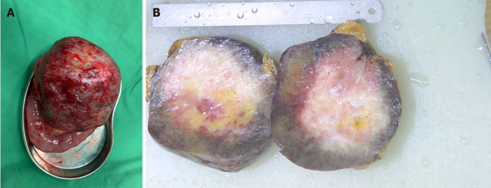
Pathology results: mesenteric see within a size of about 12 cm × 10 cm × 9 cm tumor, more open cut showed gray ash between cut red, real property, weaving, central gelatin, immunohistochemical display CD117 (-), (-) DOG-1, CD34+ (blood vessels), SMA (+), S-100 (-), SOX-10 (-), Ki-67 (+ 2%), CK (-), desmin (+), β-catenin (+) and small mesenteric fibromatosis (ligament disease) (Figure 1).
Figure 1.
Hematoxylin and eosin staining and immunohistochemistry (× 40). A: Hematoxylin and eosin staining of tumor; B: Immunohistochemistry showed positive Ki-67; C: Immunohistochemistry showed positive β-catenin.
Imaging examinations
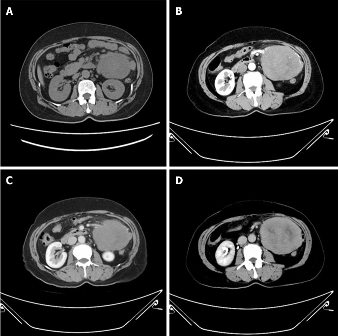
Enhanced abdominal computed tomography revealed a large circular soft tissue density shadow in the abdominal cavity of severe injury on the left side with uneven density and patchy slightly lower density shadow, which was about 106 mm × 88 mm × 110 mm. The boundary was clear, the enhanced scan was mildly to moderately enhanced and the small intestine vessels were compressed and displaced, which was considered as a possibility of left mesenchymal tumor in the middle and upper abdomen (Figure 2).
Figure 2.
Specimen of the tumor. A: A comprehensive view of the gross specimen of the tumor; B: Specimen view in section.
FINAL DIAGNOSIS
The final diagnosis was a ligamentoid fibromatosis of the small mesenteric.
TREATMENT
The patient underwent resection of a mesenteric mass. During the operation, a tumor sized 10 cm × 12 cm was seen in the mesentery at a distance of 50 cm from the flexion ligament with a complete capsule, smooth surface and no adhesion with surrounding tissues. The small intestine was closed and cut off 10 cm away from the distal and proximal ends of the tumor, and the mesenteric vessels were cut and ligated successively. The tumor was further separated by ultrasound scalpel 10 cm away from the tumor, and the tumor was completely removed with part of the small intestine (about 35 cm) (Figure 3). The superior mesenteric vessels were carefully sewn and ligated.
Figure 3.
Computed tomography. A: Abdominal computed tomography (CT) of the patient; B: Abdominal CT arterial phase; C: Abdominal CT venous phase; D: Abdominal CT delay.
OUTCOME AND FOLLOW-UP
There were no complications, and the patient was discharged 9 d later.
DISCUSSION
Ligamentoid fibromatosis usually occurs in the abdominal wall, epigastric wall, intraperitoneal cavity and mesentery. The incidence of intraperitoneal and mesenteric diseases is low, accounting for only 10%-20%. Mesenteric fibromatosis is a type of ligamentoid fibromatosis. It was first proposed by Stout[6] in 1954. Most of the lesions are located in the mesentery or retroperitoneum. The incidence of this disease is relatively low; only 2.0-4.0 per million population per year according to literature reports[7].
The cause of the disease is still controversial in the international community. It is widely believed that abdominal trauma, hormone imbalance and genetic factors can cause the disease. After comprehensive analysis, some scholars put forward three hypotheses of the etiology of the disease[8,9]: (1) Injury factors, pregnancy, childbirth, abdominal wall muscle fiber injury bleeding and hematoma formation for the occurrence of tumor to provide conditions; (2) Endocrine disorders, estrogen drugs; and (3) Chromosomal abnormalities, namely Y chromosome loss and 8/20 chromosome trisomy.
Korean scholars further discussed the genetic factors and the association between Gardner syndrome and this disease and believed that patients with Gardner syndrome have a high risk of this disease. About 1/3 of the patients with Gardner syndrome will have secondary intra-abdominal ligamentoid fibroma, and only 2% of the patients diagnosed with intra-abdominal ligamentoid fibroma can be found to have Gardner syndrome[10]. In addition, the connection between herpes virus and this disease has also been reported. It is usually divided into three types: (1) Mesenteric fibromatosis with multiple colon polyposis, bone tumors, skin cysts, Gardner syndrome, is an autosomal dominant genetic disease, has poor prognosis, high (90%-100%) postoperative recurrence rate; (2) With trauma, surgical history, hormone use, radiotherapy and other factors as inducement, postoperative recurrence rate of 10%; and (3) The cause is unknown, known as spontaneous isolated mesenteric fibromatosis, relatively rare, postoperative recurrence rate of about 10%.
From biological behavior, fibromatosis from muscle, tendon, film and fascia are rich in collagen composition. The microstructure of the tumor by pathology is benign or low-grade malignant with invasive growth. It has obvious malignant biological behavior, namely the stubborn relapse many times, but very few distant metastases. Fibromatosis hard tumors range in size from a few centimeters to fully occupying the abdominal cavity.
Most of the patients have insidious clinical symptoms without typical focal-specific symptoms. In the later stage of the disease, symptoms and signs such as pain, local discomfort, constipation, vomiting, abdominal mass, weight loss and a series of complications after internal organ compression can occur. Complications of involvement of internal organs have been reported, including small intestinal obstruction and hydronephrosis[11]. Rapidly growing tumors can also interfere with movement, compress nerves and can be found by triggering infection or invading surrounding tissues. In addition, because mesenteric fibromatosis is often secondary to Gardner syndrome and may be associated with familial adenomatous polyps and Crohn’s disease, some of the specific clinical symptoms of these diseases may also be present in affected patients.
Due to the lack of specific symptoms and signs, the diagnosis and differential diagnosis is difficult[5], and it is easy to be misdiagnosed as gastrointestinal stromal tumor, leiomyoma, leiomyosarcoma, neurofibroma, schwannoma, etc. Therefore, it is necessary to effectively make the correct diagnosis with the help of effective auxiliary examination, including abdominal ultrasound, abdominal computed tomography and abdominal magnetic resonance imaging. In the abdominal type, enhanced computed tomography is usually the first choice for the obvious nonuniform progressive enhancement during enhanced scan[12], while the abdominal wall and abdominal appearance are usually dominated by magnetic resonance imaging. Diagnostic gold standard is still the pathology examination. Microscopically, it has a large number of spindle cell hyperplasia, is rich in collagen stroma and has a large number of blood vessels. The lesion blood vessel is more muscular artery and fissure shaped with a few thin veins. The expansion of the lesion on the mesenteric, numerous myxoid matrix show a similar structure with fasciitis. Immunohistochemical characteristics were positive for -catenin (mainly nuclear) and vimentin[13-15].
At present, surgical resection is still the main method to treat mesenteric fibromatosis, and the negative cutting edge should ensure at least 2-3 cm[5]. However, the outcome of surgical resection alone is associated with a high risk of recurrence. Therefore, nonsurgical treatment also plays a role in the treatment of the disease[16]. The high recurrence rate after surgical resection may be related to the growth of the diffuse invasive tumors itself. In addition, due to the location of the tumors and the affected area, some tumors cannot be removed. In these cases, nonoperative therapy has become the first choice of treatment. It has been reported that combined therapy is effective for mesenteric fibromatosis[10]. Nonsurgical treatment has been shown to be effective in reducing the risk of recurrence. In the 2011 National Comprehensive Cancer Network guidelines for soft tissue tumors, hormone therapy (tamoxifen)[17], nonsteroidal anti-inflammatory drugs[18], low doses of interferon and systemic chemotherapy (imatinib[19] and adriamycin[20,21]) also play important roles in the treatment of mesenteric fibromatosis. In addition, postoperative local radiotherapy can also be used, but the effects and side effects of postoperative radiotherapy for mesenteric fibromatosis are still controversial in the world[16].
The recurrence rate of mesenteric fibromatosis is 25%-57%[22], and the recurrence time is more than 1 mo to 1 yr and sometimes more than 10 yrs. All these tumors are also called invasive fibromatosis. Multiple relapses may cause the lesion to involve the scope to be more extensive, but it appears cannot restrain the growth, the invasion vital organ and the crisis life. However, the overall prognosis of complete resection is good, and local recurrence should also be removed by surgery[23]. If the surgical resection is incomplete, the residual tumor can progress within a few years[24]. After the first operation, it is easy to cause adhesion in the abdominal cavity thus increasing the difficulty of the second operation. The recurrence rate and mortality after the second operation will also be greatly increased. Abdominal viscera are extensively involved and are also one of the long-term causes of death[25].
CONCLUSION
We described the surgical treatment of a ligamentoid fibromatosis of the small mesenteric. Accurate preoperative diagnosis is difficult, surgical resection is the main treatment and a variety of nonsurgical treatment methods are auxiliary. Combined treatment can effectively reduce the risk of postoperative recurrence. The prognosis is still good, and the risk of recurrence of secondary surgery is greatly increased. At present, there are few reports about this disease, and the better diagnosis and treatment methods need to be further studied.
Footnotes
Informed consent statement: All study participants, or their legal guardian, provided informed written consent prior to study enrollment.
Conflict-of-interest statement: All authors have no conflict-of-interest.
CARE Checklist (2016) statement: The authors have read the CARE Checklist (2016), and the manuscript was prepared and revised according to the CARE Checklist (2016).
Manuscript source: Unsolicited manuscript
Peer-review started: August 12, 2020
First decision: September 13, 2020
Article in press: October 1, 2020
Specialty type: Medicine, research and experimental
Country/Territory of origin: China
Peer-review report’s scientific quality classification
Grade A (Excellent): 0
Grade B (Very good): B
Grade C (Good): C
Grade D (Fair): 0
Grade E (Poor): 0
P-Reviewer: Coffin CS, Ozdemir BH S-Editor: Zhang L L-Editor: Filipodia P-Editor: Ma YJ
Contributor Information
Kai Xu, Department of General Surgery, Shougang Hospital, Peking University, Beijing 100041, China. xking55555@163.com.
Qi-Kang Zhao, Department of General Surgery, Shougang Hospital, Peking University, Beijing 100041, China.
Jing-Shan Liu, Department of General Surgery, Shougang Hospital, Peking University, Beijing 100041, China.
Dong-Hai Zhou, Department of General Surgery, Shougang Hospital, Peking University, Beijing 100041, China.
Yong-Liang Chen, Department of Hepatobiliary Surgery, Chinese People’s Liberation Army General Hospital, Beijing 100853, China.
Xing-Yi Zhu, Department of General Surgery, Shougang Hospital, Peking University, Beijing 100041, China.
Ming Su, Department of Hepatobiliary Surgery, Chinese People’s Liberation Army General Hospital, Beijing 100853, China.
Kun-Quan Huang, Department of General Surgery, Shougang Hospital, Peking University, Beijing 100041, China.
Wen Du, Department of General Surgery, Shougang Hospital, Peking University, Beijing 100041, China.
Hong-Yu Zhao, Department of General Surgery, Shougang Hospital, Peking University, Beijing 100041, China.
References
- 1.de Bree E, Zoras O, Hunt JL, Takes RP, Suárez C, Mendenhall WM, Hinni ML, Rodrigo JP, Shaha AR, Rinaldo A, Ferlito A, de Bree R. Desmoid tumors of the head and neck: a therapeutic challenge. Head Neck. 2014;36:1517–1526. doi: 10.1002/hed.23496. [DOI] [PubMed] [Google Scholar]
- 2.Duggal A, Dickinson IC, Sommerville S, Gallie P. The management of extra-abdominal desmoid tumours. Int Orthop. 2004;28:252–256. doi: 10.1007/s00264-004-0571-0. [DOI] [PMC free article] [PubMed] [Google Scholar]
- 3.Hosalkar HS, Fox EJ, Delaney T, Torbert JT, Ogilvie CM, Lackman RD. Desmoid tumors and current status of management. Orthop Clin North Am. 2006;37:53–63. doi: 10.1016/j.ocl.2005.08.004. [DOI] [PubMed] [Google Scholar]
- 4.Buitendijk S, van de Ven CP, Dumans TG, den Hollander JC, Nowak PJ, Tissing WJ, Pieters R, van den Heuvel-Eibrink MM. Pediatric aggressive fibromatosis: a retrospective analysis of 13 patients and review of literature. Cancer. 2005;104:1090–1099. doi: 10.1002/cncr.21275. [DOI] [PubMed] [Google Scholar]
- 5.Liu X, Zong S, Cui Y, Yue Y. Misdiagnosis of aggressive fibromatosis of the abdominal wall: A case report and literature review. Medicine (Baltimore) 2018;97:e9925. doi: 10.1097/MD.0000000000009925. [DOI] [PMC free article] [PubMed] [Google Scholar]
- 6.Stout AP. Juvenile fibromatoses. Cancer. 1954;7:953–978. doi: 10.1002/1097-0142(195409)7:5<953::aid-cncr2820070520>3.0.co;2-w. [DOI] [PubMed] [Google Scholar]
- 7.Heiskanen I, Järvinen HJ. Occurrence of desmoid tumours in familial adenomatous polyposis and results of treatment. Int J Colorectal Dis. 1996;11:157–162. doi: 10.1007/s003840050034. [DOI] [PubMed] [Google Scholar]
- 8.Xu HM, Han JG, Ma SZ, Zhao B, Pang GY, Wang ZJ. Treatment of massive desmoid tumour and abdominal wall reconstructed with meshes in Gardner's Syndrome. J Plast Reconstr Aesthet Surg. 2010;63:1058–1060. doi: 10.1016/j.bjps.2009.04.013. [DOI] [PubMed] [Google Scholar]
- 9.Ioannou M, Demertzis N, Iakovidou I, Kottakis S. The role of imatinib mesylate in adjuvant therapy of extra-abdominal desmoid tumors. Anticancer Res. 2007;27:1143–1147. [PubMed] [Google Scholar]
- 10.Choi JY, Kang KM, Kim BS, Kim TH. Mesenteric fibromatosis causing ureteral stenosis. Korean J Urol . 2010;51:501–504. doi: 10.4111/kju.2010.51.7.501. [DOI] [PMC free article] [PubMed] [Google Scholar]
- 11.Faria SC, Iyer RB, Rashid A, Ellis L, Whitman GJ. Desmoid tumor of the small bowel and the mesentery. AJR Am J Roentgenol. 2004;183:118. doi: 10.2214/ajr.183.1.1830118. [DOI] [PubMed] [Google Scholar]
- 12.Otero S, Moskovic EC, Strauss DC, Benson C, Miah AB, Thway K, Messiou C. Desmoid-type fibromatosis. Clin Radiol. 2015;70:1038–1045. doi: 10.1016/j.crad.2015.04.015. [DOI] [PubMed] [Google Scholar]
- 13.Fisher C, Thway K. Aggressive fibromatosis. Pathology. 2014;46:135–140. doi: 10.1097/PAT.0000000000000045. [DOI] [PubMed] [Google Scholar]
- 14.Bhattacharya B, Dilworth HP, Iacobuzio-Donahue C, Ricci F, Weber K, Furlong MA, Fisher C, Montgomery E. Nuclear beta-catenin expression distinguishes deep fibromatosis from other benign and malignant fibroblastic and myofibroblastic lesions. Am J Surg Pathol. 2005;29:653–659. doi: 10.1097/01.pas.0000157938.95785.da. [DOI] [PubMed] [Google Scholar]
- 15.Montgomery E, Torbenson MS, Kaushal M, Fisher C, Abraham SC. Beta-catenin immunohistochemistry separates mesenteric fibromatosis from gastrointestinal stromal tumor and sclerosing mesenteritis. Am J Surg Pathol. 2002;26:1296–1301. doi: 10.1097/00000478-200210000-00006. [DOI] [PubMed] [Google Scholar]
- 16.Kabra V, Chaturvedi P, Pathak KA, deSouza LJ. Mesenteric fibromatosis: a report of three cases and literature review. Indian J Cancer. 2001;38:133–136. [PubMed] [Google Scholar]
- 17.Hansmann A, Adolph C, Vogel T, Unger A, Moeslein G. High-dose tamoxifen and sulindac as first-line treatment for desmoid tumors. Cancer. 2004;100:612–620. doi: 10.1002/cncr.11937. [DOI] [PubMed] [Google Scholar]
- 18.Chao AS, Lai CH, Hsueh S, Chen CS, Yang YC, Soong YK. Successful treatment of recurrent pelvic desmoid tumour with tamoxifen: case report. Hum Reprod. 2000;15:311–313. doi: 10.1093/humrep/15.2.311. [DOI] [PubMed] [Google Scholar]
- 19.Chugh R, Wathen JK, Patel SR, Maki RG, Meyers PA, Schuetze SM, Priebat DA, Thomas DG, Jacobson JA, Samuels BL, Benjamin RS, Baker LH Sarcoma Alliance for Research through Collaboration (SARC) Efficacy of imatinib in aggressive fibromatosis: Results of a phase II multicenter Sarcoma Alliance for Research through Collaboration (SARC) trial. Clin Cancer Res. 2010;16:4884–4891. doi: 10.1158/1078-0432.CCR-10-1177. [DOI] [PubMed] [Google Scholar]
- 20.Seiter K, Kemeny N. Successful treatment of a desmoid tumor with doxorubicin. Cancer. 1993;71:2242–2244. doi: 10.1002/1097-0142(19930401)71:7<2242::aid-cncr2820710713>3.0.co;2-0. [DOI] [PubMed] [Google Scholar]
- 21.Patel SR, Evans HL, Benjamin RS. Combination chemotherapy in adult desmoid tumors. Cancer. 1993;72:3244–3247. doi: 10.1002/1097-0142(19931201)72:11<3244::aid-cncr2820721118>3.0.co;2-d. [DOI] [PubMed] [Google Scholar]
- 22.Siegel RL, Miller KD, Jemal A. Cancer statistics, 2019. CA Cancer J Clin. 2019;69:7–34. doi: 10.3322/caac.21551. [DOI] [PubMed] [Google Scholar]
- 23.Mendenhall WM, Zlotecki RA, Hochwald SN, Hemming AW, Grobmyer SR, Cance WG. Retroperitoneal soft tissue sarcoma. Cancer. 2005;104:669–675. doi: 10.1002/cncr.21264. [DOI] [PubMed] [Google Scholar]
- 24.Schulz-Ertner D, Zierhut D, Mende U, Harms W, Branitzki P, Wannenmacher M. The role of radiation therapy in the management of desmoid tumors. Strahlenther Onkol. 2002;178:78–83. doi: 10.1007/s00066-002-0900-4. [DOI] [PubMed] [Google Scholar]
- 25.Lath C, Khanna PC, Gadewar SB, Agrawal D. Inoperable aggressive mesenteric fibromatosis with ureteric fistula. Case report and literature review. Eur J Radiol. 2006;59:117–121. doi: 10.1016/j.ejrad.2005.12.036. [DOI] [PubMed] [Google Scholar]


