Zarnescu previews work by the Lerit laboratory highlighting a role for FMRP in ensuring centrocortin mRNA localization to centrosomes, which is needed for cell division and embryonic development.
Abstract
The functional importance of mRNA localization to centrosomes is unclear. Ryder et al. (2020. J. Cell Biol. https://doi.org/10.1083/jcb.202004101) identify fragile-X mental retardation protein as a regulator of centrocortin (cen) mRNA dynamics in Drosophila. Mistargeting of cen impairs division and development, indicating that cen mRNA localization to centrosomes ensures mitotic fidelity.
Finely tuned mRNA localization and translation ensure that the right amount of a specific protein is in the right place at the right time. Localized transcripts are linked to the translation of proteins that are structurally and functionally distinct from transported proteins and promote specialized, local cellular events (1). In addition, highly asymmetric cells, such as neurons, rely on mRNA localization and translation for acute responses and specialized functions such as axonal pathfinding and synaptic plasticity (2). Expanding our knowledge of the extent of mRNA localization, a high-resolution fluorescence in situ hybridization (FISH)-based screen of Drosophila embryos found that 71% of mRNAs expressed during early development exhibit specific subcellular localization patterns (3). Several mRNAs showed striking associations with centrosomes and spindles. Findings across model systems have suggested that mRNA localization to centrosomes may contribute to their complex and dynamic roles during the cell cycle. The centrosome is a dynamic, membraneless organelle comprising a pair of centrioles surrounded by a matrix of pericentriolar material (PCM; 4). Although some differences exist between vertebrates and invertebrates, the PCM is a highly dynamic entity during the cell cycle and dysregulation of PCM is linked to genomic instability and disease (4). Despite their critical importance during development and evidence that mRNAs associate with centrosomes in multiple model systems (3, 5, 6), important questions remain, including the following: (a) How do mRNA dynamics correlate with the cell cycle? (b) What are the mechanisms localizing specific mRNAs to centrosomes? (c) What is the functional significance of mRNA localization to centrosomes throughout development and in different cell types? In this issue, Ryder et al. address these questions to shed light on the functional significance of mRNA localization to centrosomes (7).
How do mRNA dynamics correlate with the cell cycle? Using single-molecule FISH (smFISH) and an automated, custom image analysis pipeline, Ryder et al. quantified the distribution of a subset of mRNAs including cyclin B (cyc B), centrocortin (cen), pericentrin-like protein (plp), small ovary (sov), and partner of inscuteable (pins). These mRNAs were previously found to localize in the proximity to spindle poles and encode proteins linked to centrosome function (3). After labeling centrosomes with GFP-Centrosomin (GFP-Cnn) and measuring the distance of individual mRNA molecules from the nearest centrosome, the authors determined the percentage of mRNA overlapping with the centrosomal surface in interphase and mitosis. A key take-away message from these detailed analyses is that cyc B, cen, plp, and pins mRNAs associated with the centrosome in a cell cycle–dependent manner, with more mRNA detected on centrosomes during interphase than in mitosis. In contrast, sov mRNA association with centrosomes was more constant throughout the cell cycle, indicating that mRNA association with centrosomes is dynamic and transcript dependent. Of note, in these experiments, plp mRNA enrichment during interphase coincided with the formation of PLP protein “flares,” suggesting a possible cotranslational mechanism, as previously shown for the zebrafish PCM component, pericentrin (8).
Interestingly, some centrosome-associated mRNAs were found as single molecules, while others were organized in granules. Consistent with a recent study (9), cen mRNA formed micron-scale granules that were enriched with centrosomes during interphase compared with metaphase (60% versus 25%) and exhibited a bias toward the mother centrosome. The latter could be explained by the mother centrosome having a larger PCM with a higher content of Cnn scaffolding protein. Ryder et al. performed the majority of their analyses at nuclear cycle (NC) 13, which has a longer interphase that facilitates sample acquisition (7). When visualized at stage NC 10, cen mRNA granules were not detected, suggesting distinct mechanisms of mRNA localization and assembly with the centrosome during development.
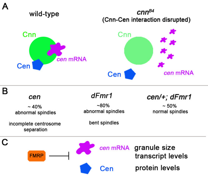
What are the mechanisms localizing specific mRNAs to centrosomes? The researchers investigated the mechanism by which cen mRNA granules are recruited to centrosomes. Using a combination of immunofluorescence and genetics, they found that when Cnn–Cen protein interactions are disrupted in the mutant cnnB4, cen mRNA granules were no longer detected. Surprisingly, Cen protein levels remained unchanged, and Ryder et al. found that cen mRNA granules were not required for steady-state levels of Cen (7). While the authors suggested that the effect of cen mRNA granule loss could be confounded by maternal Cen deposition, it is also possible that Cen protein levels remain unchanged due to protein degradation and translation cancelling each other out in the context of the cnnB4 mutation. Regardless, these experiments indicate that cen mRNA granule association with centrosomes is dependent on the interactions between Cnn and Cen proteins (Fig. 1).
Figure 1.

Model for cen mRNA recruitment and regulation. (A) cen mRNA assembles in micron-scale granules at centrosomes (marked by Cnn). Its recruitment requires intact Cnn–Cen protein interactions as evidenced by dispersal of cen mRNA granules in cnnB4, a mutant that disrupts Cnn’s association with Cen. (B) Cen levels (lower in cen and higher in dFmr1 mutants) are critical for proper spindle formation. dFmr1 mutants exhibit abnormal, bent spindles that are partially rescued by cen heterozygous mutations (cen/+; dFmr1). (C) Genetic interaction results (B) together with imaging and molecular analyses show that dFmr1 mutants exhibit larger centrosome-associated cen mRNA granules as well as higher levels of cen transcript and Cen protein, consistent with FMRP acting as a negative regulator of cen mRNA granule assembly and expression, possibly at the levels of transcript stability and translation.
Further investigations into the composition of cen mRNA granules indicated that a known translational regulator, fragile-X mental retardation protein (FMRP), but not other RNA binding proteins (i.e., ME31B, Pumilio, Egalitarian, Orb2), colocalized with cen mRNA at centrosomes. RNA immunoprecipitations showed that cen mRNA was enriched in FMRP complexes. cen mRNA granules were larger and Cen protein levels increased in dFmr1 loss-of-function mutants, consistent with FMRP acting as a negative regulator of cen mRNA stability and translation (Fig. 1). Further supporting the notion that cen mRNA is a target of FMRP, reducing cen dosage in dFmr1 mutant embryos mitigated FMRP-dependent spindle defects and lethality.
What is the functional significance of mRNA localization to centrosomes throughout development and in different cell types? Ryder et al. set out to investigate the functional consequences of cen mRNA mistargeting in Drosophila embryos (7). They generated chimeric c′UTR transgenes that target cen mRNA to the anterior pole while depleting it from the middle of the embryo. These experiments showed that the cen coding sequence was sufficient to target cen mRNA to centrosomes, consistent with previous studies (9), where they accumulated at the anterior pole and sequestered FMRP. Consequently, the loss of cen mRNA and Cen protein in the middle of the embryo caused by mistargeting to the anterior pole led to abnormal spindles in nearly half the embryos examined. cen dosage and localization to centrosomes are therefore critical for mitotic fidelity.
While Ryder et al. provide critical new insight into the mechanism and significance of cen mRNA localization to centrosomes during early Drosophila development, questions about this fascinating cellular process remain. It will be interesting to identify additional mRNAs associated with centrosomes at different developmental stages and in different cell types. What will their recruitment mechanisms and functional consequences on mitosis be? Does FMRP have other centrosomal targets? Ultimately, can these studies inform strategies to mitigate diseases caused by centrosome-linked genomic instability and mitotic failure? The quantitative imaging tools developed by Ryder et al. could help answer these questions in the future.
Acknowledgments
The Zarnescu laboratory is supported by funding from the National Institutes of Health (grant no. R01 NS091299) and the Department of Defense (grant no. W81XWH-18-1-0313).
D.C. Zarnescu is a member of the Scientific Advisory Board for the Fox Chase Chemical Diversity Center, Inc., Doylestown PA.
References
- 1.Buxbaum, A.R., et al. 2015. Nat. Rev. Mol. Cell Biol. 10.1038/nrm3918 [DOI] [PMC free article] [PubMed] [Google Scholar]
- 2.Ryder, P.V., and Lerit D.A.. 2018. Traffic. 10.1111/tra.12571 [DOI] [PMC free article] [PubMed] [Google Scholar]
- 3.Lécuyer, E., et al. 2007. Cell. 10.1016/j.cell.2007.08.003 [DOI] [Google Scholar]
- 4.Conduit, P.T., et al. 2015. Nat. Rev. Mol. Cell Biol. 10.1038/nrm4062 [DOI] [PubMed] [Google Scholar]
- 5.Alliegro, M.C., et al. 2006. Proc. Natl. Acad. Sci. USA. 10.1073/pnas.0602859103 [DOI] [Google Scholar]
- 6.Lambert, J.D., and Nagy L.M.. 2002. Nature. 10.1038/nature01241 [DOI] [Google Scholar]
- 7.Ryder, P.V., et al. 2020. J. Cell Biol. 10.1083/jcb.202004101 [DOI] [Google Scholar]
- 8.Sepulveda, G., et al. 2018. eLife. 10.7554/eLife.34959 [DOI] [Google Scholar]
- 9.Bergalet, J., et al. 2020. Cell Rep. 10.1016/j.celrep.2020.02.047 [DOI] [Google Scholar]


