The right ventricle seems to have been forgotten among heart chambers, although some studies have shown its crucial role in coronavirus disease 2019 (COVID-19). Interestingly, both its size and function are believed to be associated with cardiac complications and mortality in COVID-19.
COVID-19 is caused by severe acute respiratory syndrome coronavirus 2 (SARS-CoV-2) which spread globally after the first case was observed in Wuhan at the end of 2019.1 Recent studies suggested that COVID-19 may be accompanied by cardiac complications, including acute coronary syndrome, cardiac arrhythmia, myocarditis, pericarditis, and heart failure in nearly 20% of patients, which are ssociated with an increased risk of mortality.2 Laboratory data such as cardiac troponin as well as echocardiography parameters can be effective means of cardiac assessment in these patients. Transthoracic echocardiography (TTE) is the optimum method of cardiac imaging used in COVID-19 patients which is able to diagnose different cardiac abnormalities including haemodynamic dysfunction. Also, it is useful for the prediction of future cardiac morbidity in these patients.3
Reduced right ventricular (RV) activity is a good predictor for heart failure and cardiac mortality.4 The effect of COVID-19 on the right ventricle activity is in the main unknown. It seems that the pathophysiological pathways of COVID-19 including increased afterload after acute respiratory distress syndrome, pulmonary embolism, cytokine-negative inotropic effects, and renin–angiotensin system dysfunction are possible mechanisms for RV dysfunction in COVID-19 patients.5 A summary of some studies which have been conducted to assess RV function in COVID-19 patients is presented in Table 1.
Table 1.
Studies related to RV function in COVID-19
Li et al. performed an echocardiographic investigation in 120 cases of COVID-19. They assessed RV fractional area change, tricuspid annular plane systolic excursion, and Doppler tricuspid tissue annular velocity. In their study, RV function was categorized by right ventricular longitudinal strain (RVLS). It was concluded that patients with the lowest RVLS tertile were more susceptible to cardiac injury, and a higher mortality rate was observed in this group. In addition, patients’ mortality was associated with RVLS, RV fractional area change, and tricuspid annular plane systolic excursion.6
In the study of Szekely et al., the most common cardiac pathology in COVID-19 was RV dilatation (39%) which was more than both left ventricular (LV) diastolic dysfunction (16%) and LV systolic dysfunction (10%). In fact, it was concluded that LV systolic function is preserved in these patients, although LV diastolic function and RV function are impaired.2
In a retrospective study by Argulian et al., echocardiography parameters of 105 cases of COVID-19 were studied. RV dilation was observed in 32 patients (31%). Furthermore, RV hypokinesis and moderate to severe tricuspid regurgitation were more common in patients with RV dilation. Interestingly, there were no differences in LV size and function in patients with RV dilation in comparison with normal patients.7
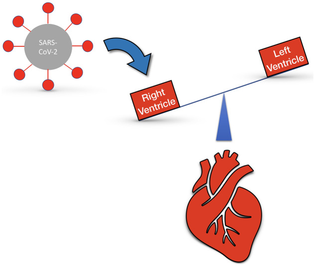
As mentioned above, RV size and function play a crucial role in the pathogenesis of cardiovascular complications in COVID-19 patients (Figure 1). Due to the severity of pulmonary involvement in COVID-19 patients, the right heart-associated parameters including RV size and function, tricuspid valve regurgitation, and pulmonary artery pressure should be considered in the cardiac evaluation of these patients. Considering the propensity of cardiologists for evaluating LV and diastolic function, the right ventricle may be forgotten. In addition, echocardiography can be used to detect any signs of RV failure in COVID-19 patients.
Figure 1.

SARS-CoV-2 may involve the right ventricle more than expected.


Conflict of interest: none declared.
References
- 1. Kasal DAB, De Lorenzo A, Tibiriçá E. COVID-19 and microvascular disease: pathophysiology of SARS-Cov-2 infection with focus on the renin–angiotensin system. Heart Lung Circ 2020; doi: 10.1016/j.hlc.2020.08.010. [DOI] [PMC free article] [PubMed] [Google Scholar]
- 2. Szekely Y, Lichter Y, Taieb P, Banai A, Hochstadt A, Merdler I, Gal Oz A, Rothschild E, Baruch G, Peri Y. The spectrum of cardiac manifestations in coronavirus disease 2019 (COVID-19)—a systematic echocardiographic study. Circulation 2020;142:342–353. [DOI] [PMC free article] [PubMed] [Google Scholar]
- 3. Mahmoud-Elsayed HM, Moody WE, Bradlow WM, Khan-Kheil AM, Senior J, Hudsmith LE, Steeds RP. Echocardiographic findings in patients with COVID-19 pneumonia. Can J Cardiol 2020;36:1203–1207. [DOI] [PMC free article] [PubMed] [Google Scholar]
- 4. Khani M, Hosseintash M, Foroughi M, Naderian M, Khaheshi I. Assessment of the effect of off-pump coronary artery bypass (OPCAB) surgery on right ventricle function using strain and strain rate imaging. Cardiovasc Diagn Ther 2016;6:138–143. [DOI] [PMC free article] [PubMed] [Google Scholar]
- 5. Park JF, Banerjee S, Umar S. In the eye of the storm: the right ventricle in COVID-19. Pulm Circ 2020;10:doi: 10.1177/2045894020936660. [DOI] [PMC free article] [PubMed] [Google Scholar]
- 6. Li Y, Li H, Zhu S, Xie Y, Wang B, He L, Zhang D, Zhang Y, Yuan H, Wu C. Prognostic value of right ventricular longitudinal strain in patients with COVID-19. JACC: Cardiovasc Imaging 2020;doi: 10.1016/j.jcmg.2020.04.014. [DOI] [PMC free article] [PubMed] [Google Scholar]
- 7. Argulian E, Sud K, Vogel B, Bohra C, Garg VP, Talebi S, Lerakis S, Narula J. Right ventricular dilation in hospitalized patients with COVID-19 infection. JACC: Cardiovasc Imaging 2020;doi: 10.1016/j.jcmg.2020.05.010. [DOI] [PMC free article] [PubMed] [Google Scholar]


