Abstract
Purpose
Topical carbonic anhydrase inhibitors (CAIs) can influence retinal fluid distribution, but their role in treating central serous chorioretinopathy (CSCR) has not been studied. We examined the efficacy of a topical CAI (dorzolamide) in treating chronic CSCR.
Methods
Prospective, nonrandomized, controlled intervention study of patients with chronic CSCR of at least 3 months duration. Observed controls (n = 15) were recruited consecutively from 2016 to 2017; treated cases (n = 18) were recruited from 2018 to 2019. Controls were observed without active intervention, whereas treated cases were treated with topical dorzolamide for 3 months. The study end points were change in central macular thickness (CMT), change in best corrected visual acuity (BCVA), and proportion of eyes achieving complete resolution of subretinal fluid (SRF). All end points were at 3 months.
Results
Treated patients who received topical CAI had greater reduction in CMT (−145.6 µm, 95% confidence interval [CI] −170.5 to −120.7) compared to observed controls (−45.1 µm, 95% CI −65.3 to −25.1) at the main study end point of 3 months (P = 0.015). A higher proportion of treated patients achieved complete resolution of SRF compared to observed controls (77.8% vs. 40.0%, P = 0.04) at 3 months. However, change in BCVA at 3 months was similar in both groups (P = 0.12).
Conclusions
Topical CAI resulted in more rapid reduction of CMT compared to observation. These results, if confirmed in other studies, suggest topical CAI may be a viable treatment option for patients with chronic CSCR.
Translational Relevance
Topical CAI is used to treat a number of retinal disorders, and may be a novel treatment option for chronic CSCR.
Keywords: central serous chorioretinopathy, carbonic anhydrase, treatment, cohort
Introduction
Central serous chorioretinopathy (CSCR) is the fourth most common cause of retinal vision loss.1–3 It is characterized by accumulation of fluid in the subretinal space. Acute CSCR usually resolves within 3 months, whereas chronic CSCR persists beyond 3 months and can continue for years.1,2 Chronic CSCR can be a sight threatening disease with legal blindness ensuing in over 10% of patients.4 The underlying pathophysiology is believed to involve retinal pigment epithelium (RPE) dysfunction resulting in reversal of normal fluid flow across the retinal layers.2,3 This RPE dysfunction may be precipitated by an increase in endogenous or exogenous corticosteroids.5,6
A number of treatments have been proposed for chronic CSCR.1,3 The best evidence is for indocyanine green angiography guided photodynamic therapy (PDT), which is supported by randomized clinical trial evidence7–9 and results in fluid resolution in more than half of patients by 6 to 8 weeks.10 PDT is effective but requires specialized equipment, and there is a small risk of complications.11 Other interventions that have been studied include thermal laser,12 micropulse diode laser,10 mineralocorticosteroid receptor antagonists,13–15 anti-vascular endothelial growth factor (VEGF) intravitreal injections,16 and use of other oral agents, such as rifampicin17 and mifepristone.18 There is limited evidence for these and other interventions.3
Carbonic anhydrase inhibitors (CAIs) may improve subretinal fluid absorption through the RPE.19–21 Our group22 and others23 have previously shown that topical CAIs are effective at managing subretinal and intraretinal fluid accumulation in retinal diseases with RPE dysfunction, such as retinitis pigmentosa and x-linked retinoschisis.24 A small study of oral CAIs,25 and another of combined topical CAI and nonsteroidal anti-inflammatory agents,26 has shown promising findings in managing acute CSCR. However, the efficacy of CAIs in chronic CSCR is unknown. Further, topical dorzolamide has greater CAI activity in eye tissues than oral acetazolamide,27 suggesting it may have greater efficacy. We therefore hypothesized that topical CAIs could be effective in treating chronic CSCR and conducted a prospective, nonrandomized, open-label, controlled study to investigate the efficacy and safety of topical dorzolamide in treatment of chronic CSCR.
Methods
Patients were prospectively recruited from a tertiary retinal practice in Sydney, Australia, between June 2016 and December 2019. Observed controls were consecutively recruited between June 2016 and December 2017 and offered observation without any active intervention, whereas cases were consecutively recruited from January 2018 to December 2019 and offered topical intervention with dorzolamide. Both observed controls and treated patients were instructed to cease any exogenous precipitating factors, such as steroid use. Patients who declined the intervention during the case recruitment period were eligible to be controls if they consented for their data to be prospectively collected. Patients and controls were age matched. Patients provided signed informed consent and the study was conducted according to the tenants of the Declaration of Helsinki. Ethics approval was provided by the University of Sydney human research ethics committee.
Inclusion criteria for the study followed currently accepted criteria for chronic CSCR2,3,5 and were (1) consultant confirmed diagnosis of chronic CSCR confirmed on fundus fluorescein angiography (FFA) and indocyanine green angiography (ICGA). This required the presence of one or more regions of active leakage (focal or diffuse) at the level of the RPE on FFA, which corresponded to one or more regions of choroidal hyperfluorescence on ICGA; (2) subretinal fluid (SRF) on spectral domain optical coherence tomography (SD-OCT); (3) subjective visual symptoms of reduced acuity, micropsia, metamorphopsia, or other symptoms attributed to CSCR for at least 3 months (12 weeks) duration prior to entering the study; (4) treatment naïve with no previous treatments for CSCR; (4) absence of other ocular pathology, such as choroidal neovascularization, polypoidal choroidal vasculopathy, diabetic retinopathy, diabetic macular edema, or retinal vein occlusion; and (5) no ocular surgery in the last 6 months. If FFA/ICGA could not be performed due to patient allergy or other contraindications, a typical CSCR appearance on SD-OCT with enhanced depth imaging (EDI)28 and classic fundus autofluorescence signs2 was accepted for diagnosing chronic CSCR. Recurrent patients with CSCR were eligible provided the current episode was over 3 months in duration. Acute CSCR was defined as onset of symptoms and resolution of SRF within 3 months.
All treated patients and observed controls had medical and ocular history documented, best corrected visual acuity (BCVA) and intraocular pressure (IOP) measured, and slit-lamp clinical examination conducted at each visit. The mandatory visits were at baseline, 1 month, and 3 months, with other visits as per the consultants’ routine management. Color fundus photographs, FFA, and ICGA were performed on the Zeiss Visucam 524 (Zeiss, Switzerland). SD-OCT with EDI and fundus autofluorescence were performed on the Heidelberg Spectralis Scanning Laser Ophthalmoscope (Heidelberg Engineering GmbH, Heidelberg, Germany). The HRA2/Spectralis Family Acquisition Module 5.4.7.0 was used. Patients underwent the “fast macular” scan preset, which uses a 25-line horizontal raster scan covering 20 degrees × 20 degrees centered on the fovea. The raster scans comprised 25 B-scans with 768 A-scans per B-scan. To ensure the same region was scanned at each visit, a proprietary software algorithm was used to align images (TruTrack Active Eye Tracking; Heidelberg Engineering GmbH). Central macular thickness (CMT) was read off the central subfield thickness display, which covers the central 1 mm2 of the 9 subfields early treatment diabetic retinopathy study (ETDRS) grid macula map.
All outcomes were measured at 1 and 3 months. The primary outcome measure was change in CMT from baseline to 3 months. The main secondary outcomes measures were proportion of eyes with complete resolution of SRF at 3 months, and change in BCVA from baseline to 3 months. Data on adverse events was also collected. Treated cases received the open label intervention of topical dorzolamide applied 4 times a day to the affected eye with punctal occlusion for 10 seconds. The dosing schedule was chosen based on prior experience with using topical dorzolamide to treat retinal conditions.22,23 Topical CAI therapy was continued at a dose of 4 times a day for 3 months, after which it was reduced to 3 times a day for 1 week, then 2 times a day for 1 week, then once a day for a week, after which topical therapy was ceased. Observed controls underwent close observation without active intervention. Rescue treatment of PDT with verteporfin was allowed if the patient's visual acuity worsened by 10 ETDRS letters or more due to the CSCR, or if CMT increased by >50%. If rescue treatment for chronic CSCR was required in either patients or controls, the patient exited the study at the time the standard treatment of PDT with verteporfin was delivered.29
Statistical Analyses
Baseline characteristics of patients were compared using the Student's t-test and χ2 tests. Outcomes at 1 and 3 months were compared with mixed models. Multivariable models adjusting for age and duration of CSCR were constructed. Chi-square test was used to compare the percentage of patients who achieved complete resolution of SRF at 3 months. Statistical analyses were performed using SAS version 9.1 (SAS Institute, Cary, NC, USA). P values, adjusted means, and 95% confidence intervals (CIs) are reported.
Results
This study recruited 18 patients who were treated with dorzolamide in one eye, and 15 patients who provided control data. At baseline, treated patients and observed controls were similar in terms of age, gender distribution, and underlying precipitant of CSCR (Table 1). The average age of treated patients was 51.3 years (SD ±12.7 years) whereas that of observed controls was 47.0 years (±13.4). The most common underlying precipitant was work and/or personal stress, whereas 20% of observed controls and 17% of treated patients had exogenous steroid exposure, which was ceased. Eighty-seven percent of observed controls and 89% of treated patients had FFA/ICGA confirmed CSCR (P = 0.90). Mean duration of CSCR was similar in both groups prior to enrollment (8.8 ± 5.9 vs. 5.8 ± 5.7 months for observed controls and treated patients, P = 0.19).
Table 1.
Baseline Characteristics of Patients With Central Serous Chorioretinopathy
| Characteristics | Observed Controls (N = 15) | Treated Patients (N = 18) | P Value |
|---|---|---|---|
| Age, y (±SD) | 51.3 (±12.7) | 47.0 (±13.4) | 0.39 |
| Male, % | 87 | 72 | 0.31 |
| Right eyes, % | 53 | 44 | 0.99 |
| FFA/ICGA confirmed | 87% | 89% | 0.90 |
| Mean duration of CSCR prior to enrollment, months (range) | 8.8 (3–24) | 5.8 (3–24) | 0.19 |
| Initial BCVA, mean (±SD) | 77.9 (±11.3) | 78.5 (±13.4) | 0.91 |
| Initial CMT, mean µm (SD) | 370.5 (±97.4) | 427.8 (±107.4) | 0.07 |
| Initial IOP, mm Hg (SD) | 16.8 (±2.1) | 16.7 (±1.9) | 0.88 |
| Initial choroidal thickness, mean µm (SD) | 404.0 (±97.4) | 437.5 (±88.3) | 0.37 |
BCVA, best corrected visual acuity; CMT, central macular thickness; CSCR, central serous chorioretinopathy; FFA, fundus fluorescein angiography; ICGA, indocyanine green angiography; IOP, intraocular pressure; SD, standard deviation.
Initial baseline BCVA was similar in observed controls and treated cases (77.9 ETDRS letters and 78.5, respectively, P = 0.91; Table 2). There was no significant difference in change in BCVA in observed controls and treated patients at 1 or 3 months (Table 2). Initial CMT at baseline was similar in treated controls and observed patients (370.5 µm and 427.8 µm, respectively, P = 0.07). At 1 and 3 months, treated patients had a greater reduction in CMT compared to observed controls (Table 2). At 3 months, patients who received topical CAI had a greater reduction in CMT (−145.6 µm, 95% CI −170.5 to −120.7) compared to observed controls (−45.1 µm, 95% CI −65.3 to −25.1, P = 0.015). A higher proportion of treated cases achieved complete resolution of SRF compared to observed controls (77.8% vs. 40.0%, P = 0.04). IOP was significantly reduced in treated cases at 1 and 3 months compared to controls (3 month change in IOP: −2.4 mm Hg vs. +0.9 mm Hg, respectively, P = 0.003). There was no significant change in choroidal thickness in either group at 1 or 3 months (Table 2).
Table 2.
Change in Outcomes in Treated Cases and Observed Controls
| Observed Controls (95% Confidence Intervals) | Treated Cases (95% Confidence Intervals) | P Value | |
|---|---|---|---|
| Change in BCVA at: | |||
| 1 mo | −0.6 (−1.6 to 0.4) | 1.2 (0.07 to 2.3) | 0.15 |
| 3 mo | −0.3 (−1.7 to 1.1) | 2.2 (0.8 to 3.6) | 0.12 |
| Change in CMT at: | |||
| 1 mo | −24.1 (−34.0 to −14.2) | −94.3 (−131.4 to −57.7) | 0.008 |
| 3 mo | −45.1 (−65.3 to −25.1) | −145.6 (−170.5 to −120.7) | 0.015 |
| % achieved complete resolution at 3 mo | 6 (40.0%) | 14 (77.8%) | 0.04 |
| Change in IOP at: | |||
| 1 mo | 1.1 (0.2 to 1.9) | −2.9 (−4.0 to −1.8) | 0.0009 |
| 3 mo | 0.9 (−0.06 to 1.9) | −2.4 (−3.5 to −1.3) | 0.003 |
| Change in choroidal thickness at: | |||
| 1 mo | 0.8 (−4.4 to 6.0) | −13.3 (−25.1 to −1.5) | 0.20 |
| 3 mo | −0.6 (−6.3 to 5.1) | −13.4 (−25.1 to −1.7) | 0.24 |
BCVA, best corrected visual acuity; CMT, central macular thickness; IOP, intraocular pressure; SD, standard deviation.
Table 3 shows the results of multivariable modeling of outcomes adjusted for age and duration of CSCR prior to enrollment. After adjustment, there was no difference in change in BCVA at 1 or 3 months between treated patients and observed controls. The reduction in CMT remained significantly greater in treated patients than observed controls at both 1 and 3 months after adjustment, with a reduction in CMT of −138.0 µm (95% CI −147.1 to −129.3) in treated patients, compared to −51.7 µm (95% CI −60.2 to −43.2, P = 0.03) in observed controls at 3 months. No clinically significant ocular or systemic adverse effects were reported. No patients required rescue treatment with PDT during the study period.
Table 3.
Multivariable Adjusted Change in Outcome Variables
| Observed Controls (95% Confidence Interval) | Treated Cases (95% Confidence Interval) | P Value | |
|---|---|---|---|
| Change in BCVA at: | |||
| 1 mo | −0.8 (−1.1 to −0.5) | 1.4 (1.1 to 1.7) | 0.10 |
| 3 mo | −0.4 (−0.8 to −0.03) | 2.3 (1.8 to 2.7) | 0.12 |
| Change in CMT at: | |||
| 1 mo | −27.4 (−32.5 to −22.3) | −90.6 (−96.2 to −85.0) | 0.01 |
| 3 mo | −51.7 (−60.2 to −43.2) | −138.0 (−147.1 to −129.3) | 0.03 |
Values are adjusted for age and duration of CSCR prior to enrolment.
BCVA, best corrected visual acuity; CMT, central macular thickness.
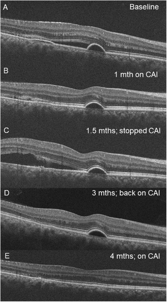
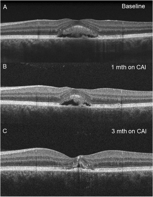
Figure 1 shows the response of a patient to topical CAI therapy with a good response at month 1. The patient ceased therapy and within 2 weeks (6 weeks after baseline) the CSCR recurred. Treatment was restarted and the CSCR mostly resolved by 3 months, and treatment was weaned over the next month. The CSCR was fully resolved by 4 months. Figure 2 shows the response of a patient to topical CAIs, with reduced SRF at month 1 and resolution by month 3, following which the treatment was weaned over month 4.
Figure 1.

OCT images from the right eye of a 38-year-old male patient showing response of central serous chorioretinopathy to topical carbonic anhydrase inhibitor (CAI) therapy. (A) Initial OCT appearance at baseline with two small pigment epithelial detachments and subretinal fluid. (B) Resolution of most of the subretinal fluid after 1 month of CAI. (C) Recurrence of subretinal fluid at 6 weeks (1.5 months) after baseline when the patient stopped treatment and represented with symptom recurrence. (D) CAI therapy was reinstated and the subretinal fluid and pigment epithelial detachment had almost fully resolved by 3 months. CAI treatment was weaned. (E) At 4 months following presentation, the subretinal fluid and pigment epithelial detachment had resolved fully and CAI therapy was ceased. The patient had no further recurrences.
Figure 2.

OCT images from the left eye of a 43-year-old female patient showing response of central serous chorioretinopathy to topical carbonic anhydrase inhibitor (CAI) therapy. (A) Initial OCT appearance at baseline with central subretinal fluid. (B) Reduction in subretinal fluid after 1 month on topical CAI therapy. (C) Resolution of subretinal fluid after 3 months on topical CAI therapy. The patient was weaned off CAI over the next month with no recurrence.
Discussion
In this study, we show that a topical CAI (dorzolamide) produced superior anatomic outcomes compared to observation in treating chronic CSCR. Patients who received topical dorzolamide had a larger reduction in CMT and were more likely to achieve complete resolution of SRF at 3 months. However, there was no difference in BCVA gains compared to observation in controls. The intervention was well-tolerated with no ocular or systemic adverse effects reported. To our knowledge, this is the first report to show that topical CAI may be a potential new treatment option for CSCR.
These results are consistent with the known effects of CAI on subretinal fluid. Oral CAIs have been shown in two studies to accelerate reduction of subretinal fluid in CSCR.25,26 CAIs, both oral and topical, are also effective in reducing cystoid macular edema in similar conditions where there is a breakdown in outer blood-retinal-barrier function of the RPE, such as in retinitis pigmentosa22,23 and x-linked retinoschisis.24 The mechanism through which CAIs exert their beneficial effect is unclear but may be related to the restoration of normal direction of fluid flow across the RPE.19,21,30 The underlying dysfunction in CSCR may be a localized reversal of fluid transfer from the RPE into the subretinal space rather than vice versa.1–3 We hypothesize that topical CAIs inhibit the RPE-membrane bound carbonic anhydrase enzyme IV, causing acidification and decreased pH in the subretinal space.19 As RPE polarity is influenced by pH gradients, this restores the normal polarity of RPE and the normal trans-RPE direction of fluid flow out of the subretinal space and into the choroidal circulation. This reduces the subretinal fluid in CSCR.21,30 The RPE dysfunction may be triggered by changes in choroidal vessel permeability and CSCR is now believed to be part of the spectrum of pachychoroid disease.31 This is a newly described entity characterized by attenuation of the choriocapillaris and dilated deep choroidal veins that are associated with progressive dysfunction of the overlying RPE and occurs in choroidal diseases, such as CSCR and polypoidal choroidal vasculopathy.31
The anatomic improvement with topical CAI is modest compared with PDT. Studies have shown PDT treatment results in 91 to 100% complete SRF resolution after 6 months,8,32 compared to 77% in our study. PDT also improves BCVA by 4 to 5 ETDRS letters,7,10 which was not observed with topical CAIs. Nonetheless, topical therapy with CAIs may be a noninvasive and safe initial option for patients with CSCR that has not resolved after 3 months, after which more invasive options, such as PDT could be considered.
PDT works by causing occlusion of the choriocapillaris and reducing choroidal hyperpermeability and extravascular leakage.7,10 In contrast, topical CAI improves the pumping function of RPE leading to increased egress of SRF into the choroidal circulation. Although SRF resolved more rapidly with topical CAIs in our study, BCVA was not significantly different. This may be related to the small study sample, which limited power to detect a difference. BCVA in treated patients improved by +2.2 ETDRS letters (95% CI 0.8 to 3.6) at 3 months, compared to a loss of −0.3 letters (95% CI −1.7 to 1.1) in observed controls (P = 0.12). Our study had 33.5% power to detect a difference of this magnitude, suggesting larger sample sizes are needed to detect differences in BCVA.
A strength of this study is the availability of a comparison group as there is a high rate of spontaneous resolution in CSCR. The results should be interpreted while considering a few limitations. The major limitation is that treatment allocation was not randomized and there may be unknown confounders. We attempted to control for confounders by age matching and adjusting for age and duration of CSCR prior to entering the study. We examined short-term outcomes, so long-term outcomes (e.g. recurrence rates), are unknown. Further follow-up of this cohort may provide this additional data. The study sample size is relatively small. Nonetheless, this sample was sufficiently powered to detect an effect, so larger samples may provide more accurate estimates but would not change the overall conclusion of the study. The findings in this study are thus best viewed as providing pilot data for subsequent larger, prospective studies, and randomized controlled trials. Finally, the definition of chronic CSCR3 is evolving and some of the patients may have had acute CSCR, which has a higher likelihood of spontaneous resolution. We followed existing guidelines on diagnosing chronic CSCR as duration of over 3 months and characteristic FFA and ICGA signs,2,3,5 but future studies may also consider including autofluorescence changes to further tighten diagnostic criteria for chronic CSCR.
In conclusion, use of topical CAIs resulted in more rapid reduction of CMT and a higher proportion of patients with complete resolution of SRF in chronic CSCR compared to observation. These results, if confirmed in other studies, suggest topical CAI may be a viable, novel treatment option for patients with chronic CSCR.
Acknowledgments
Disclosure: G. Liew, None; I.-V. Ho, None; S. Ong, None; B. Gopinath, None; P. Mitchell, None
References
- 1. Quin G, Liew G, Ho IV, Gillies M, Fraser-Bell S. Diagnosis and interventions for central serous chorioretinopathy: review and update. Clin Exp Ophthalmol. 2013; 41: 187–200. [DOI] [PubMed] [Google Scholar]
- 2. Liew G, Quin G, Gillies M, Fraser-Bell S. Central serous chorioretinopathy: a review of epidemiology and pathophysiology. Clin Exp Ophthalmology. 2013; 41: 201–214. [DOI] [PubMed] [Google Scholar]
- 3. van Rijssen TJ, van Dijk EHC, Yzer S, et al.. Central serous chorioretinopathy: towards an evidence-based treatment guideline. Prog Retin Eye Res. 2019; 73: 100770. [DOI] [PubMed] [Google Scholar]
- 4. Mrejen S, Balaratnasingam C, Kaden TR, et al.. Long-term visual outcomes and causes of vision loss in chronic central serous chorioretinopathy. Ophthalmology. 2019; 126: 576–588. [DOI] [PubMed] [Google Scholar]
- 5. Schellevis RL, Altay L, Kalisingh A, et al.. Elevated steroid hormone levels in active chronic central serous chorioretinopathy. Invest Ophthalmol Vis Sci. 2019; 60: 3407–3413. [DOI] [PubMed] [Google Scholar]
- 6. Arndt C, Sari A, Ferre M, et al.. Electrophysiological effects of corticosteroids on the retinal pigment epithelium. Invest Ophthalmol Vis Sci. 2001; 42: 472–475. [PubMed] [Google Scholar]
- 7. van Rijssen TJ, van Dijk EHC, Scholz P, et al.. Focal and diffuse chronic central serous chorioretinopathy treated with half-dose photodynamic therapy or subthreshold micropulse laser: PLACE Trial Report No. 3. Am J Ophthalmol. 2019; 205: 1–10. [DOI] [PubMed] [Google Scholar]
- 8. Liu HY, Yang CH, Yang CM, Ho TC, Lin CP, Hsieh YT. Half-dose versus half-time photodynamic therapy for central serous chorioretinopathy. Am J Ophthalmol. 2016; 167: 57–64. [DOI] [PubMed] [Google Scholar]
- 9. Chan WM, Lai TY, Lai RY, Liu DT, Lam DS. Half-dose verteporfin photodynamic therapy for acute central serous chorioretinopathy: one-year results of a randomized controlled trial. Ophthalmology. 2008; 115: 1756–1765. [DOI] [PubMed] [Google Scholar]
- 10. van Dijk EHC, Fauser S, Breukink MB, et al.. Half-dose photodynamic therapy versus high-density subthreshold micropulse laser treatment in patients with chronic central serous chorioretinopathy: the PLACE Trial. Ophthalmology. 2018; 125: 1547–1555. [DOI] [PubMed] [Google Scholar]
- 11. Lai FH, Ng DS, Bakthavatsalam M, et al.. A multicenter study on the long-term outcomes of half-dose photodynamic therapy in chronic central serous chorioretinopathy. Am J Ophthalmol. 2016; 170: 91–99. [DOI] [PubMed] [Google Scholar]
- 12. Robertson DM, Ilstrup D. Direct, indirect, and sham laser photocoagulation in the management of central serous chorioretinopathy. Am J Ophthalmol. 1983; 95: 457–466. [DOI] [PubMed] [Google Scholar]
- 13. Rahimy E, Pitcher JD 3rd, Hsu J, et al.. A randomised double blind placebo controlled pilot study of eplerenone for treatment of central serous chorioretinopathy. Retina (Philadelphia, PA). 2018; 38: 962–969. [DOI] [PubMed] [Google Scholar]
- 14. Lee JH, Lee SC, Kim H, Lee CS. Comparison of short term efficacy between oral spironolactone and photodynamic therapy for central serous chorioretinopathy. Retina (Philadelphia, PA). 2019; 39: 127–133. [DOI] [PubMed] [Google Scholar]
- 15. Bousquet E, Zhao M, Daruich A, Behar-Cohen F. Mineralocorticoid antagonists in the treatment of central serous chorioretinopathy: review of the pre-clinical and clinical evidence. Exp Eye Res. 2019; 187: 107754. [DOI] [PubMed] [Google Scholar]
- 16. Chung YR, Kim JW, Song JH, Park A, Kim MH. Twelve month efficacy of intravitreal bevacizumab for chronic recurrent central serous chorioretinopathy. Retina (Philadelphia, PA). 2019; 39: 134–142. [DOI] [PubMed] [Google Scholar]
- 17. Shulman S, Goldenberg D, Schwartz R, et al.. Oral Rifampin treatment for longstanding chronic central serous chorioretinopathy. Graefes Arch Clin Exp Ophthalmol. 2016; 254: 15–22. [DOI] [PubMed] [Google Scholar]
- 18. Nielsen JS, Jampol LM.. Oral mifepristone for chronic central serous chorioretinopathy. Retina (Philadelphia, PA). 2011; 31: 1928–1936. [DOI] [PubMed] [Google Scholar]
- 19. Wolfensberger TJ, Mahieu I, Jarvis-Evans J, et al.. Membrane-bound carbonic anhydrase in human retinal pigment epithelium. Invest Ophthalmol Vis Sci. 1994; 35: 3401–3407. [PubMed] [Google Scholar]
- 20. Wolfensberger TJ. The role of carbonic anhydrase inhibitors in the management of macular edema. Doc Ophthalmol. 1999; 97: 387–397. [DOI] [PubMed] [Google Scholar]
- 21. Cox SN, Hay E, Bird AC. Treatment of chronic macular edema with acetazolamide. Arch Ophthalmol (Chicago, Ill: 1960). 1988; 106: 1190–1195. [DOI] [PubMed] [Google Scholar]
- 22. Liew G, Moore AT, Webster AR, Michaelides M. Efficacy and prognostic factors of response to carbonic anhydrase inhibitors in management of cystoid macular edema in retinitis pigmentosa. Invest Ophthalmol Vis Sci. 2015; 56: 1531–1536. [DOI] [PubMed] [Google Scholar]
- 23. Grover S, Fishman GA, Fiscella RG, Adelman AE. Efficacy of dorzolamide hydrochloride in the management of chronic cystoid macular edema in patients with retinitis pigmentosa. Retina (Philadelphia, PA). 1997; 17: 222–231. [DOI] [PubMed] [Google Scholar]
- 24. Zhang L, Reyes R, Lee W, et al.. Rapid resolution of retinoschisis with acetazolamide. Doc Ophthalmol. 2015; 131: 63–70. [DOI] [PMC free article] [PubMed] [Google Scholar]
- 25. Pikkel J, Beiran I, Ophir A, Miller B. Acetazolamide for central serous retinopathy. Ophthalmology. 2002; 109: 1723–1725. [DOI] [PubMed] [Google Scholar]
- 26. Wuarin R, Kakkassery V, Consigli A, et al.. Combined topical anti-inflammatory and oral acetazolamide in the treatment of central serous chorioretinopathy. Optom Vis Sci. 2019; 96: 500–506. [DOI] [PubMed] [Google Scholar]
- 27. Terashima H, Suzuki K, Kato K, Sugai N. Membrane-bound carbonic anhydrase activity in the rat corneal endothelium and retina. Jpn J Ophthalmol. 1996; 40: 142–153. [PubMed] [Google Scholar]
- 28. Yang L, Jonas JB, Wei W. Optical coherence tomography-assisted enhanced depth imaging of central serous chorioretinopathy. Invest Ophthalmol Vis Sci. 2013; 54: 4659–4665. [DOI] [PubMed] [Google Scholar]
- 29. Mehta PH, Meyerle C, Sivaprasad S, Boon C, Chhablani J. Preferred practice pattern in central serous chorioretinopathy. Br J Ophthalmol. 2017; 101: 587–590. [DOI] [PubMed] [Google Scholar]
- 30. Wolfensberger TJ, Chiang RK, Takeuchi A, Marmor MF. Inhibition of membrane-bound carbonic anhydrase enhances subretinal fluid absorption and retinal adhesiveness. Graefes Arch Clin Exp Ophthalmol. 2000; 238: 76–80. [DOI] [PubMed] [Google Scholar]
- 31. Cheung CMG, Lee WK, Koizumi H, Dansingani K, Lai TYY, Freund KB. Pachychoroid disease. Eye (London, England). 2019; 33: 14–33. [DOI] [PMC free article] [PubMed] [Google Scholar]
- 32. Cheng CK, Chang CK, Peng CH. Comparison of photodynamic therapy using half-dose of verteporfin or half-fluence of laser light for the treatment of chronic central serous chorioretinopathy. Retina (Philadelphia, PA). 2017; 37: 325–333. [DOI] [PubMed] [Google Scholar]


