Abstract
Objective:
The aim of this study is to introduce a novel program of panoramic radiography that shows 41 multilayer images from the buccal to lingual aspects in a region of interest, and to evaluate the effectiveness of this program for detecting proximal caries.
Methods:
In total, 480 premolars and molars on 30 panoramic radiographs taken with the multilayer imaging program were included in this study. The presence or absence of caries in 960 proximal surfaces was assessed by three experienced oral and maxillofacial radiologists as a consensus-based gold-standard. Two general dentists evaluated and scored proximal caries on 980 surfaces on panoramic radiographs with and without the multilayer imaging program. The two general dentists’ scores were compared with the gold-standard, and were analyzed for sensitivity, specificity, positive predictive value, negative predictive value, and area under the receiver operating characteristic (ROC) curve with and without using the program. The area under the ROC curve was analyzed using STATA/SE 13.1.
Results:
When the multilayer imaging program was used for panoramic radiography, the inter- and intraobserver agreement of the two general dentists improved. All values, including the area under the ROC curve, were higher when the multilayer imaging program was used than when it was not used. The area under the ROC curve showed a statistically significant improvement only in Observer 1, whose diagnostic ability was poorer than that of Observer 2.
Conclusions:o
This multilayer imaging program might help the inexperienced dentist to improve the diagnostic accuracy of proximal caries. If further studies would be performed in various clinical application, it could be useful when intraoral radiography taking is difficult for reasons such as mouth-opening limitations and the gag reflex of the patients.
Keywords: Radiography, Panoramic; Dental caries; Diagnosis, Oral
Introduction
Dental caries is one of the most common diseases in dentistry. The delayed diagnosis of dental caries may lead to pulp infection, periapical abscess, and tooth loss, which in turn increases treatment costs and time. The detection of caries is usually based on a clinical examination and radiographic evaluation.1 Occlusal caries are easily detectable through clinical examinations, whereas it is nearly impossible to identify proximal caries unless cavitation has occurred. Therefore, the radiographic evaluation of proximal caries is especially important.
Panoramic radiography is widely used as a primary radiological diagnostic technique in dentistry.2–4 It is primarily considered as a screening method for the entire dentition, maxilla, or mandible. However, the images do not provide high resolution, and additional intraoral radiography is required for some dental diseases, especially caries detection.
Since dental X-ray equipment has been digitized, extensive efforts have been made to develop special programs to enhance the diagnostic capability using special image acquisition and processing techniques. However, the development of imaging techniques in panoramic radiography was relatively tardy compared to other imaging modalities such as cone-beam CT (CBCT).
Recently, a novel image acquisition technique of panoramic radiography was developed that provides sequential multilayer imaging, from the buccal to lingual aspects. This imaging technique provides multilayer view of buccolingual depth, similar to the panoramic reconstructed view of CBCT images. Using 41 sequential imaging layers within the region of interest (ROI), it is possible to observe the buccolingual relationship between the tooth and its surrounding structure. This novel technique enables the localization of impacted supernumerary teeth and adjacent permanent teeth, discrimination between buccal and lingual root canals with regard to endodontic treatment, and examination of the proximal area of the crown without overlap of adjacent surfaces. However, the efficiency of this imaging program for dental diseases has not yet been studied in clinical trials. Thus, the present study aimed to introduce a novel imaging program of panoramic radiography that shows 41 multilayer images from the buccal to lingual aspects in an ROI, and to evaluate the effectiveness of this program for detecting proximal caries.
Methods and materials
Data and image acquisition
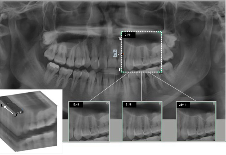
The randomized panoramic radiographs of 348 patients (age range, 20–74 years; 196 female, 152 male) who visited Yonsei University Dental Hospital for the purpose of dental disease diagnosis and treatment from October 2017 to February 2018 were randomly included. This study was approved by the Institutional Review Board of Yonsei University Dental Hospital (IRB no. 2-2017-0008). All images were obtained with a Pax-i plus device (Vatech Co., Hwaseung Si, Korea) with the following conditions: 70 kVp, 5 mA, and 10.1 s, according to the manufacturer guidelines. All image acquisitions were performed with the special program described above, which is named Insight Navi (Vatech Co., Hwaseung Si, Korea). This multilayer imaging program provides a total of 41 multilayer images from the buccal to lingual aspects within the ROI as a post-processing option using the tomosynthetic technique (Figure 1). Tomosynthesis provides depth information from a series of two-dimensional projection series obtained under different geometry and angles, giving 2.5-dimensional information. The reconstruction method is generally quite simple, consisting of a shifting and adding of the constituent projection images to bring structures of a given plane into registration or focus.5
Figure 1.
Multilayer imaging program. When a region of interest (white dot box) is set in a panoramic image, a total of 41 sequential images are automatically provided from the buccal (B) to lingual (L) aspects. Upon scrolling using the mouse, the 18th image showed a distinct outline of the left first premolar enamel. The 25th image showed a distinct outline of the left second premolar enamel.
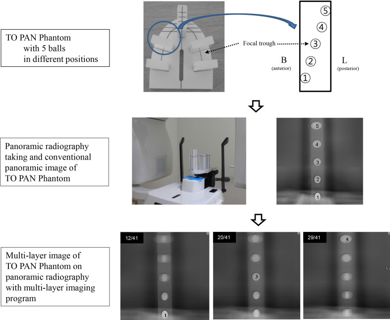
The program was applied to a rectangular block containing five metal balls in the buccolingual direction (TO PAN phantom; Leeds Test Objects Ltd, North Yorkshire, UK), with the block placed on a standard dental arch plate (Figure 2). The sequential multilayer images in the buccolingual direction showed distinct visibility of the individual balls on each layer. For example, the buccal ball (number 1) was most clearly shown in the 12th image layer, while the lingual ball (number 5) was clearly visible in the 29th layer (Figure 2).
Figure 2.
Conventional panoramic image and multilayer image of the TO PAN Phantom (a phantom containing five metal balls in the buccolingual direction). The 12th image (out of 41) distinctly shows the buccal ball (Number 1), the 20th image sharply shows the center ball (Number 3), and the 29th image distinctly shows the lingual ball (Number 5).
Inclusion criteria:
Patients with premolar and molar teeth with an unrestored proximal surface
Patients who had undergone periapical and bitewing radiography of the premolars and molars using a parallel technique at the same time as panoramic radiography
Exclusion criteria:
Patients with loss of premolar and molar teeth
Poor image quality (blurring, magnification, shrinkage, asymmetry, etc.)
The panoramic radiographs of 30 of the 348 patients were selected according to the above criteria (age range, 20–59 years; 19 female, 11 male). A total of 960 proximal surfaces of premolars (240 teeth) and molars (240 teeth) were assessed for proximal caries on panoramic radiography. The evaluation was done using brightness/contrast adjustment tools on two monitors (Totoku Electric Co., Nagano, Japan) in a dark and quiet room.
Evaluation of the presence of proximal caries by radiologists as the gold-standard
Three oral and maxillofacial radiologists with 7, 9, and 20 years of experience, respectively, assessed the presence or absence of dental caries for 960 proximal surfaces on panoramic radiography, periapical radiography, and bitewing radiography. The presence or absence of proximal caries was defined by consensus among the three radiologists.
Proximal caries evaluation by two general dentists
Two general dentists who had recently graduated from dental school evaluated the presence of proximal caries on panoramic radiography without the multilayer imaging program. Each observer scored the presence or absence of proximal caries at a 2 week interval. The scoring criteria were defined with reference to a previous study,6 as below:
Score of 1: Caries definitely not present
Score of 2: Caries probably not present
Score of 3: Uncertain
Score of 4: Caries probably present
Score of 5: Caries definitely present
After a minimum interval of 1 month as a wash-out period, the same evaluation was performed by the same two general dentists on panoramic radiography with the multi layer imaging program. The images were evaluated twice at 2 week intervals.
Statistical analysis
Caries presence scores of 4 and 5 were considered as a positive diagnosis, and caries scores of 1, 2, and 3 were considered as a negative diagnosis. κ analysis was used to assess the inter- and intraobserver agreement of the two general dentists for the detection of proximal caries. The data of the two general dentists with or without the multilayer imaging program were compared with the consensus-based gold-standard by radiologists. The data were analyzed for the area under the receiver operating characteristic (ROC) curve, sensitivity, specificity, positive predictive value (PPV), and negative predictive value (NPV). The ROC curves were compared using the methods proposed by DeLong et al.7 Statistical analysis was performed using the software STATA/SE 13.1 (STATA Corporation, College Station, TX).
Results
The assessment of proximal caries presence by consensus of the three radiologists for 960 proximal surfaces revealed no caries on 903 surfaces (94.1%) and caries on 57 surfaces (5.9%) (Table 1). 18 (31.6%) of the 57 surfaces with caries were on premolars, and 39 (68.4%) were on molars.
Table 1.
The distribution of proximal caries by consensus of the three radiologists
| Presence of proximal caries N (%) |
Absence of proximal caries N (%) |
Total | |
|---|---|---|---|
| Premolar | 18 (3.8%) | 462 (96.2%) | 480 |
| Molar | 39 (8.1%) | 441 (91.9%) | 480 |
| Total | 57 (5.9%) | 903 (94.1%) | 960 |
N, number
When the two general dentists used the multilayer imaging program, the intraobserver agreement was 0.960 and 0.696, respectively, which was much higher than the corresponding values of 0.643 and 0.536 when the program was not used (Table 2). The interobserver agreement of the two general dentists was higher with the aid of the program (κ = 0.461) than without the program (κ = 0.384) (Table 3). The area under the ROC curve was also higher when the program was applied for caries detection for both observers. Both sensitivity and specificity were superior when the program was used for each observer (Table 4). When the multilayer imaging program was used, all values, including the area under the ROC curve, increased in comparison to when the program was not used. The increase of the area under the ROC curve upon use of the program was statistically significant in Observer 1, but not in Observer 2.
Table 2.
Intraobserver agreement for two general dentists: κ index (95% CI)
| Without program | With program | |
|---|---|---|
| Observer 1 | 0.643 | 0.960 |
| Observer 2 | 0.536 | 0.696 |
CI, confidence interval
Table 3.
Interobserver agreement for two general dentists: κ index (95% CI)
| Without program | With program | |
|---|---|---|
| Observer 1–Observer 2 | 0.384 | 0.461 |
CI, confidence interval
Table 4.
Accuracy of the two general dentists with and without the multilayer imaging program
| Observer 1 | Observer 2 | |||||
|---|---|---|---|---|---|---|
| Without program (95% CI) |
With program (95% CI) |
p-value | Without program (95% CI) |
With program (95% CI) |
p-value | |
| Area under the ROC curve | 0.792 (0.738–0.846) |
0.923 (0.885–0.960) |
<0.001* | 0.833 (0.782–0.883) |
0.862 (0.818–0.906) |
0.148 |
| Sensitivity | 0.793 (0.666–0.888) |
0.914 (0.810–0971) |
0.828 (0.706–0.714) |
0.879 (0.767–0.950) |
||
| Specificity | 0.790 (0.762–0.817) |
0.931 (0.713–0.947) |
0.838 (0.812–0.862) |
0.845 (0.819–0.868) |
||
| PPV | 0.196 (0.169–0.226) |
0.461 (0.399–0.524) |
0.247 (0.214–0.238) |
0.267 (0.233–0.304) |
||
| NPV | 0.983 (0.973–0.990) |
0.994 (0.986–0.997) |
0.987 (0.977–0.993) |
0.991 (0.982–0.995) |
||
ROC, receiver operating characteristic; PPV, positive predictive value; NPV, negative predictive value; CI, confidence interval
*p<0.05.
Discussion
Panoramic radiography has the disadvantages of low resolution, image blurring, distortion, superimposition of additional structures, and overlapping of tooth crowns.8–10 In order to improve the diagnostic ability of panoramic radiographic images themselves without exposing patients to additional radiation, various specialized techniques have been introduced. Some devices adjust the rotational arc of the X-ray source–receptor movement for individual patients, thereby optimising the shape and size of the focal trough for the anatomy of each patient.11 An extraoral bitewing technique is offered by a few devices, such as the Planmeca ProMax 3D (Planmeca, Helsinki, Finland), which provides images with less overlap of adjacent teeth by adjusting the projection angle of the X-ray beam. Previous studies have reported that the extraoral bitewing technique was superior to conventional panoramic radiography for detecting proximal caries.6,12–14
Digital X-ray tomosynthesis methods used for chest imaging, mammography, and angiography have also been applied to panoramic radiography.15–17 Tomosynthesis is accomplished by shifting and adding a series of projection images obtained from conventional panoramic radiography and provides limited three-dimensional (2.5-dimensional) information, by depth.5 Tomosynthetically reconstructed images are offered as an option by a few devices, such as the Orthophos SL (Dentsply Sirona, Deutschland, Bensheim, Germany).17 Tomosynthesis applied to panoramic radiography with the option of post-processing provides information about depth and is useful in various clinical situations, including supernumerary teeth. In this study, we used a device equipped with a multilayer imaging program combined with tomosynthesis. This multilayer imaging program that provides 41 consecutive images in the buccolingual direction provided information on the relative buccolingual position of anatomical structures. This program provides limited three-dimensional information through sequential depth images that can be viewed by scrolling the mouse (Figure 1). However, few studies have investigated its effectiveness. Recently, Rahmel et al reported its usefulness in the diagnosis of artificial root resorption on panoramic radiography, showing 41 layers similar to the equipment used in this study.18
In this study, we evaluated the effectiveness of panoramic radiography with the application of tomosynthesis in the diagnosis of proximal dental caries. On panoramic radiography, the diagnostic ability of general dentists for proximal caries is very low. According to Pakbaznejad Esmaeili et al., 61% of proximal caries found by oral and maxillofacial radiologists in 100 panoramic radiographs were not found by general dentists.19 Many proximal caries remain unobserved by general dentists, which exacerbates patients’ conditions.
The results of this study show that the diagnostic capability of proximal caries detection was somewhat improved by using the multilayer imaging program. Two general dentists showed higher sensitivity and specificity with the multilayer imaging program than without it. When interpreting proximal caries on panoramic radiography, the overlapping of the proximal surfaces of adjacent teeth is the most significant problem. The program produced serial images from the buccal to lingual aspects within a single ROI, enabling the enamel outline of each tooth to be clearly seen without overlapping of the proximal surfaces of the adjacent tooth. As a result, the use of multilayer images reduced the masking of incipient proximal caries. When advanced proximal caries are present on only one side of two adjacent teeth, they may overlap, such that dental caries appear to be present on the proximal surfaces of other healthy teeth. Using a multilayer imaging program reduced the likelihood of these false positives. The reproducibility of proximal caries detection was also increased by using the multilayer imaging program. The κ values of intra- and interobserver agreement for the two general dentists was higher when using a multilayer imaging program than when not using it. The interobserver agreement improved, yet the value was relatively low. As was stated above regarding a previous study,19 this was probably due to the widely varying ability of general dentists to detect proximal caries.
All diagnostic performance metrics improved when using the program, but the statistical analysis showed that the area under the ROC curve showed a statistically significant improvement in Observer 1, but not in Observer 2. Since all diagnostic performance metrics of Observer 1 were lower than those of Observer 2 on panoramic radiography without the program, it is thought that the lower the diagnostic ability of a general dentist, the more effective the program. Additionally, between-user differences in software usability may have influenced this difference between the observers. An observer who is more comfortable using the new program might obtain better results. However, since there were only two general dentists in this study, further studies with more observers should be performed to investigate this possibility.
The use of panoramic radiography with a multilayer imaging program was shown to be useful for proximal caries detection, but additional intraoral radiography is still needed. Intraoral bitewing radiography is most recommended for the diagnosis of proximal caries. According to Kamburoglu et al, intraoral bitewing radiography was superior to conventional panoramic radiography and panoramic radiography with an extraoral bitewing in diagnosing proximal caries of premolar and molar teeth.12
This study has limitations in terms of the total sample number, observer number, and the number of dental caries, so further studies should be carried out including more samples, observers, and appropriately distributed samples of proximal caries. Panoramic radiography with a multilayer imaging program showed the outline of adjacent teeth well, but image distortion occurred in areas located in the front or rear, away from the focal trough, which is a fundamental limitation of panoramic radiography. It is likely that this multilayer imaging program will replace CBCT in some cases, as CBCT has a higher radiation dose and is more costly.
Conclusion
In conclusion, panoramic radiography with a multilayer imaging program was more effective than conventional panoramic radiography for detection of proximal caries. This program is expected to reduce the number of additional intraoral radiographs and may be useful for patients in whom it is difficult to take intraoral radiographs. Furthermore, this program is expected to be useful for a variety of clinical applications beyond dental caries detection, and additional studies are needed to investigate these possibilities.
Contributor Information
Kug Jin Jeon, Email: dentjeon@yuhs.ac.
Sang-Sun Han, Email: sshan@yuhs.ac.
Hoi In Jung, Email: JUNGHOIIN@yuhs.ac.
REFERENCES
- 1.Gordan VV, Riley JL, Carvalho RM, Snyder J, Sanderson JL, Anderson M, et al. Methods used by dental practice-based research network (DPBRN) dentists to diagnose dental caries. Oper Dent 2011; 36: 2–11. doi: 10.2341/10-137-CR [DOI] [PMC free article] [PubMed] [Google Scholar]
- 2.Barrett AP, Waters BE, Griffiths CJ. A critical evaluation of panoramic radiography as a screening procedure in dental practice. Oral Surg Oral Med Oral Pathol 1984; 57: 673–7. doi: 10.1016/0030-4220(84)90292-5 [DOI] [PubMed] [Google Scholar]
- 3.Rushton VE, Horner K. The use of panoramic radiology in dental practice. J Dent 1996; 24: 185–201. doi: 10.1016/0300-5712(95)00055-0 [DOI] [PubMed] [Google Scholar]
- 4.Choi J-W. Assessment of panoramic radiography as a national oral examination tool: review of the literature. Imaging Sci Dent 2011; 41: 1–6. doi: 10.5624/isd.2011.41.1.1 [DOI] [PMC free article] [PubMed] [Google Scholar]
- 5.Dobbins JT, Godfrey DJ. Digital X-ray tomosynthesis: current state of the art and clinical potential. Phys Med Biol 2003; 48: R65–106. doi: 10.1088/0031-9155/48/19/R01 [DOI] [PubMed] [Google Scholar]
- 6.Gaalaas L, Tyndall D, Mol A, Everett ET, Bangdiwala A. Ex vivo evaluation of new 2D and 3D dental radiographic technology for detecting caries. Dentomaxillofac Radiol 2016; 45: 20150281. doi: 10.1259/dmfr.20150281 [DOI] [PMC free article] [PubMed] [Google Scholar]
- 7.DeLong ER, DeLong DM, Clarke-Pearson DL. Comparing the areas under two or more correlated receiver operating characteristic curves: a nonparametric approach. Biometrics 1988; 44: 837–45. doi: 10.2307/2531595 [DOI] [PubMed] [Google Scholar]
- 8.Akkaya N, Kansu O, Kansu H, Cagirankaya LB, Arslan U. Comparing the accuracy of panoramic and intraoral radiography in the diagnosis of proximal caries. Dentomaxillofac Radiol 2006; 35: 170–4. doi: 10.1259/dmfr/26750940 [DOI] [PubMed] [Google Scholar]
- 9.Flint DJ, Paunovich E, Moore WS, Wofford DT, Hermesch CB. A diagnostic comparison of panoramic and intraoral radiographs. Oral Surg Oral Med Oral Pathol Oral Radiol Endod 1998; 85: 731–5. doi: 10.1016/S1079-2104(98)90043-9 [DOI] [PubMed] [Google Scholar]
- 10.Akarslan ZZ, Akdevelioğlu M, Güngör K, Erten H. A comparison of the diagnostic accuracy of bitewing, periapical, unfiltered and filtered digital panoramic images for approximal caries detection in posterior teeth. Dentomaxillofac Radiol 2008; 37: 458–63. doi: 10.1259/dmfr/84698143 [DOI] [PubMed] [Google Scholar]
- 11.Kim S, Ra JB. Dynamic focal plane estimation for dental panoramic radiography. Med Phys 2019; 46: 4907–17. doi: 10.1002/mp.13823 [DOI] [PubMed] [Google Scholar]
- 12.Kamburoglu K, Kolsuz E, Murat S, Yüksel S, Ozen T. Proximal caries detection accuracy using intraoral bitewing radiography, extraoral bitewing radiography and panoramic radiography. Dentomaxillofac Radiol 2012; 41: 450–9. doi: 10.1259/dmfr/30526171 [DOI] [PMC free article] [PubMed] [Google Scholar]
- 13.Abdinian M, Razavi SM, Faghihian R, Samety AA, Faghihian E. Accuracy of digital Bitewing radiography versus different views of digital panoramic radiography for detection of proximal caries. J Dent 2015; 12: 290–7. [PMC free article] [PubMed] [Google Scholar]
- 14.Abu El-Ela WH, Farid MM, Mostafa MSE-D. Intraoral versus extraoral bitewing radiography in detection of enamel proximal caries: an ex vivo study. Dentomaxillofac Radiol 2016; 45: 20150326. doi: 10.1259/dmfr.20150326 [DOI] [PMC free article] [PubMed] [Google Scholar]
- 15.Ogawa K, Langlais RP, McDavid WD, Noujeim M, Seki K, Okano T, et al. Development of a new dental panoramic radiographic system based on a tomosynthesis method. Dentomaxillofac Radiol 2010; 39: 47–53. doi: 10.1259/dmfr/12999660 [DOI] [PMC free article] [PubMed] [Google Scholar]
- 16.Noujeim M, Prihoda T, McDavid WD, Ogawa K, Yamakawa T, Seki K, et al. Pre-Clinical evaluation of a new dental panoramic radiographic system based on tomosynthesis method. Dentomaxillofac Radiol 2011; 40: 42–6. doi: 10.1259/dmfr/73312141 [DOI] [PMC free article] [PubMed] [Google Scholar]
- 17.Katsumata A, Ogawa K, Inukai K, Matsuoka M, Nagano T, Nagaoka H, et al. Initial evaluation of linear and spatially oriented planar images from a new dental panoramic system based on tomosynthesis. Oral Surg Oral Med Oral Pathol Oral Radiol Endod 2011; 112: 375–82. doi: 10.1016/j.tripleo.2011.04.024 [DOI] [PubMed] [Google Scholar]
- 18.Rahmel S, Schulze RKW. Accuracy in detecting artificial root resorption in panoramic radiography versus Tomosynthetic panoramic radiographs. J Endod 2019; 45: 634–9. doi: 10.1016/j.joen.2019.01.009 [DOI] [PubMed] [Google Scholar]
- 19.Pakbaznejad Esmaeili E, Pakkala T, Haukka J, Siukosaari P. Low reproducibility between oral radiologists and general dentists with REGARDS to radiographic diagnosis of caries. Acta Odontol Scand 2018; 76: 346–50. doi: 10.1080/00016357.2018.1460490 [DOI] [PubMed] [Google Scholar]


