Abstract
Background:
Candida auris is an emerging and often multidrug-resistant fungal pathogen with an exceptional ability to persist on hospital surfaces. These surfaces can act as a potential source of transmission. Therefore, effective disinfection strategies are urgently needed. We investigated the efficacy of ultraviolet C light (UV-C) disinfection for C. auris isolates belonging to 4 different clades.
Methods:
In vitro testing of C. auris isolates was conducted using 106 colony-forming units (CFU) spread on 20-mm diameter steel carriers and exposed to a broad-spectrum UV-C light source for 10, 20, and 30 minutes at a 1.5 m (5 feet) distance. Post-UV survivors on the coupons were subsequently plated. Colony counts and log reductions were recorded, calculated, and compared to untreated control carriers. Identification of all isolates were confirmed by MALDI-TOF and morphology was visualized by microscopy.
Results:
We observed an increased susceptibility of C. auris to UV-C in 8 isolates belonging to clades I, II and IV with increasing UV exposure time. The range of log kill (0.8–1.19) was highest for these isolates at 30 minutes. But relatively no change in log kill (0.04–0.35) with increasing time in isolates belonging to clade III were noted. Interestingly, C. auris isolates susceptible to UV-C were mostly nonaggregating, but the isolates that were more resistant to UV exposure formed aggregates.
Conclusions:
Our study suggests variability in susceptibility to UV-C of C. auris isolates belonging to different clades. More studies are needed to assess whether a cumulative impact of prolonged UV-C exposure provides additional benefit.
Candida auris, first reported in 2009, is an emerging fungal pathogen that is often multidrug resistant, difficult to identify, and has an ability to persist in the hospital environment.1-3 Also, C. auris has been reported to cause hospital outbreaks with high mortality rates, especially among critically ill patients.4 The emergence and detection of C. auris on multiple continents simultaneously has led to identification of 4 distinct clonal lineages via whole-genome sequencing: clade I (South Asian), clade II (East Asian), clade III (African), and clade IV (South American).5 Interestingly, despite intraclonal heterogeneity, very few single-nucleotide polymorphism (SNP) differences exist within each clade. In addition, it has been illustrated that some isolates form aggregates potentially conferring protection from the environment.6
Presently, no specific guidelines has been established for C. auris disinfection on hospital surfaces. The Centers for Disease Control and Prevention (CDC) recommends the use of Environmental Protection Agency (EPA)–registered hospital-grade disinfectants that are effective against Clostridioides difficile (C. diff) spores. Surface disinfection protocols with chemical disinfectants have shown variable outcomes.7,8 Therefore, to prevent the transmission of C. auris from hospital surfaces, more effective strategies in conjunction with daily chemical disinfection of patient-occupied rooms are urgently needed.
Mobile, automated broad-spectrum ultraviolet-C light (UV-C) decontamination systems using pulsed-xenon (PX-UV) or other mercury-based sources have reduced the recovery of methicillin-resistant Staphylococcus aureus (MRSA), Clostridioides difficile, and vancomycin-resistant Enterococci (VRE) from glass carriers and frequently touched surfaces in hospitals.9 Candida spp, including C. auris, appear to be significantly less affected by UV-C decontamination than MRSA.10 A recent study demonstrated that UV-C has been effective at reducing colony counts of C. auris, especially with longer exposure time and closer proximity to UV source.11 However, whether there are notable differences between isolates belonging to different clades in their response to UV-C exposure at increasing time intervals remains unknown. We further evaluated whether the susceptibility to UV-C depends on the aggregate-forming capability of C. auris clade(s).
Methods
We evaluated the efficacy of a pulsed-xenon (PX) UV-C room decontamination device (Xenex Disinfection, San Antonio, TX) against 10 C. auris isolates. These 10 isolates of C. auris (AR# 0381-0390) were obtained from the CDC & FDA Antibiotic Isolate Bank. The PX-UV device contained a xenon gas flash bulb that operates at 2 Hz and emits a broad spectrum of radiation covering the UV-C spectrum of 200–280 nm as well as the visible light spectrum. For each pathogen, 10 µL aliquots (3 biological replicates) containing 106 colony-forming units (CFU) in phosphate-buffered saline (PBS) with 5% fetal bovine serum (FBS) were spread over 20 mm diameter steel coupons and allowed to air dry for 30 minutes in a laminar flow hood. The steel coupons were then placed perpendicular to the PX-UV lamps of the device and at distance of 1.5 m (5 feet). A set of three biological replicates were exposed to UV-C at 10, 20, and 30 minutes, respectively. Our control groups were not exposed to UV-C and were plated last to account for any desiccation. To quantify viable organisms, treated and untreated control coupons were submersed in 10 mL PBS and vortexed vigorously, and serial dilutions were plated on Sabouraud dextrose agar (SDA, Remel, Lenexa, KS) and were incubated at 37°C for 72 hours. Colonies were then counted, and log reductions were calculated. The experiments were repeated 3 times (technical replicates) to mitigate any variability of the protocol or day-to-day weather conditions such as humidity or airflow.
We used the method of Borman et al6 for aggregation assay. First, each C. auris isolate was grown on a Sabouraud dextrose agar plate at 37°C for 48 hours. Two closely related yeast strains, such as C. krusei (AR#0397) and C. lusitiniae (AR# 0398), were used as controls. A cell suspension was prepared with a C. auris colony from each of the 10 isolates and vortexed for 30 seconds. Each isolate was resuspended in PBS to achieve a McFarland standard of 4–5 (for a final cell density of 1×10⁸ cells/mL). Then, 30 μL of this suspension was loaded on a glass slide and examined under a microscope (Olympus BX53 laden with Olympus camera DP73).
Statistical methods
Raw plate data of counts of colony-forming units killed were converted to a log value, and log kill was determined from the initial concentration at time zero. Mean log kill and standard deviations were calculated for each isolate at 10, 20, and 30 minutes. A Bayesian linear regression model with a hierarchical structure for clade, isolate number, and experiment was used to estimate the slope of log kill over time for each clade. Results are reported as the estimated slope of log kill per minute with 95% uncertainty intervals. All analyses were conducted in R version 3.5.3 software (R Foundation for Statistical Computing, Vienna, Austria) utilizing the ‘brms’ package for modeling and the ‘ggplot2’ package for plotting.
Results
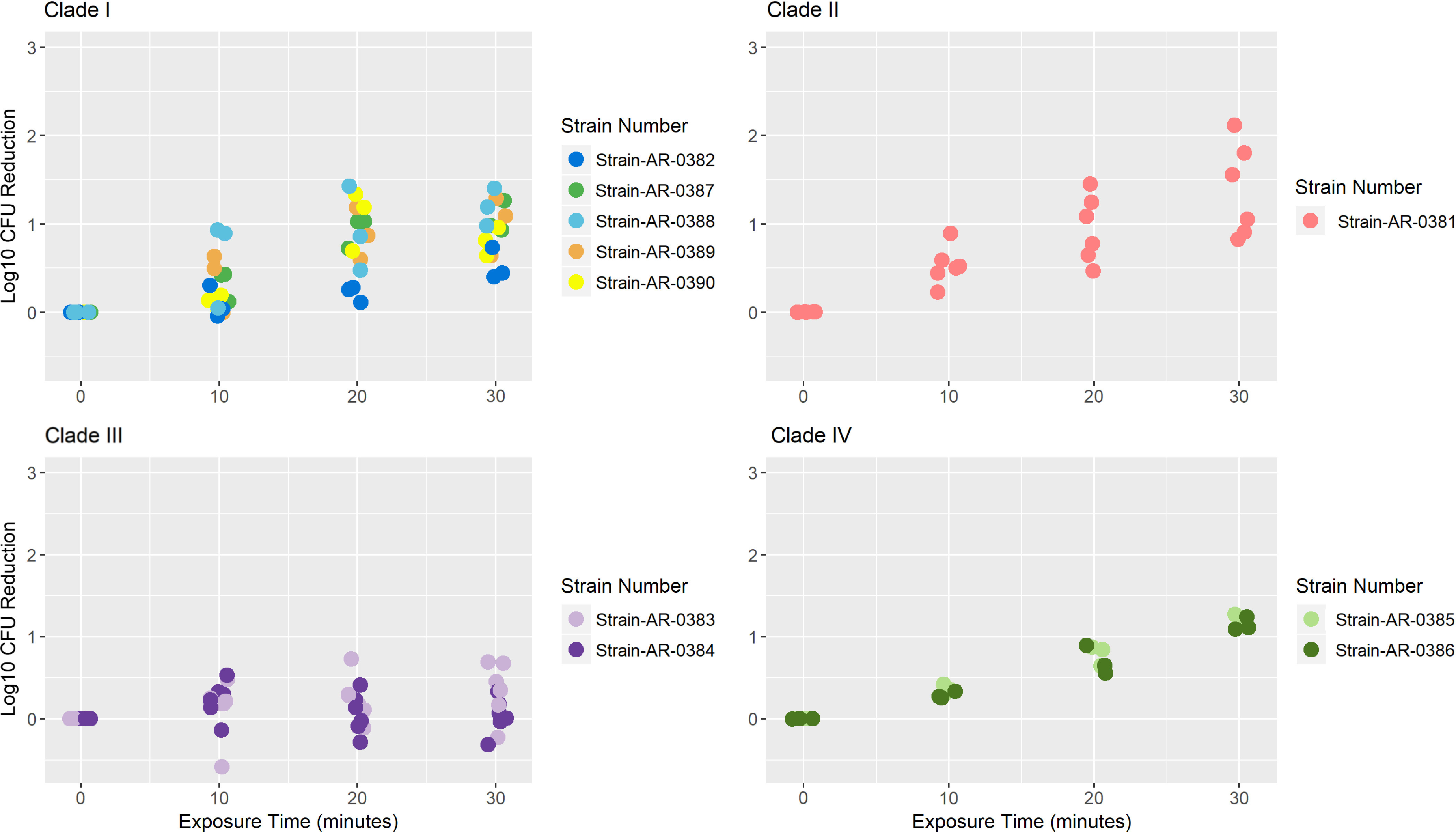
Mean log kill at 10, 20, and 30 minutes for each of the 10 C. auris isolates are shown in Table 1. The raw mean log-kill at 30 minutes for clade 1 was 0.92 (SD, 0.31), for clade II was 1.38 (SD, 0.53), for clade III was 0.19 (SD, 0.32), and for clade IV was 1.18 (SD, 0.07). Also, 5 clade I isolates (AR#0382, AR#0387, AR#0388, AR#0389, AR#0390) were tested with 0.5 log10 (AR#0382) to 1.2 log10 (AR#0388) reduction at 30 minutes of exposure. One clade II isolate AR#0381 was tested, and it demonstrated a 1.38 log10 reduction at 30 minutes of UV-C exposure. Two clade III isolates (AR#0383 & AR#0384) were tested with 0.04 (AR#0384) to 0.35 (AR#0383) log10 reduction at 30 minutes of UV-C exposure, showing the worst response to UV-C exposure among all C. auris clades. Further, the 2 clade IV strains tested, AR#0385 and AR#0386, showed almost similar reductions in CFU count after UV-C exposure (1.22 and 1.15, respectively). The log-kill data were plotted for each clade and were colored by isolates (Fig. 1).
Table 1.
Mean Log-Kill at 10, 20, and 30 Minutes for Each Candida auris Strain
| C. auris Clade | Strain No. | 10 Minutes, Mean Log Kill (SD) | 20 Minutes, Mean Log Kill (SD) | 30 Minutes, Mean Log Kill (SD) |
|---|---|---|---|---|
| Clade I | Strain-AR-0382 | 0.10 (0.18) | 0.21 (0.09) | 0.52 (0.18) |
| Clade I | Strain-AR-0387 | 0.32 (0.17) | 0.93 (0.17) | 1.06 (0.18) |
| Clade I | Strain-AR-0388 | 0.62 (0.50) | 0.92 (0.48) | 1.19 (0.21) |
| Clade I | Strain-AR-0389 | 0.37 (0.34) | 0.88 (0.30) | 1.01 (0.33) |
| Clade I | Strain-AR-0390 | 0.12 (0.09) | 1.07 (0.34) | 0.80 (0.16) |
| Clade II | Strain-AR-0381 | 0.53 (0.22) | 0.94 (0.38) | 1.38 (0.53) |
| Clade III | Strain-AR-0383 | 0.12 (0.36) | 0.25 (0.28) | 0.35 (0.35) |
| Clade III | Strain-AR-0384 | 0.23 (0.22) | 0.06 (0.25) | 0.04 (0.22) |
| Clade IV | Strain-AR-0385 | 0.36 (0.05) | 0.79 (0.12) | 1.22 (0.06) |
| Clade IV | Strain-AR-0386 | 0.29 (0.04) | 0.70 (0.17) | 1.15 (0.08) |
Fig. 1.

The effect of UV-C exposure on different clades of C. auris. Log reductions for each of the C. auris isolates belonging to clades I-IV are shown here. Log reductions were calculated by subtracting viable organisms recovered after exposure to UV versus controls (no UV exposure) for each time points (10, 20, 30 minutes).
The model estimated slope of the log kill over time pooled across all clades and isolates was 0.03 (−0.004 to 0.06) log-kill per minute of UV exposure. The model-estimated slopes of the log kill per minute of UV over time were 0.03 (0.02–0.05) for clade I, 0.04 (0.02–0.06) for clade II, 0.01 (−0.004 to 0.03) for clade III, and 0.04 (0.02–0.06) for clade IV.
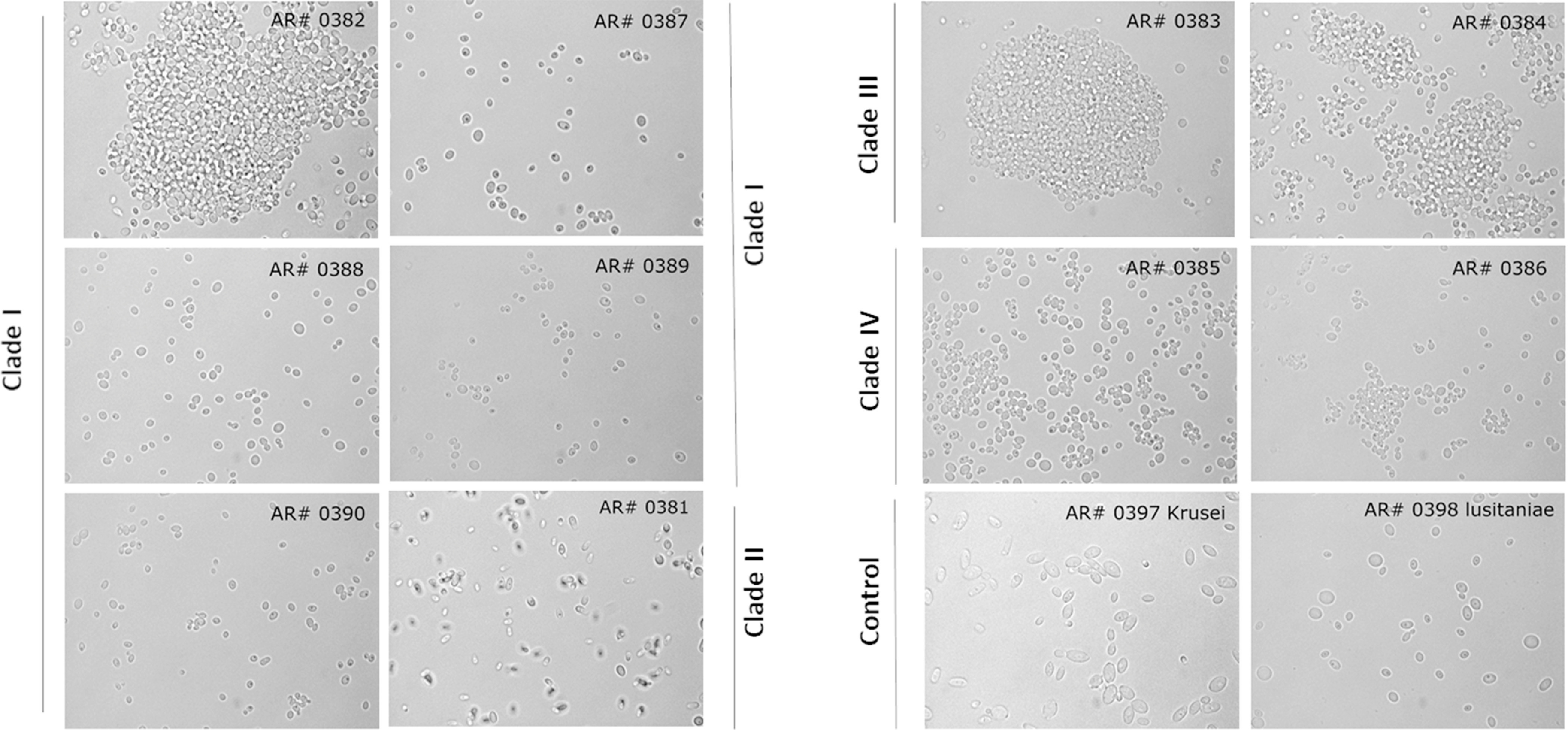
We evaluated the cellular morphology of all C. auris isolates using a microscope. Strains from clade III (isolates AR# 0383 and AR# 0384) formed aggregates (Fig. 2), whereas strains from all other clades (except AR#0382 from clade I) were non–aggregate forming.
Fig. 2.

Micrographs of different C. auris isolates belonging to different clades. Images taken under a microscope (x100 magnification) for all 10 isolates of C. auris belonging to four different clades I-IV in PBS suspensions. C. krusei and C. lusitaniae were used as controls.
Discussion
High virulence and pathogenicity of C. auris coupled with reduced susceptibility to antifungals have already caused several outbreaks worldwide.12 Various chemical disinfectants such as 27.5% hydrogen peroxide with 5.8% peracetic acid (OxyCide, Ecolab, St Paul, MN) or 10% sodium hypochlorite (Clorox, Oakland, CA), and accelerated hydrogen peroxide (Oxivir TB, Diversey, Charlotte, NC) have shown >5 log10 reduction, while other commonly used quaternary ammonium compound–based disinfectants are less effective.8 C. auris has been shown to persist especially on moist hospital surfaces such as sinks for a prolonged period.13 Due to the possibility that some hospital surfaces may be inadvertently missed during manual cleaning combined with the sporadic frequency of manual cleaning events, the addition of more efficient no-touch disinfection devices to the overall cleaning process may be beneficial. We found that C. auris isolates are susceptible to UV-C; however, clade differences in susceptibility to UV-C must also be considered. It is known that the complex cell structure and the glucan cell wall layer of eukaryotes render them less susceptible to UV light compared to prokaryotes.10 In our study, we observed relatively lower log kill levels for C. auris isolates at a high concentrations of 106 cells/mL than have been reported in other studies.10,11 These differences in results could be related to 2 factors: First, one study used glass slides instead of steel carriers, and different surfaces and the different spread of inoculum on them might affect the outcome.11 Previous studies have shown that the larger the surface on which the inoculum is spread, the higher the kill rate by UV-C.10 Second, the intensities of the UV-C bulbs were different based on the manufacturer used between these experiments. Interestingly, when a lower concentration of 105 cells were used on the carriers, we achieved higher log kill, similar to the aforementioned studies. Because the concentration-dependent effect of the UV susceptibility of various clades was not the focus of this study, we have not included these data.
Our data also demonstrate that UV-C exposure had little discernable effect on log kill of isolates belonging to clade III, even after 30 minutes, which exceeded the manufacturer’s recommended UV disinfection protocol. Therefore, shorter duration of UV-C exposure would not be as effective for isolates belonging to clade III compared to the other clades. It is likely that these isolates demonstrate resistance to UV-C because they form aggregates. These aggregates can confer a protective effect and thus prevent the penetration of UV-C light to the core of the aggregate due to a stacking effect. A study of 50 isolates from the South African lineage (clade III) produced similar cell aggregates, which likely aided in biofilm formation.14,15 Interestingly, 1 isolate (AR#0382) belonging to clade I, which demonstrated the lowest log kill at 30 minutes, also formed an aggregate. Our findings are consistent with those of Szekely et al,16 who found that some isolates of C. auris belonging to clade I and almost all belonging to clade III were phenotypically different (ie, formed aggregates) than the other clades.
In conclusion, our findings suggest that it is unlikely that using the same dose and duration of UV would be effective against all isolates of C. auris to the same extent. A previous study only used 1 C. auris isolate and precluded comparison among different clades.17 We acknowledge that even when the disc carriers were placed at a 1.5-m (5-ft) distance perpendicular to the UV device, adequate log kill was not achieved among all the clades studied here at a higher concentration, possibly due to a stacking effect. Therefore, further study is warranted to address this issue. Apart from chemical disinfectants, which have been shown to be efficacious for disinfection, the addition of automated UV-C devices may be complementary and may provide some incremental benefit in achieving superior environmental disinfection. One limitation of this study is that it includes a relatively small number of isolates from each clade. This study generates awareness about the problems of C. auris persistence on hospital surfaces and does not provide recommendations for the use of UV devices. Future studies using these UV devices in a real hospital setting where C. auris is prevalent may provide additional information that will help control the spread of this pathogen.
Acknowledgments
We thank Rana Radwan for her help with these experiments. The views expressed in this article are those of the author(s) and do not necessarily represent the views of the Department of Veterans’ Affairs.
Conflicts of interest
All authors report no conflicts of interest relevant to this article.
Financial support
This work was supported by the Central Texas Veterans’ Health Care System.
References
- 1. Satoh K, Makimura K, Hasumi Y, Nishiyama Y, Uchida K, Yamaguchi H. Candida auris sp nov, a novel ascomycetous yeast isolated from the external ear canal of an inpatient in a Japanese hospital. Microbiol Immunol 2009;53:41–44. [DOI] [PubMed] [Google Scholar]
- 2. Candida auris fungal diseases. Centers for Disease Control and Prevention website. https://www.cdc.gov/fungal/candida-auris/index.html Updated 2019. Accessed August 11, 2020.
- 3. Piedrahita CT, Cadnum JL, Jencson AL, Shaikh AA, Ghannoum MA, Donskey CJ. Environmental surfaces in healthcare facilities are a potential source for transmission of Candida auris and other Candida species. Infect Control Hosp Epidemiol 2017;38:1107–1109. [DOI] [PubMed] [Google Scholar]
- 4. Kumar J, Eilertson B, Cadnum JL, et al. Environmental contamination with Candida species in multiple hospitals including a tertiary-care hospital with a Candida auris outbreak. Pathog Immun 2019;4:260–270. [DOI] [PMC free article] [PubMed] [Google Scholar]
- 5. Rhodes J, Fisher MC. Global epidemiology of emerging Candida auris . Curr Opin Microbiol 2019;52:84–89. [DOI] [PubMed] [Google Scholar]
- 6. Borman AM, Szekely A, Johnson EM. Comparative pathogenicity of United Kingdom isolates of the emerging pathogen Candida auris and other key pathogenic Candida species. mSphere 2016;1(4). doi: 10.1128/mSphere.00189-16. [DOI] [PMC free article] [PubMed] [Google Scholar]
- 7. Kean R, Sherry L, Townsend E, et al. Surface disinfection challenges for Candida auris: an in-vitro study. J Hosp Infect 2018;98:433–436. [DOI] [PubMed] [Google Scholar]
- 8. Cadnum JL, Shaikh AA, Piedrahita CT, et al. Effectiveness of disinfectants against Candida auris and other Candida species. Infect Control Hosp Epidemiol 2017;38:1240–1243. [DOI] [PubMed] [Google Scholar]
- 9. Nerandzic MM, Thota P, Sankar CT, et al. Evaluation of a pulsed xenon ultraviolet disinfection system for reduction of healthcare-associated pathogens in hospital rooms. Infect Control Hosp Epidemiol 2015;36:192–197. [DOI] [PubMed] [Google Scholar]
- 10. Cadnum JL, Shaikh AA, Piedrahita CT, et al. Relative resistance of the emerging fungal pathogen Candida auris and other Candida species to killing by ultraviolet light. Infect Control Hosp Epidemiol 2018;39:94–96. [DOI] [PubMed] [Google Scholar]
- 11. de Groot T, Chowdhary A, Meis JF, Voss A. Killing of Candida auris by UV-C: importance of exposure time and distance. Mycoses 2019;62:408–412. [DOI] [PMC free article] [PubMed] [Google Scholar]
- 12. Tsay S, Welsh RM, Adams EH, et al. Notes from the field: ongoing transmission of Candida auris in healthcare facilities—United States, June 2016–May 2017. Morb Mortal Wkly Rep 2017;66:514–515. [DOI] [PMC free article] [PubMed] [Google Scholar]
- 13. Jencson AL, Cadnum JL, Piedrahita C, Donskey CJ. Hospital sinks are a potential nosocomial source of Candida infections. Clin Infect Dis 2017;65:1954–1955. [DOI] [PubMed] [Google Scholar]
- 14. Bidaud AL, Chowdhary A, Dannaoui E. Candida auris: an emerging drug resistant yeast—a mini-review. J Mycol Med 2018;28:568–573. [DOI] [PubMed] [Google Scholar]
- 15. Corsi-Vasquez G, Ostrosky-Zeichner L. Candida auris: what have we learned so far? Curr Opin Infect Dis 2019;32:559–564. [DOI] [PubMed] [Google Scholar]
- 16. Szekely A, Borman AM, Johnson EM. Candida auris isolates of the Southern Asian and South African lineages exhibit different phenotypic and antifungal susceptibility profiles in vitro. J Clin Microbiol 2019;57(5). doi: 10.1128/JCM.02055-18. [DOI] [PMC free article] [PubMed] [Google Scholar]
- 17. Maslo C, du Plooy M, Coetzee J. The efficacy of pulsed-xenon ultraviolet light technology on Candida auris . BMC Infect Dis 2019;19:540. [DOI] [PMC free article] [PubMed] [Google Scholar]


