Abstract
At two meetings of a Central European board of multiple sclerosis (MS) experts in 2018 and 2019 factors influencing daily treatment choices in MS, especially practice guidelines, biomarkers and burden of disease, were discussed. The heterogeneity of MS and the complexity of the available treatment options call for informed treatment choices. However, evidence from clinical trials is generally lacking, particularly regarding sequencing, switches and escalation of drugs. Also, there is a need to identify patients who require highly efficacious treatment from the onset of their disease to prevent deterioration. The recently published European Committee for the Treatment and Research in Multiple Sclerosis/European Academy of Neurology clinical practice guidelines on pharmacological management of MS cover aspects such as treatment efficacy, response criteria, strategies to address suboptimal response and safety concerns and are based on expert consensus statements. However, the recommendations constitute an excellent framework that should be adapted to local regulations, MS center capacities and infrastructure. Further, available and emerging biomarkers for treatment guidance were discussed. Magnetic resonance imaging parameters are deemed most reliable at present, even though complex assessment including clinical evaluation and laboratory parameters besides imaging is necessary in clinical routine. Neurofilament-light chain levels appear to represent the current most promising non-imaging biomarker. Other immunological data, including issues of immunosenescence, will play an increasingly important role for future treatment algorithms. Cognitive impairment has been recognized as a major contribution to MS disease burden. Regular evaluation of cognitive function is recommended in MS patients, although no specific disease-modifying treatment has been defined to date. Finally, systematic documentation of real-life data is recognized as a great opportunity to tackle unresolved daily routine challenges, such as use of sequential therapies, but requires joint efforts across clinics, governments and pharmaceutical companies.
Keywords: biomarkers, burden of disease, cognitive dysfunction, magnetic resonance imaging, multiple sclerosis, neurofilament
Introduction
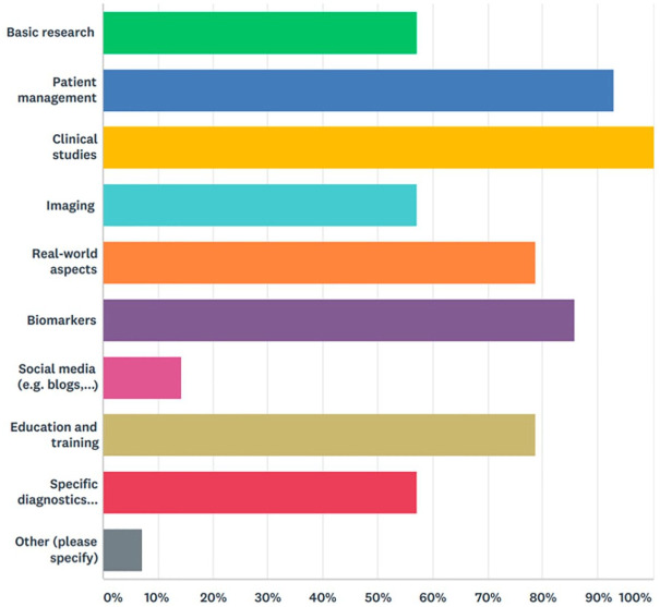
The Central European Multiple Sclerosis (MS) Expert Board was founded in 2007 with the aim of improving the management of MS patients in the Central European area. At annual board meetings, renowned MS experts from Austria, the Czech Republic, Hungary, Poland, Slovakia and Slovenia discuss practical aspects, including local diagnostic and therapeutic algorithms, educational requirements, data gaps and support for argumentation in discussions with health authorities. The experts represent mainly the main academic MS centers in their respective countries and are, thus, engaged in both MS patient care and (basic and clinical) MS research (Figure 1). Here we summarize the content of the two most recent board meetings, held on 27 January 2018 and 26 January 2019 in Vienna, Austria. According to the given clinical and scientific fields of interest of the participants, lectures and debates focused on the implications of the recently published clinical practice guidelines, treatment decisions in light of existing and potential biomarkers, and consequences of the burden of MS and costs of illness.
Figure 1.
Multiple sclerosis related clinical and scientific fields of interests of expert panel participants (N = 23, multiple answers possible).
European Committee for the Treatment and Research in Multiple Sclerosis/European Academy of Neurology clinical practice guideline
In January 2018 the first European clinical practice guidelines on the pharmacological management of MS patients were published by the European Committee for the Treatment and Research in Multiple Sclerosis (ECTRIMS) and the European Academy of Neurology (EAN).1,2 The availability of guidelines that can be perceived as a European consensus was deemed essential for both physicians and authorities. They contain a total of 21 recommendations and cover treatment efficacy, response criteria, strategies to address suboptimal response and safety concerns, including pregnancy.
All of the disease-modifying therapies (DMTs) approved by the European Medicine Agency at the time of publication are taken into account but not prioritized due to insufficient evidence by formal head-to-head studies. There is a focus on early treatment of clinically isolated syndrome, treatment in patients with established relapsing and progressive MS, monitoring of treatment response, and stopping or switching treatment. The guidelines recommend an attempt to specify a provisional disease course as soon as the diagnosis is made in a patient. This should be closely followed up, timely re-evaluated and re-classified as needed. Regular magnetic resonance imaging (MRI) monitoring is justified based on the fact that early disease activity predicts the risk of future disability. Due to the lack of high-quality evidence on MRI monitoring of DMT efficacy, the guidelines recommend annual scans. As re-occurrence of disease activity can be expected upon discontinuation of effective agents, treatment switches to other DMTs should be considered. Among the 21 recommendations three were rated as strong, nine as weak and nine based on expert consensus.
The expert panel emphasized that the ECTRIMS/EAN guidelines represent the optimum of what can be achieved considering the available evidence, and that the recommendations constitute an excellent framework that should be adapted to local regulations, MS center capacities and infrastructure. Given the cost constraints, implementation might be problematic in some Central European countries with economically driven limited access to MS drugs; details have been described already by the expert panel.3 Fortunately, these problems have been diminishing in the region in recent years. However, based on these European recommendations, national discussions with local health authorities might improve access.
MS patients are increasingly being treated at specialized centers, as office-based general neurologists cannot always handle the complexity of MS treatment and monitoring. However, specialized MS centers do not have to be necessarily restricted to hospitals but office-based MS care centers should also make specific commitments to certain standards, retain responsibility for pharmacovigilance and issue clear monitoring protocols. Thorough education is an important aspect in this context of local or outsourced care.
Biomarkers for supporting treatment decisions
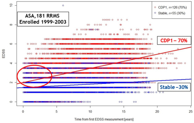
The heterogeneous nature of MS gives rise to different phenotypes. Approximately one-third of patients experience only slowly progressing changes, whereas the remaining two-thirds deteriorate severely without treatment according to the ASA trial (Figure 2). ASA (Avonex Steroid Azathioprine) is an investigator initiated clinical trial that enrolled 181 patients with early active relapsing–remitting MS between 1999 and 2003. Most of the patients have been followed in the same clinical and MRI settings since 1999.4 Studies and registries have shown that early diagnosis and optimized treatment are a key determinant of long-term outcomes.5–7 However, not all patients might require immediate therapy after diagnosis.8 Another aspect under debate is treatment failure; here, established scoring systems have been proposed for interferons, but not yet for new drugs.9 In stable disease, shared treatment discontinuation represents an option only if complex monitoring is provided every 6 months, but this approach will require further validation.10
Figure 2.
Confirmed disability progression EDSS: one-third of patients shows relatively stable disease over time.3
ASA, Avonex Steroid Azathioprine Study,; CDP, confirmed disability progression; EDSS, Expanded Disability Status Scale; RRMS, relapsing–remitting multiple sclerosis.
Treatment algorithms would be welcome, but data are lacking in many settings, and physicians are often left without evidence-based guidance. The possibility of mild and stable disease courses should be kept in mind11 when considering treatment switches and escalation therapies. Modern drugs can improve the course of MS but might also cause serious adverse events.12,13 Thus, a more personalized approach to identify an individual’s prognosis is essential to identify patients who benefit from timely drug intervention.
MRI
MRI is acknowledged as the best available biomarker in MS at present. It reflects both inflammation and tissue damage and supports diagnosis as well as prognosis assessment, disease activity monitoring and pharmacovigilance. This underlines the importance of a close cooperation between neurologists and (neuro-) radiologists. Although not formally proven, MRI monitoring of simple parameters indicative of inflammation (T2 lesion load, contrast-enhanced T1 lesions) is used in everyday clinical practice. It enables changes in therapy immediately once MRI pattern is changed significantly, but also prevents overreactions in patients whose MRI findings are stable.
Sequential MRI scans appear also be used to monitor MS related neuroaxonal damage in terms of total brain atrophy14 or atrophy of certain regions such as the corpus callosum or the thalamus15 – however, this requires rigorous standardized protocols, which are so far only available and used in specialized MRI centers, thus limiting yet the meaningfulness of brain atrophy measure on individual levels in routine practice.16 To better understand variability of brain volume changes, real-world data might provide helpful insights here. A comparison of longitudinal MRI scans across 1565 Czech MS patients and 131 healthy controls showed wide inter-individual variability in both groups, although volume loss over more than 4 years of follow-up was generally greater in MS patients. In many instances, regional atrophy appeared to provide better differentiation between the two groups, but more work is needed to improve accuracy and to be able to set up reliable cut-off values between normal and pathological brain volume loss.17
Cross-sectional results showed promising correlations between brain atrophy and cognitive function. A cut-off of >3.5 cm3 for T2 lesion volume gave an odds ratio (OR) of 5.1 for cognitive impairment, while a cut-off of <0.85 cm3 for brain parenchymal fraction resulted in an OR of 2.9. Use of both cut-offs provided accuracy of 75%, specificity of 83% and sensitivity of 51%.18 Another example of how quantitative MRI data can be used in clinical practice is results from the 12 year follow-up in the ASA cohort. Here, combination of particular clinical [relapses and Expanded Disability Status Scale (EDSS) change] and MRI (lesions and corpus callosum volume change) parameters in the first year was able to predict disability progression. The strength of prediction was improved when particular parameters were used in a specific composite score.19
Overall, brain volume changes over time are relevant and may drive treatment modifications in the future; however, formal establishment of evidence by proving that treatment intervention (or switch) changes the risk of further brain atrophy is urgently needed.
Spinal cord imaging should not be neglected as spinal cord is affected in many patients, and a close and dominant relationship between spinal cord pathology and clinical disability is well established.20 Particularly in early MS, assessment of the spinal cord volume could be helpful.16,21 However, the identification of new lesions is more challenging in the spinal cord than in the brain, as tools for quantification are lacking.16
Despite enormous developments in MRI techniques clinicians are still confronted with the “clinico-radiological paradox”.22,23 Thus, combinations of imaging and non-imaging biomarkers, for example, with neurofilament-light chains (NFLs),24 are indispensable and may therefore hopefully attenuate the paradox for future MS routine care. Among emerging non-imaging biomarkers for disease activity in MS, NFLs are considered the most promising candidate to translate into clinical routine practice.
Neurofilament levels
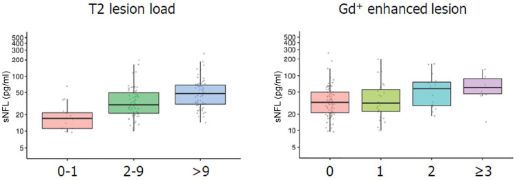
NFL can be measured using the most advanced and sensitive Single Molecule Array (Simoa®) method in cerebrospinal fluid (CSF), serum and plasma. NFL levels appear to reflect acute and chronic axonal damage, thus showing an association with relapses and EDSS progression.25–28 Data suggest that NFL levels might also predict brain atrophy over 15 years;12 however, this still needs to be established beyond doubt. Further, correlations have been demonstrated between NFL levels and other brain MRI measures (i.e. T2-lesion load, gadolinium-enhanced lesions; Figure 3), spinal cord atrophy and treatment response.25,29,30 Significant increases in NFL levels will most likely correspond to disease activity if other reasons are excluded. Increased NFL levels might be an important hint of ongoing inflammation that is not reflected by clinical signs or even MRI, although this has to be proven by prospective studies.
Figure 3.
Correlation between neurofilament-light chain levels and T2 lesion load/gadolinium-enhanced magnetic resonance imaging lesions.25
Gd, gadolinium; sNFL, serum neurofilament-light.
For practical purposes, NFL levels in the CSF have been shown to correlate well with corresponding serum/plasma levels, even though they are considerably lower. Serum may be more promising than plasma, as NFL levels and detection sensitivity are higher compared with plasma.30 Despite the increased NFL concentration in the CSF, 14% and 22% of the respective paired serum/plasma samples showed normal values.30 Therefore, monitoring intervals of 2–3 months appear reasonable if blood is examined in order to capture disease activity. Concomitant disorders (e.g. physical activity, trauma or small vessel disease31) may confound NFL levels and therefore need to be excluded as a reason for increased levels.
The panel experts agree that the clinical utility of NFL levels has not been fully determined yet. Evidently this is a marker suitable for monitoring rather than for diagnostics. Plasma and serum levels reflect treatment responses and disease reactivation. It might therefore be assumed that patients whose NFL levels do not normalize with treatment are at risk of current/ongoing disease activity. However, the available evidence does not yet justify treatment decisions in the individual patient. The significance of certain levels at different disease stages still needs to be defined. Dynamics play an important role, thus, the pattern might be more relevant than the absolute levels, especially in progressive cases. Also, costs and availability of routine measures might restrict the access to NFL assessment in many countries.
Issues left to elucidate include the sensitivity of measurements in CSF and blood, the implementation of cut-off levels including age-adjusted ranges, and the harmonization of assessment methods. Studies investigating these questions are ongoing. Ring-experimental studies among laboratories are required to specify the added value of NFL levels and to ensure the reliability and validity of measurements. There is a need for prospective studies that involve the collection of serum and plasma samples including their correlations with MRI and clinical findings.
Combinations of markers
For clinical routine, the experts recommended complex monitoring at specialized MS centers incorporating clinical evaluation (e.g. progression, relapses, cognition, patient-reported outcomes), MRI and laboratory parameters. Disability requires comprehensive regular assessments. Besides quantitative aspects, the quality of relapses (e.g. severity of relapses, incompleteness of remission) and the clinical and MRI topography of lesions deserve further attention.
An issue is the lack of standardization of disease biomarkers, especially composite ones, including the “No Evidence of Disease Activity” concept.32 Also, the question remains of which biomarkers to include in a composite system and how to measure, validate and interpret them. Biomarker dimensions might replace or support each other, or they might be independent.
Standardization is of particular importance with respect to MRI assessment. This implies the use of the same scanner and standardization of protocols and read-outs by (neuro-) radiologists. The findings should be systematically documented and entered into one database.
Implications for treatment
The panel expressed the opinion that there is no doubt about the usefulness of additional data constituting more rational and personalized treatment choices in MS patients. Certain drugs might limit the use of subsequent agents by facilitating adverse events in the long-term.12 The type and timing of escalation plays an enormous role in clinical practice. Investigation of sequential drug use and long-term safety outcomes are of utmost importance; such information can be gained from registry data since head-to-head and sequential treatment trials are unlikely to be conducted. The modes of drug action should be taken into consideration in the process of drug switching and treatment sequencing.12,13
It was agreed and advocated that early treatment and escalation therapy are necessary in patients with active disease as timely initiation of or switch to higher-efficacy agents are likely to improve long-term clinical outcomes.5,6 Natalizumab was the first drug to change significantly the course of disease in MS patients. However, treatment switches frequently take place rather late in clinical practice, although a trend to early escalation therapy has been already observed over the last years. The panel stated that the management should generally be more pro-active. Also, the idea of de-escalation has been raised in the face of a growing number of long-term treated patients. It refers to patients with clinical and MRI stable disease for many years, but, even more important, also to patients as they get older. In patients with advanced age, immunological function and clinical effectiveness of DMTs are unclear yet as the risk of adverse events might be increasing.33
The experts encouraged further investigations about how to procure, release and share these data. Support is required from pharmaceutical companies and government agencies with respect to collecting data routinely in a systematic way. Merging of databases can specifically promote this research. Of course, because of differences across MRI protocols and other measures, real-life data frequently lack standardization. Therefore, another option should be collaboration with pharmaceutical companies that often harbor big clinical data obtained in clinical trials.
Insights from the Cost of Illness Study
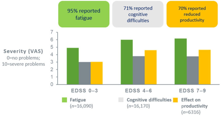
The observational, cross-sectional, retrospective MS Cost of Illness Study that was conducted in 16,808 patients from 16 countries showed that health-related quality of life decreased in MS patients with increase in their disability over time, while costs likewise increased in all 16 countries.34 Patterns of care were affected by healthcare structures, and almost all costs increased during relapses. DMTs represented a large proportion of the total cost in early disease, whereas other costs were increasing as the disease progressed. Dependence on others (i.e. informal care) greatly increased in severe MS. Fatigue was found to be an important problem across all severities and in all countries, affecting 95% of patients (Figure 4). Seventy-one percent reported cognitive impairment, and productivity at work was reduced in 70% of patients. Overall, the MS Cost of Illness Study confirmed the detrimental effect of MS on patients, their families and society. Healthcare consumption was influenced by the system, availability of services and medical tradition rather than by the disease itself.
Figure 4.
MS Cost of Illness Study: prevalence of fatigue, cognitive difficulties, and reduced productivity across all EDSS levels in multiple sclerosis patients.34
EDSS, Expanded Disability Status Scale; VAS, Visual Analogue Scale.
The analysis includes three sub-studies, the first of which showed a steep decline in utility at higher EDSS levels, which might suggest room for improvement with respect to treatment.35 Although the utility scores at different EDSS levels varied across countries, possibly reflecting attitudes towards disability, the shapes of the curves were similar. The second sub-study evaluated the effect of MS symptoms on work capacity and productivity while at work. Here, 79% of patients reported limitations in their productivity due to MS. After control for EDSS, both cognition and fatigue showed a strong and independent correlation with reduced work capacity.36 Dynamics were more relevant than the absolute levels of cognition and fatigue. The experts noted that the incorporation of Visual Analogue Scale assessments of these two parameters into routine practice might be helpful. The third sub-study assessed the effectiveness of DMTs on relapses and disability measured with EDSS in the Czech Republic, comparing results from 2007 and 2015.37 The analysis showed that the number of relapses was associated with a higher risk of progression. Treatment with second generation DMTs was associated with a lower risk of both relapses and progression to EDSS 4.
Management of cognitive deficits
Cognitive problems in MS are due to both synaptic network disruptions and brain atrophy. Certain brain regions known to be connected to cognitive domains tend to develop volume loss in MS patients (Table 1).38–46 During early disease, it is mainly clinically silent T2 lesions that impact cognition.47 More than 70% of patients have cognitive impairment across all stages of their disease.39 Patients with primary progressive MS show more severe cognitive decline than those with relapsing–remitting MS.48 Cognition is closely linked to employment status that represents an important and solid outcome.
Table 1.
| Regional brain volume loss | Associated cognitive domain |
|---|---|
| Cortex | • Verbal fluency and attention/concentration38,39
• Learning and memory40 |
| Corpus callosum | • Processing speed and rapid problem solving41
• Verbal fluency42 |
| Thalamus | • Information processing and attention43 |
| Hippocampus | • Memory, word-list learning44,45 |
| Parieto-occipital lobes | • Verbal learning and complex visual integration46 |
Cognitive impairment indicates disease activity or progression and should of course be prevented. However, pure cognitive relapses are controversial at this time. Moreover, transient cognitive decline has been observed at times of increased CNS inflammation. In a clinical trial, the cognitive performance worsened together with the emergence of contrast-enhanced MRI lesions but improved again when the contrast enhancement declined or resolved.49 This indicates that acute inflammation plays a role in cognitive dysfunction.
Diagnostics
Cognition is a complex process, which also necessitates complex evaluation. Various neuropsychological test batteries, including the Symbol Digit Modalities Test (SDMT),50 the Paced Auditory Serial Addition Test51 and the Brief International Cognitive Assessment for MS,52 have been established. As most of the panel experts use one or either of these cognitive tests in their clinical routine setting it has been agreed that certain practice effects need to be considered regarding SDMT, which should therefore not be used more often than every 6 months in the individual patient, as well as some pitfalls that relate to the fact that depression, fatigue and motor hand dysfunction can affect neuropsychological test results. The proportion of patients with cognitive impairment decreases if more rigorous and pragmatic tools are applied. Recently, the iPad-based self-administered CogEval™ tool, which was developed for screening of cognitive function in patients with MS, has been introduced. It provides a Processing Speed Test based on SDMT that uses attention, psychomotor speed, visual processing and working memory domains. The assessment takes 2 min and showed advantages over SDMT in a validation study.53
Despite their own routine practice in mainly academic MS centers, the panelists recognized that cognitive testing is usually not commonly performed on a regular basis in MS. Many neurologists lack practical experience in measures of cognitive functions. Even many MS centers lack neuropsychologists for more professional assessments and, of special importance, cognitive training. If a cognitive decline is observed during routine neurological examination, it should be mandatory to refer patients to full neuropsychological evaluation performed by well-trained psychologists. Of course, this requires more awareness in the care-giving community and appropriate resources given its time-consumption of cognitive examining. The experts assumed that MS-related cognitive dysfunction might be underestimated by neurologists in general as it eludes accurate assessment without standardization.
Aspects of treatment of cognitive dysfunction
Treatability of cognitive decline is subject to debate. No DMT has been approved specifically for the therapy of cognitive impairment in MS, and no evidence exists that shows clear improvement in cognition after DMT treatment. However, DMTs and symptomatic therapy might have some beneficial effects, as well as lifestyle modification and rehabilitation. Interferon beta gave rise to improvement of different domains of cognition compared with placebo.54 Also, reports suggest benefits of 1-year and 3-year natalizumab treatment.55,56 Contradictory results have been obtained for the symptomatic treatment of cognitive dysfunction in MS with various other drugs, including potassium channel blockers, dopaminergic modulators, acetylcholinesterase inhibitors, stimulants and fampridine.57–59 Rather than switching pharmaceutical agents, advising the patient on lifestyle modifications might be helpful. The experts admitted that this should be a general approach as treatments cannot be expected to improve cognition per se and, on the other hand, side effects of drugs might enhance cognitive dysfunction, for example, acetylcholinesterase inhibitors.
Recommendations on cognitive screening and management in MS have been recently published.60 As a minimum requirement, the authors recommended early baseline screening with SDMT or similarly validated tests at a time when the patient is clinically stable. Annual re-assessments should follow with the same instrument to detect acute disease activity, to assess for treatment effects or relapse recovery, to evaluate progression of cognitive impairment, and/or to screen for new-onset cognitive problems. Once impairment becomes evident, the patient should receive more comprehensive assessment.
Conclusion
The first European clinical practice guidelines on pharmacological management of MS have been published by ECTRIMS and EAN in 2018. It is acknowledged that these guidelines establish benchmarks, which should be adapted to local regulations and resources.
Predictive biomarkers guiding individual treatment decisions, including treatment initiation or switches, would be highly needed but are still awaited to date. MRI is currently the best biomarker for MS. Brain atrophy or atrophy of certain regions of the central nervous system including the spinal cord might prove relevant in the future. Combining MRI with other biomarkers is deemed essential. NFL levels are most promising to provide additional information but their clinical utility in individual patients needs to be established in the near future.
Immunological considerations, including matters of immunosenescence, will gain importance for forward planning and especially for the sequencing of drugs. Generally, attitudes have moved towards earlier initiation and change of treatment. Real-world data based on registries can contribute to evaluating the effectiveness and safety of treatment sequences.
Cognitive dysfunction in MS patients is an emerging topic. There is agreement that cognition function deserves more attention in routine practice and should be routinely tested. However, no evidence-based data are available that support switches of DMTs in patients with cognitive decline so far. Nevertheless, cognitive impairment as an important part of the socio-economic disease burden urges to be prevented using several approaches, including drug treatment and lifestyle modification.
Acknowledgments
All authors were involved in reviewing the manuscript critically for important intellectual content and provided final approval of the submitted version. We thank Dr. Judith Moser for drafting the manuscript. Biogen courteously reviewed the draft and provided feedback to the authors. Authors had full editorial control and provided approval to all final content.
Footnotes
Conflict of interest statement: Thomas Berger has participated in meetings sponsored by and received honoraria (lectures, advisory boards, consultations) from pharmaceutical companies marketing treatments for multiple sclerosis: Almirall, Bayer, Biogen, Biologix, Bionorica, Genzyme, MedDay, Merck, Novartis, Octapharma, Roche, Sanofi/Genzyme, TG Pharmaceuticals, TEVA-ratiopharm and UCB. His institution has received financial support in the last 12 months by unrestricted research grants (Biogen, Merck, Novartis, Sanofi/Genzyme) and for participation in clinical trials in multiple sclerosis sponsored by Biogen, Merck, Novartis, Roche, Sanofi/Genzyme, and TEVA.
Monika Adamczyk-Sowa has participated in meetings sponsored by and received honoraria (lectures, advisory boards, consultations) from pharmaceutical companies marketing treatments for multiple sclerosis: Bayer, Biogen, Merck, Novartis, Roche, Sanofi/Genzyme, TEVA.
Tunde Csepany has participated in meetings sponsored by and received honoraria (lectures, advisory boards, consultations) from pharmaceutical companies marketing treatments for multiple sclerosis: Biogen, Merck, Novartis, Roche, Sanofi/Genzyme, TEVA.
Tanja Hojs Fabjan received speaker honoraria and consultant fees from Bayer, Biogen, Lek, Novartis, Roche, Sanofi/Genzyme and for participations in trials in multiple sclerosis sponsored by Bayer, Biogen, Roche.
Dana Horakova received compensation for travel, speaker honoraria, and consultant fees from Bayer, Biogen, Merck, Novartis, Roche, Sanofi/Genzyme and Teva, as well as support for research activities from Biogen. She is also supported by the Czech Ministry of Education research project PROGRES Q27/LF1.
Zsolt Illes has participated in meetings sponsored by and received honoraria (lectures, advisory boards, consultations) from pharmaceutical companies marketing treatments for multiple sclerosis: Biogen, Merck, Novartis, Roche, Sanofi/Genzyme, TEVA, and received research grants from Biogen, Merck and Sanofi/Genzyme in the last 12 months.
Eleonóra Klímová has participated in meetings sponsored by and received honoraria (lectures, advisory boards, consultations) from pharmaceutical companies marketing treatments for multiple sclerosis: Biogen, Merck, Novartis, Roche, Sanofi/Genzyme and TEVA.
Gisela Kobelt has provided consulting and speaking services to Almirall, Bayer, Biogen, Merck, Novartis, Oxford PharmaGenesis, Roche, Sanofi/Genzyme, and Teva.
Fritz Leutmezer has participated in meetings sponsored by and received honoraria (lectures, advisory boards, consultations) from pharmaceutical companies marketing treatments for multiple sclerosis: Biogen, Celgene, MedDay, Merck, Novartis, Roche, Sanofi/Genzyme and TEVA.
Konrad Rejdak has participated in meetings sponsored by and received honoraria (lectures, advisory boards, consultations) from pharmaceutical companies marketing treatments for multiple sclerosis: Bayer, Biogen, Merck, Novartis, Roche, Sanofi/Genzyme, TEVA.
Saša Šega Jazbec received travelling grants and speaking honoraria from Biogen, Merck, Roche, Sanofi/Genzyme and Teva.
Csilla Rozsa has participated in meetings sponsored by and received honoraria (lectures, advisory boards) for multiple sclerosis: Biogen, Merck, Novartis, Roche, Sanofi/Genzyme and TEVA.
Johann Sellner has participated in meetings sponsored by and received honoraria (lectures, advisory boards, consultations) from pharmaceutical companies marketing treatments for multiple sclerosis: Alexion, Biogen, Celgene, MedDay, Merck, Novartis, Roche, Genzyme/Sanofi, TEVA-ratiopharm. His institution has received financial support in the last 12 months by unrestricted research grants (Biogen, Merck, Sanofi/Genzyme) and for participation in clinical trials sponsored by Merck and Roche.
Krzysztof Selmaj received honoraria for consulting and speaking from Biogen, Celgene, Merck, Novartis, Roche and TG Therapeutics.
Jarmila Szilasiova has participated in meetings sponsored by and received honoraria (lectures, advisory boards, consultations) from pharmaceutical companies marketing treatments for multiple sclerosis: Biogen, Merck, Novartis, Roche, Sanofi/Genzyme and TEVA.
Manuela Vaneckova has participated in meetings sponsored by and received honoraria (lectures, advisory boards, consultations) from pharmaceutical companies marketing treatments for multiple sclerosis: Biogen, Novartis, Roche, Sanofi/Genzyme, TEVA, and received research grants from Biogen and Roche in the last 12 months; supported by the Czech Ministry of Education research project PROGRES Q27/LF1 and RVO-VFN64165.
Peter Turcˇáni has participated in meetings sponsored by and received honoraria (lectures, advisory boards, consultations) from pharmaceutical companies marketing treatments for multiple sclerosis: Bayer, Biogen, Merck, Novartis, Roche, Sanofi/Genzyme, Teva. His institution has received financial support in the last 12 months by unrestricted research grants (Merck, Novartis, Roche, Sanofi/Genzyme) and for participation in clinical trials in multiple sclerosis sponsored by Biogen, Merck, Novartis, Roche, Sanofi/Genzyme, and Teva.
László Vécsei has participated in meetings sponsored by and received honoraria (lectures, advisory boards, consultations) for multiple sclerosis: Biogen, Merck, Novartis, Roche, Sanofi/Genzyme and TEVA.
Eva Kubala Havrdová: honoraria/research support from Biogen, Merck, Novartis, Roche, and Teva; advisory boards for Actelion, Biogen, Celgene, Merck, Novartis, and Sanofi/Genzyme; supported by the Czech Ministry of Education research project PROGRES Q27/LF1.
Funding: The authors disclosed receipt of the following financial support for the research, authorship, and/or publication of this article: This advisory expert meeting as well as the first drafting of the manuscript by J Moser was funded by Biogen.
ORCID iD: Thomas Berger  https://orcid.org/0000-0001-5626-1144
Contributor Information
Thomas Berger, Department of Neurology, Medical University of Vienna, Waehringer Guertel 18–20, Vienna 1090, Austria.
Monika Adamczyk-Sowa, Department of Neurology, Faculty of Medical Sciences in Zabrze, Medical University of Silesia in Katowice, Poland.
Tünde Csépány, Department of Neurology, Faculty of Medicine, University of Debrecen, Debrecen, Hungary.
Franz Fazekas, Department of Neurology, Medical University of Graz, Graz, Austria.
Tanja Hojs Fabjan, Department of Neurology, University Medical Centre Maribor, Maribor, Slovenia.
Dana Horáková, Department of Neurology and Center of Clinical Neuroscience, First Faculty of Medicine, Charles University and General University Hospital, Prague, Czech Republic.
Alenka Horvat Ledinek, Department of Neurology, University Clinical Centre Ljubljana, Ljubljana, Slovenia.
Zsolt Illes, Department of Neurology, University of Southern Denmark, Odense, Denmark.
Gisela Kobelt, European Health Economics AB, Stockholm, Sweden.
Saša Šega Jazbec, Department of Neurology, University Clinical Centre Ljubljana, Ljubljana, Slovenia.
Eleonóra Klímová, Department of Neurology, University of Prešov and Teaching Hospital of J. A. Reiman, Prešov, Slovakia.
Fritz Leutmezer, Department of Neurology, Medical University of Vienna, Vienna, Austria.
Konrad Rejdak, Department of Neurology, Medical University of Lublin, Lublin, Poland.
Csilla Rozsa, Department of Neurology, Jahn Ferenc Dél-pesti Hospital, Budapest, Hungary.
Johann Sellner, Department of Neurology, Landesklinikum Mistelbach-Gänserndorf, Mistelbach, Austria, and Department of Neurology, Christian Doppler Medical Center, Paracelsus Medical University, Salzburg, Austria.
Krzysztof Selmaj, Department of Neurology, University of Warmia-Mazury, Olsztyn, Poland.
Pavel Štouracˇ, Department of Neurology, Masaryk University, Brno, Czech Republic.
Jarmila Szilasiová, Department of Neurology, P. J. Šafárik University Košice and University Hospital of L. Pasteur Košice, Slovakia.
Peter Turcˇáni, Department of Neurology, Comenius University, Bratislava, Slovakia.
Marta Vachová, Centrum Teplice, Teplice, Czech Republic.
Manuela Vanecková, Department of Radiology, MRI Unit, First Faculty of Medicine, Charles University and General University Hospital, Prague, Czech Republic.
László Vécsei, Department of Neurology and MTA-SZTE Neuroscience Research Group, University of Szeged, Szeged, Hungary.
Eva Kubala Havrdová, Department of Neurology and Center of Clinical Neuroscience, First Faculty of Medicine, Charles University and General University Hospital, Prague, Czech Republic.
References
- 1. Montalban X, Gold R, Thompson AJ, et al. ECTRIMS/EAN guideline on the pharmacological treatment of people with multiple sclerosis. Eur J Neurol 2018; 25: 215–237. [DOI] [PubMed] [Google Scholar]
- 2. Montalban X, Gold R, Thompson AJ, et al. ECTRIMS/EAN guideline on the pharmacological treatment of people with multiple sclerosis. Mult Scler 2018; 18: 1–25. [DOI] [PubMed] [Google Scholar]
- 3. Berger T, Adamczyk-Sowa M, Csepany T, et al. Management of multiple sclerosis patients in central European countries: current needs and potential solutions. Ther Adv Neurol Disord 2018; 11: 1756286418759189. [DOI] [PMC free article] [PubMed] [Google Scholar]
- 4. Havrdova E, Zivadinov R, Krasensky J, et al. Randomized study of interferon beta-1a, low-dose azathioprine, and low-dose corticosteroids in multiple sclerosis. Mult Scler 2009; 15: 965–976. [DOI] [PubMed] [Google Scholar]
- 5. Ontaneda D, Tallantyre E, Kalincik T, et al. Early highly effective versus escalation treatment approaches in relapsing multiple sclerosis. Lancet Neurol 2019; 18: 973–980. [DOI] [PubMed] [Google Scholar]
- 6. He A, Merkel B, Brown JWL, et al. Timing of high-efficacy therapy for multiple sclerosis: a retrospective observational cohort study. Lancet Neurol 2020; 19: 307–316. [DOI] [PubMed] [Google Scholar]
- 7. Brown JWL, Coles A, Horakova D, et al. Association of initial disease-modifying therapy with later conversion to secondary progressive multiple sclerosis. JAMA Neurol 2019; 321: 175–187. [DOI] [PMC free article] [PubMed] [Google Scholar]
- 8. Bsteh G, Hegen H, Dosser C, et al. To treat or not to treat: sequential individualized treatment evaluation in relapsing multiple sclerosis. Mult Scler Rel Dis 2020; 39: 101908. [DOI] [PubMed] [Google Scholar]
- 9. Gasperini C, Prosperini L, Tintoré M, et al. Unraveling treatment response in multiple sclerosis: a clinical and MRI challenge. Neurology 2019; 92: 180–192. [DOI] [PMC free article] [PubMed] [Google Scholar]
- 10. Bsteh G, Feige J, Ehling R, et al. Discontinuation of disease-modifying therapies in multiple sclerosis – clinical outcome and prognostic factors. Mult Scler 2017; 23: 1241–1248. [DOI] [PubMed] [Google Scholar]
- 11. Sorensen PS, Sjelleberg F, Hartung HP, et al. The apparently milder course of multiple sclerosis: changes in the diagnostic criteria, therapy and natural history. Brain 2020; 143: 2637–2652. [DOI] [PubMed] [Google Scholar]
- 12. Pardo G, Jones DE. The sequence of disease-modifying therapies in relapsing multiple sclerosis: safety and immunological considerations. J Neurol 2017; 264: 2351–2374. [DOI] [PMC free article] [PubMed] [Google Scholar]
- 13. Green AJ. Potential benefits of early aggressive treatment in multiple sclerosis. JAMA Neurol 2019; 76: 254–256. [DOI] [PubMed] [Google Scholar]
- 14. Rocca MA, Battaglini M, Benedict RH, et al. Brain MRI atrophy quantification in MS: from methods to clinical application. Neurology 2017; 88: 403–413. [DOI] [PMC free article] [PubMed] [Google Scholar]
- 15. Eshaghi A, Marinescu RV, Young AL, et al. Progression of regional grey matter atrophy in multiple sclerosis. Brain 2018; 141: 1665–1677. [DOI] [PMC free article] [PubMed] [Google Scholar]
- 16. Sastre-Garriga J, Pareto D, Battaglini M, et al. MAGNIMS consensus recommendations on the use of brain and spinal cord atrophy measures in clinical practice. Nat Rev Neurol 2020; 16: 171–182. [DOI] [PMC free article] [PubMed] [Google Scholar]
- 17. Uher T, Vaneckova M, Krasensky J, et al. Pathological cut-offs of global and regional brain volume loss in multiple sclerosis. Mult Scler 2019; 25: 541–553. [DOI] [PubMed] [Google Scholar]
- 18. Uher T, Vaneckova M, Sormani MP, et al. Identification of multiple sclerosis patients at highest risk of cognitive impairment using an integrated brain magnetic resonance imaging assessment approach. Eur J Neurol 2017; 24: 292–301. [DOI] [PubMed] [Google Scholar]
- 19. Uher T, Vaneckova M, Sobisek L, et al. Combining clinical and magnetic resonance imaging markers enhances prediction of 12-year disability in multiple sclerosis. Mult Scler 2017; 23: 51–61. [DOI] [PubMed] [Google Scholar]
- 20. Bonacchi R, Pagani E, Meani A, et al. Clinical relevance of multiparametric MRI assessment of cervical cord damage in multiple sclerosis. Radiology 2020; 296: 605–615. [DOI] [PubMed] [Google Scholar]
- 21. Hagström IT, Schneider R, Bellenberg B, et al. Relevance of early cervical cord volume loss in the disease evolution of clinically isolated syndrome and early multiple sclerosis: a 2-year follow-up study. J Neurol 2017; 264: 1402–1412. [DOI] [PubMed] [Google Scholar]
- 22. Barkhof F. The clinic-radiological paradox in multiple sclerosis revisited. Curr Opin Neurol 2002; 15: 239–245. [DOI] [PubMed] [Google Scholar]
- 23. Kerbrat A, Gros C, Badji A, et al. Multiple sclerosis lesions in motor tracts from brain to cervical cord: spatial distribution and correlation with disability. Brain 2020; 143: 2089–2105. [DOI] [PMC free article] [PubMed] [Google Scholar]
- 24. Magliozzi R, Howell OW, Nicholas R, et al. Inflammatory intrathecal profiles and cortical damage in multiple sclerosis. Ann Neurol 2018; 83: 739–755. [DOI] [PubMed] [Google Scholar]
- 25. Disanto G, Barro C, Benkert P, et al. Serum neurofilament light: a biomarker of neuronal damage in multiple sclerosis. Ann Neurol 2017; 81: 857–870. [DOI] [PMC free article] [PubMed] [Google Scholar]
- 26. Barro C, Benkert P, Disanto G, et al. Serum neurofilament as a predictor of disease worsening and brain and spinal cord atrophy in multiple sclerosis. Brain 2018; 141: 2382–2391. [DOI] [PubMed] [Google Scholar]
- 27. Bhan A, Jacobsen C, Myhr KM, et al. Neurofilaments and 10-year follow-up in multiple sclerosis. Mult Scler 2018; 24: 1301–1307. [DOI] [PubMed] [Google Scholar]
- 28. Williams T, Zetterberg H, Chataway J. Neurofilaments in progressive multiple sclerosis: a systematic review. J Neurol. Epub ahead of print 23 May 2020. DOI: 10.1007/s00415-020-09917-x. [DOI] [PMC free article] [PubMed] [Google Scholar]
- 29. Petzold A, Steenwijk MD, Eikelenboom JM, et al. Elevated CSF neurofilament proteins predict brain atrophy: a 15-year follow-up study. Mult Scler 2016; 22: 1154–1162. [DOI] [PubMed] [Google Scholar]
- 30. Sejbaek T, Nielsen HH, Penner N, et al. Dimethyl fumarate decreases neurofilament light chain in CSF and blood of treatment naïve relapsing MS patients. J Neurol Neurosurg Psych 2019; 90: 1324–1330. [DOI] [PMC free article] [PubMed] [Google Scholar]
- 31. Gattringer T, Pinter D, Enzinger C, et al. Serum neurofilament light is sensitive to active cerebral small vessel disease. Neurology 2017; 89: 2108–2114. [DOI] [PMC free article] [PubMed] [Google Scholar]
- 32. Hegen H, Bsteh G, Berger T. No evidence of disease activity - is it an appropriate surrogate in multiple sclerosis? Eur J Neurol 2018; 25: 1107-e101. [DOI] [PMC free article] [PubMed] [Google Scholar]
- 33. Paghera S, Sottini A, Previcini V, et al. Age-related lymphocyte output during disease-modifying therapy in multiple sclerosis. Drugs Aging 2020; 37: 739–746. [DOI] [PubMed] [Google Scholar]
- 34. Kobelt G, Thompson A, Berg J, et al. New insights into the burden and costs of multiple sclerosis in Europe. Mult Scler 2017; 23: 1123–1136. [DOI] [PMC free article] [PubMed] [Google Scholar]
- 35. Eriksson J, Kobelt G, Gannedahl M, et al. Association between disability, cognition, fatigue, EQ-5D-3L domains, and utilities estimated with different western European value sets in patients with multiple sclerosis. Value Health 2019; 22: 231–238. [DOI] [PubMed] [Google Scholar]
- 36. Kobelt G, Langdon D, Jonsson L. The effect of self-assessed fatigue and subjective cognitive impairment on work capacity: the case of multiple sclerosis. Mult Scler 2019; 25: 740–749. [DOI] [PMC free article] [PubMed] [Google Scholar]
- 37. Kobelt G, Jönsson L, Pavelcova M, et al. Real-life outcome in multiple sclerosis in the Czech Republic. Mult Scler Int 2019; 2019: 7290285. [DOI] [PMC free article] [PubMed] [Google Scholar]
- 38. Amato MP, Bartolozzi ML, Zipoli V, et al. Neocortical volume decrease in relapsing-remitting MS patients with mild cognitive impairment. Neurology 2004; 63: 89–93. [DOI] [PubMed] [Google Scholar]
- 39. Rovaris M, Barkhof F, Calabrese M, et al. MRI features of benign multiple sclerosis: toward a new definition of this disease phenotype. Neurology 2009; 72: 1693–1701. [DOI] [PubMed] [Google Scholar]
- 40. Bagnato F, Salman Z, Kane R, et al. T1 cortical hypointensities and their association with cognitive disability in multiple sclerosis. Mult Scler 2010; 16: 1203–1212. [DOI] [PubMed] [Google Scholar]
- 41. Rao SM, Leo GJ, Haughton VM, et al. Correlation of magnetic resonance imaging with neuropsychological testing in multiple sclerosis. Neurology 1989; 39: 161–166. [DOI] [PubMed] [Google Scholar]
- 42. Giorgio A, De Stefano N. Cognition in multiple sclerosis: relevance of lesions, brain atrophy and proton MR spectroscopy. Neurol Sci 2010; 31(Suppl. 2): S245–S248. [DOI] [PubMed] [Google Scholar]
- 43. Houtchens MK, Benedict RH, Killiany R, et al. Thalamic atrophy and cognition in multiple sclerosis. Neurology 2007; 69: 1213–1223. [DOI] [PubMed] [Google Scholar]
- 44. Sicotte NL, Kern KC, Giesser BS, et al. Regional hippocampal atrophy in multiple sclerosis. Brain 2008; 131: 1134–1141. [DOI] [PubMed] [Google Scholar]
- 45. Planche V, Koubiyr I, Romero JE, et al. Regional hippocampal vulnerability in early multiple sclerosis: dynamic pathological spreading from dentate gyrus to CA1. Hum Brain Mapp 2018; 39: 1814–1824. [DOI] [PMC free article] [PubMed] [Google Scholar]
- 46. Filippi M, Rocca A. MRI and cognition in multiple sclerosis. Neurol Sci 2010; 31(Suppl. 2): S231–S234. [DOI] [PubMed] [Google Scholar]
- 47. Wybrecht D, Reuter F, Pariollaud F, et al. New brain lesions with no impact on physical disability can impact cognition in early multiple sclerosis: a ten-year longitudinal study. PLoS One 2017; 12: e0184650. [DOI] [PMC free article] [PubMed] [Google Scholar]
- 48. Ruet A, Deloire M, Charré-Morin J, et al. Cognitive impairment differs between primary progressive and relapsing-remitting MS. Neurology 2013; 80: 1501–1508. [DOI] [PubMed] [Google Scholar]
- 49. Bellmann-Strobl J, Wuerfel J, Aktas O, et al. Poor PASAT performance correlates with MRI contrast enhancement in multiple sclerosis. Neurology 2009; 73: 1624–1627. [DOI] [PubMed] [Google Scholar]
- 50. Strober L, De Luca J, Benedict RHB, et al. Symbol digit modalities test: a valid clinical trial endpoint for measuring cognition in multiple sclerosis. Mult Scler 2019; 25: 1781–1790. [DOI] [PMC free article] [PubMed] [Google Scholar]
- 51. Renner A, Baetge SJ, Filser M, et al. Characterizing cognitive deficits and potential predictors in multiple sclerosis: a large nationwide study applying BICAMS in standard clinical care. J Neuropsychol 2020; 14: 347–369. [DOI] [PubMed] [Google Scholar]
- 52. Niccolai C, Portaccio E, Goretti B, et al. A comparison of the brief international cognitive assessment for multiple sclerosis and the brief repeatable battery in multiple sclerosis patients. BMC Neurol 2015; 15: 204. [DOI] [PMC free article] [PubMed] [Google Scholar]
- 53. Rao SM, Losinski G, Mourany L, et al. Processing speed test: validation of a self-administered, iPad®-based tool for screening cognitive dysfunction in a clinic setting. Mult Scler 2017; 23: 1929–1937. [DOI] [PubMed] [Google Scholar]
- 54. Fischer JS, Priore RL, Jacobs LD, et al. Neuropsychological effects of interferon beta-1a in relapsing multiple sclerosis. Multiple Sclerosis Collaborative Research Group. Ann Neurol 2000; 48: 885–892. [PubMed] [Google Scholar]
- 55. Rorsman I, Petersen C, Nilsson PC. Cognitive functioning following one-year natalizumab treatment: a non-randomized clinical trial. Acta Neurol Scand 2018; 137: 117–124. [DOI] [PubMed] [Google Scholar]
- 56. Mattioli F, Stampatori C, Bellomi F, et al. Natalizumab significantly improves cognitive impairment over three years in MS: pattern of disability progression and preliminary MRI findings. PLoS One 2015; 10: e0131803. [DOI] [PMC free article] [PubMed] [Google Scholar]
- 57. Amato MP, Langdon D, Montalban X, et al. Treatment of cognitive impairment in multiple sclerosis: position paper. J Neurol 2013; 260: 1452–1468. [DOI] [PubMed] [Google Scholar]
- 58. Morrow SA, Rosehart H, Johnson AM. The effect of Fampridine-SR on cognitive fatigue in a randomized double-blind crossover trial in patients with MS. Mult Scler Relat Disord 2017; 11: 4–9. [DOI] [PubMed] [Google Scholar]
- 59. Broicher SD, Filli L, Geisseler O, et al. Positive effects of fampridine on cognition, fatigue and depression in patients with multiple sclerosis over 2 years. J Neurol 2018; 265: 1016–1025. [DOI] [PubMed] [Google Scholar]
- 60. Kalb R, Beier M, Benedict RH, et al. Recommendations for cognitive screening and management in multiple sclerosis care. Mult Scler 2018; 24: 1665–1680. [DOI] [PMC free article] [PubMed] [Google Scholar]


