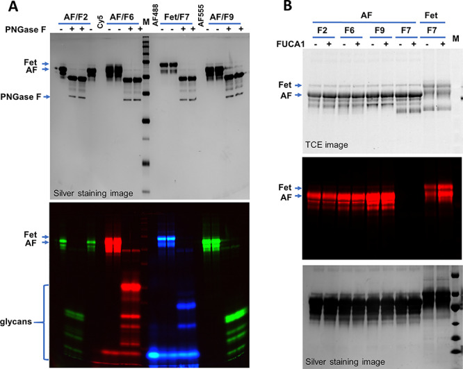
Fig. 2.

Characterizing the labeled glycans on fetuin (Fet) and asialofetuin (AF) with PNGase F and FUCA1. (A) PNGase F treatment of the labeled samples. AF was labeled by FUT2 (F2), FUT6 (F6) and FUT9(F9) with Alexa Fluor® 555 (green) or Cy5 (red). Fet was labeled by FUT7 (F7) with Alexa Fluor® 488 (blue). Labeled samples were then treated with PNGase F to release the glycans. (B) Effect of FUCA1 on the labeling. Samples without or with FUCA1 treatment were labeled by the indicated enzymes with Cy5. All samples were separated on 15% SDS–PAGE and imaged by silver staining, TCE staining and fluorescent imaging as indicated. M, western blot molecular marker.
