Abstract
Background
Myocarditis is a rare but life-threatening adverse event of cancer treatments with immune checkpoint inhibitors (ICIs). Recent guidelines recommend the use of high doses of corticosteroids as a first-line treatment, followed by intensified immunosuppressive therapy (IIST) in the case of unfavorable evolution. However, this strategy is empirical, and no studies have specifically addressed this issue. Therefore, we aimed to investigate and compare the clinical course, management and outcome of ICI-induced myocarditis patients requiring or not requiring IIST.
Methods
This case–control study included all patients consecutively admitted to The Mediterranean University Center of Cardio-Oncology (Aix-Marseille University, France) for the diagnosis of ICI-induced myocarditis according to Bonaca’s criteria and treated with or without IIST. In addition, we searched PubMed and included patients from previously published case reports treated with IIST in the analysis. The clinical, biological, imaging, treatment, all-cause death and cardiovascular death data of patients who required IIST were compared with those of patients who did not.
Results
A total of 60 patients (69±12 years) were included (36 were treated with IIST and 24 were not). Patients requiring IIST were more likely to have received a combination of ICIs (39% vs 8%, p=0.01), and developed the first symptoms/signs of myocarditis earlier after the onset of ICI therapy (median, 18 days vs 60 days, p=0.002). They had a significantly higher prevalence of sustained ventricular arrhythmia, complete atrioventricular block, cardiogenic shock and troponin elevation. Moreover, they were more likely to have other immune-related adverse events simultaneously (p<0.0001), especially myositis (p=0.0002) and myasthenia gravis (p=0.009). Patients who required IIST were more likely to die from any cause (50% vs 21%, p=0.02). Among them, patients who received infliximab were more likely to die from cardiovascular causes (OR, 12.0; 95% CI 2.1 to 67.1; p=0.005).
Conclusion
The need for IIST was more common in patients who developed myocarditis very early after the start of ICI therapy, as well as when hemodynamic/electrical instability or neuromuscular adverse events occurred. Treatment with infliximab might be associated with an increased risk of cardiovascular death.
Keywords: immunotherapy
Background
Immune checkpoint inhibitors (ICIs) are monoclonal antibodies that restore the immune response of CD8+ and CD4+T cells against tumor tissue by blocking the inhibitory action of ligand/receptor interactions. They include programmed death-1 checkpoint inhibitor (PD-1i), PD ligand-1 checkpoint inhibitor (PD-L1i), cytotoxic T-lymphocyte-associated protein-4 inhibitor (CTLA-4i), and lymphocyte-activation gene 3 inhibitor (LAG-3 i).1 Although these drugs represent a major advance in the treatment of many cancers, they are associated with several immune-related adverse events (irAEs) that may lead to mitigated overall therapeutic efficacy.2–4 ICI-induced myocarditis is one of the most feared irAEs, as it is associated with a case fatality rate of approximately 40%.5 It exposes patients to a risk of acute heart failure and sudden death due to ventricular arrythmia, pulseless electrical activity or complete atrioventricular block.6–11 Histological studies have shown myocyte necrosis associated with CD4+ and CD8+T cell infiltration similar to that observed during acute cell rejection of transplanted hearts.6 12 Thus, recent American and European guidelines have recommended the discontinuation of ICIs, treatment with high doses of corticosteroids as first-line therapy, and intensified immunosuppressive therapy (IIST) as soon as evolution is unfavorable. It is then recommended to consider other immunosuppressive drugs, such as infliximab, mycophenolate mofetil (MMF), antithymocyte globulin (ATG) or tacrolimus.13–15 However, these guidelines are based on expert consensus without strong evidence, and no studies have analyzed these immunosuppressive therapeutic strategies. In addition, the use of other immunosuppressive therapies, such as abatacept, alemtuzumab, tocilizumab, intravenous Ig and plasma exchange, have been recently described in a few case reports.16–18
In an effort to provide more data on the utilization of IIST, we aimed to investigate and compare the clinical course, management, and outcome of ICI-induced myocarditis patients requiring or not requiring IIST in a case–control study.
Methods
Study design and participants
We conducted a retrospective case–control study. From March 1 2015 to March 1 2020, the medical records of consecutive patients with a clinical suspicion of ICI-induced myocarditis were reviewed from the databases of The University Mediterranean Center of Cardio-Oncology in the North Hospital (Aix-Marseille University, France), a referral teaching hospital. During this period, patients were referred to our center when physicians had suspected myocarditis on the basis of clinical, biological or imaging cardiovascular evidence. From January 2018, all patients receiving ICI therapy in our center were also followed according to a standardized protocol. It included a cardio-oncology clinical visit with an ECG, transthoracic echocardiogram (TTE), and ultrasensitive troponin measurement (I then T from January 2019) before the beginning of treatment. Then, troponin measurement and ECG were performed before each ICI administration. In the case of cardiovascular symptoms/signs, recent ECG changes, or troponin increase >99th percentile of the upper reference limit provided by the manufacturing companies, patients were referred to our center for workup. Additional investigations, including cardiovascular MR (CMR) or endomyocardial biopsy, were performed according to the physician’s decision. Myocarditis management was left to the physician’s discretion. Only patients with a diagnosis of definite, probable, or possible myocarditis according to Bonaca’s criteria19 were included in the study. Patients were divided into two groups based on whether they were receiving an IIST defined as immunosuppressive therapy other than corticosteroids. All patients had given their consent on admission to permit the use of their personal medical data for research purposes.
In an effort to increase the size of the IIST patient group, we also included patients from previously published case reports who were treated for ICI-induced myocarditis with IIST. To identify these cases, we searched PubMed for articles using all combinations between group A and group B search terms with a cut-off date of April 2020. The group A search terms included the following: “myocarditis”, “cardiotoxicity”, “heart failure”, “adverse events”, “cardiac”, “cardiovascular”, “heart”, “pericarditis”, “myocardium” and “myocardial”. The group B search terms included the following: “immunotherapy”, “immunotherapies”, “checkpoint blockade”, “checkpoint inhibitors”, “CTLA-4”, “PD-1”, “PD-L1” and “LAG-3”. This search was limited to English articles. We also searched the reference lists of articles identified by this search strategy and selected additional references that we judged to be relevant. We finally selected the cases reporting in detail the patient characteristics, the evolution of the disease, and IIST as defined above. We ensured that each patient was included only once. Among the selected cases, we excluded those for which data were considered insufficient to formally draw a conclusion on definite, probable, or possible ICI-induced myocarditis according to Bonaca’s criteria.19 The patients of our center and those previously published were confirmed by two independent researchers.
The Strengthening the Reporting of Observational Studies in Epidemiology statement was followed to ensure the quality of data reported in this study.
Data collection
Data of interest were extracted retrospectively from electronic medical records and case report publications. These included standard demographic, cardiovascular risk factor, other irAEs, ECG, TTE, CMR and biomarker data. Myocarditis-CMR diagnosis was defined as the presence of two out of two 2018-Lake Louise major criteria,20 or one out of two criteria associated with a plausible clinical scenario. Positive troponin was defined as a serum level >99th percentile of the URL associated with a dynamic evolution. Cancer-specific covariates included the type of cancer, ICI therapy and prior cancer treatment. Myocarditis-specific covariates also included clinical presentation, physical examination, CMR data, biopsy data and myocarditis treatments. Myocarditis complications were analyzed, and each myocarditis episode was graded according to the guidelines of The American Society of Clinical Oncology.13 For patients admitted to our center, the diagnosis of immune-related neuromuscular disorders was made by experienced neurologists by integrating the results of their clinical evaluation, serum CK levels, the presence of autoantibodies, the results of electroneuromyography and the neuromuscular biopsy. For reported cases, this diagnosis was considered in the analysis if the authors reported it in their description.
IIST and outcome
Indications for IIST were classified as (1) hemodynamic, defined as the development, persistence or aggravation of heart failure syndrome or decreased left ventricular ejection fraction despite corticosteroid therapy; (2) electrical, defined as the development, persistence, or aggravation of ventricular arrhythmia or severe cardiac conduction abnormality despite corticosteroid therapy; (3) biological, defined as no decrease or increase in troponin levels after more than 3 days of corticosteroid therapy; (4) other, defined as the presence of severe noncardiac irAEs or a decision by physicians to initiate IIST in the absence of the other previous indications.
Study outcomes were deaths from any cause and myocarditis-related cardiovascular deaths.
For patients included in our center, the vital status was determined within the year after the day of admission. For patients previously published in case reports, the time from admission to death was always specified in the publication. Therefore, we also considered only deaths occurring within the year after the day of admission. Since we did not have the date of the vital status for some patients who survived the myocarditis episode, we considered them to be alive at 1 year.
Statistical analysis
Continuous variables are expressed as means±SDs or medians (IQR) and compared using an unpaired t-test or Mann-Whitney U test. Categorical variables are expressed as frequencies (percentages) and compared using the χ2 or Fisher exact test. ORs and 95% CIs were estimated by logistic regression. All tests of significance were two sided, and a p value of 0.05 was considered significant. Statistical analysis was performed using SPSS V.20.0 (SPSS).
Results
Study population
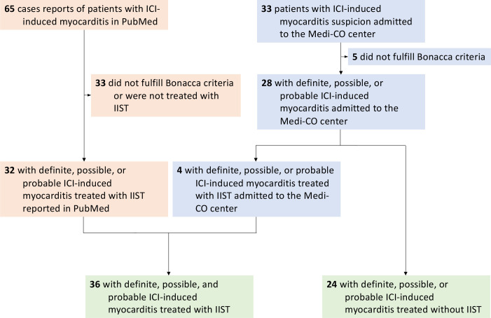
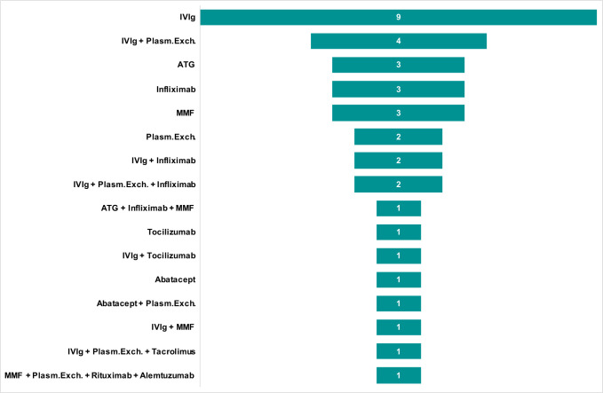
From March 1 2015 to January 2020, a total of 33 consecutive patients in whom ICI-induced myocarditis was suspected were admitted to our center. Of these patients, five were excluded because they did not fulfill Bonaca’s criteria for the diagnosis of myocarditis. Of the 28 on-site patients, four (14%) required IIST. Additionally, our research identified 65 published case reports of ICI-induced myocarditis. Of them, 33 were excluded because they did not fulfill Bonaca’s criteria and/or were not treated with IIST (online supplemental table S1). A total of 60 patients (69±12 years) were finally analyzed in this study (36 were treated with IIST and 24 were not, figure 1). The type of IIST is shown in figure 2. It involved intravenous Ig (n=20), ATG (n=4), infliximab (n=8), tocilizumab (n=2), rituximab (n=1), MMF (n=6), abatacept (n=2), alemtuzumab (n=1), tacrolimus (n=1) and plasma exchange therapy (n=11). Combinations of these therapies were used in eight patients (22%). The reasons for IIST were hemodynamic (n=10), electrical (n=2), biological (n=7) and others (n=15).
Figure 1.
Study flow chart. ICI, immune checkpoint inhibitor; IIST, intensified immunosuppressive therapy.
Figure 2.
Intensified immunosuppressive therapy. ATG, antithymocyte globulin; ICI, immune checkpoint inhibitor; IIST, intensified immunosuppressive therapy; IV, intravenous; MMF, mycophenolate mofetil; Plasm.Exch., plasma exchange therapy.
jitc-2020-001887supp001.pdf (35.5KB, pdf)
Baseline characteristics before myocarditis
The baseline characteristics of the two groups of patients are shown in table 1. In comparison with patients treated without IIST, IIST patients had a lower prevalence of smoking history and non-small-cell lung cancer. Other demographic characteristics and other types of cancer did not differ significantly between the two groups. Regarding immunotherapy, IIST patients were more likely to have received a combination of ICIs, especially PD-1i with CTLA-4i (39% vs 8%, p=0.01).
Table 1.
Baseline characteristics
| IIST (n=36) |
No IIST (n=24) |
P value |
|
| Age, years | 69±11 | 69±11 | 0.88 |
| Female | 16 (44) | 9 (38) | 0.59 |
| CV risk factors and diseases | |||
| Current or prior smoking* | 4 (17) | 11 (46) | 0.03 |
| Hypertension* | 9 (38) | 14 (22) | 0.15 |
| Diabetes mellitus* | 6 (25) | 2 (8) | 0.25 |
| Dyslipidemia* | 5 (21) | 2 (8) | 0.42 |
| Coronary artery disease* | 2 (8) | 3 (13) | 1.0 |
| Stroke* | 1 (4) | 0 (0) | 1.0 |
| Atrial fibrillation* | 1 (4) | 3 (13) | 0.61 |
| Heart failure* | 0 (0) | 2 (8) | 0.49 |
| COPD* | 0 (0) | 3 (13) | 0.23 |
| Cancer | |||
| Melanoma | 19 (53) | 9 (38) | 0.25 |
| Non-small-cell lung cancer | 6 (17) | 11 (46) | 0.01 |
| Renal cell carcinoma | 4 (11) | 1 (4) | 0.64 |
| Gastric carcinoma | 1 (3) | 1 (4) | 1.0 |
| Glioblastoma | 1 (3) | 0 (0) | 1.0 |
| Myelodysplastic syndrome | 2 (6) | 0 (0) | 0.51 |
| Mesothelioma | 1 (3) | 0 (0) | 1.0 |
| Thymoma | 1 (3) | 0 (0) | 1.0 |
| Head and neck | 0 (0) | 2 (8) | 0.16 |
| Uterus | 1 (3) | 0 (0) | 1.0 |
| Prior chemotherapy† | 15 (60) | 15 (63) | 0.86 |
| Overall types of ICI | |||
| Nivolumab (PD-1i) | 23 (64) | 14 (58) | 0.67 |
| Pembrolizumab (PD-1i) | 9 (25) | 2 (8) | 0.17 |
| Sintilimab (PD-1i) | 1 (3) | 0 (0) | 1.0 |
| Atezolizumab (PD-L1i) | 0 (0) | 4 (17) | 0.02 |
| Durvalumab (PD-L1i) | 2 (6) | 0 (0) | 0.51 |
| Ipilimumab (CTLA-4i) | 16 (44) | 4 (17) | 0.03 |
| Tremelimumab (CTLA-4i) | 1 (3) | 0 (0) | 1.0 |
| Relatlimab (LAG-3i) | 0 (0) | 3 (13) | 0.06 |
| Any PD-1i/PD-L1i | 34 (94) | 23 (96) | 1.0 |
| Any PD-1i | 33 (92) | 17 (71) | 0.07 |
| Any PD-L1i | 1 (3) | 6 (25) | 0.009 |
| Any CTLA-4i | 17 (47) | 4 (17) | 0.03 |
| Any LAG-3i | 0 (0) | 3 (13) | 0.06 |
| Single ICI agent | |||
| Nivolumab | 9 (25) | 9 (38) | 0.3 |
| Pembrolizumab | 7 (19) | 2 (8) | 0.29 |
| Atezolizumab | 0 (0) | 4 (17) | 0.02 |
| Ipilimumab | 2 (6) | 1 (4) | 1.0 |
| Durvalumab | 0 (0) | 1 (4) | 0.4 |
| Sintilimab | 1 (3) | 0 (0) | 1.0 |
| Combination ICI | |||
| Any PD-1i/PD-L1i + CTLA-4i | 15 (42) | 3 (13) | 0.01 |
| Nivolumab+ipilimumab | 14 (39) | 2 (8) | 0.01 |
| Durvalumab+tremelimumab | 1 (3) | 0 (0) | 1.0 |
| Nivolumab+relatlimab | 0 (0) | 1 (4) | 0.4 |
| Durvalumab+tremelimumab | 1 (3) | 0 (0) | 1.0 |
| Nivolumab+ipilimumab+relatlimab | 0 (0) | 1 (4) | 0.4 |
Values are mean±SD, n (%).
*Twenty-four of the 36 IIST patients had this information.
†Twenty-five of the 36 IIST patients had this information.
COPD, chronic obstructive pulmonary disease; CTLA-4i, cytotoxic T-lymphocyte-associated protein-4 inhibitor; CV, cardiovascular; ICI, immune checkpoint inhibitor; IIST, intensified immunosuppressive therapy; PD-1i/PD-L1i, programmed death-1 checkpoint inhibitor/programmed death-1 checkpoint inhibitor.
Myocarditis presentation, clinical course and management
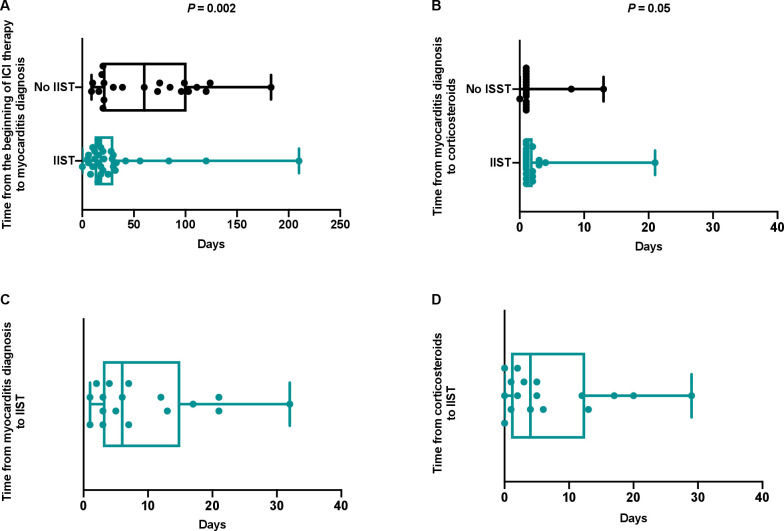
The median time from first ICI administration to the onset of myocarditis was 21 days (IQR 15–63 days). It was significantly shorter in patients who subsequently required IIST (18 days (IQR 12–30 days) vs 60 days (IQR 20–201 days), p=0.002) (figure 3). These patients were more likely to have a more severe form of myocarditis (table 2). Indeed, they had a significantly higher prevalence of sustained ventricular arrhythmia, complete atrioventricular block, cardiogenic shock and troponin elevation. Moreover, they were more likely to have other irAEs simultaneously (p<0.0001), especially myositis (p=0.0002) and myasthenia gravis (p=0.009).
Figure 3.
Times between beginning of immune checkpoint inhibitor therapy, onset of myocarditis, corticosteroid therapy and intensified immunosuppressive therapy. ICI, immune checkpoint inhibitor; IIST, intensified immunosuppressive therapy.
Table 2.
Myocarditis presentation and clinical course
| IIST (n=36) | No IIST (n=24) | P value | |
| Time from beginning ICI to onset myocarditis, days* | 18 (12–30) | 60 (20–101) | 0.002 |
| Clinical presentation | |||
| Chest pain | 4 (11) | 3 (13) | 1.0 |
| Shortness of breath | 22 (61) | 11 (46) | 0.24 |
| Palpitations | 5 (14) | 1 (4) | 0.39 |
| Pericardial effusion | 1 (3) | 0 (0) | 1.0 |
| Asymptomatic troponin elevation | 7 (19) | 8 (33) | 0.22 |
| ECG on admission | |||
| Atrial fibrillation | 8 (22) | 3 (13) | 0.50 |
| Ventricular tachycardia | 4 (11) | 1 (4) | 0.64 |
| Complete atrioventricular block† | 12 (34) | 0 (0) | 0.002 |
| Complete left bundle branch block† | 3 (9) | 2 (8) | 1.0 |
| Complete right bundle branch block† | 8 (23) | 2 (8) | 0.29 |
| T wave or ST segment abnormality† | 8 (23) | 3 (13) | 0.51 |
| LVEF on admission, %‡ | 50±14 | 55±12 | 0.12 |
| CMR | |||
| Performed | 20 (56) | 20 (83) | 0.03 |
| Myocarditis-CMR diagnosis | 18 (90) | 19 (95) | 1.0 |
| Elevated troponin‡ | 32 (89) | 17 (71) | 0.02 |
| Endomyocardial biopsy | |||
| Performed | 10 (28) | 0 (0) | 0.004 |
| Positive for myocarditis | 9 (90) | – | – |
| Myocarditis diagnosis | |||
| Definite | 25 (69) | 14 (58) | 0.35 |
| Probable | 6 (17) | 3 (13) | |
| Possible | 5 (14) | 7 (29) | |
| Myocarditis grade§ | |||
| Grade 1 | 0 (0) | 0 (0) | 0.017 |
| Grade 2 | 0 (0) | 4 (17) | |
| Grade 3 | 0 (0) | 1 (4) | |
| Grade 4 | 36 (100) | 19 (79) | |
| Myocarditis-related complications | |||
| Cardiogenic shock | 7 (19) | 0 (0) | 0.03 |
| Ventricular tachycardia or complete atrioventricular block | 15 (42) | 1 (4) | 0.001 |
| Sustained ventricular arrhythmia | 8 (22) | 1 (4) | 0.04 |
| Complete atrioventricular block | 15 (42) | 1 (4) | 0.002 |
| Other irAEs | |||
| Any irAEs | 29 (81) | 6 (25) | <0.0001 |
| Myositis | 24 (67) | 4 (17) | 0.0002 |
| Myasthenia gravis | 12 (33) | 1 (4) | 0.009 |
| Dermatitis | 4 (11) | 0 (0) | 0.14 |
| Thyroiditis | 3 (8) | 1 (4) | 1.0 |
| Polyradiculoneuritis | 2 (6) | 0 (0) | 0.51 |
| Arthritis | 0 (0) | 1 (4) | 0.40 |
| Uveitis | 1 (3) | 0 (0) | 1.0 |
| Corticosteroids | 33 (92) | 21 (88) | 0.60 |
| Time from first irAEs to corticosteroids, days¶ | 1 (1–2) | 1 (1–1) | 0.71 |
| Initial supportive therapy | |||
| Mechanical ventilation | 12 (33) | 2 (8) | 0.03 |
| Diuretics** | 4 (13) | 6 (25) | 0.31 |
| Beta-blockers‡ | 6 (19) | 7 (29) | 0.16 |
| Angiotensin-converting enzyme inhibitors* | 3 (9) | 8 (33) | 0.04 |
| Inotropic agents or vasopressors | 7 (19) | 0 (0) | 0.04 |
| Mechanical assist device | 3 (8) | 0 (0) | 0.27 |
| Pacing | 12 (33) | 0 (0) | 0.0008 |
| Study outcomes | |||
| Death from any cause | 18 (50) | 5 (21) | 0.02 |
| Death from cardiovascular cause | 7 (19) | 1 (4) | 0.13 |
Values are mean±SD, median (IQR), or n (%).
*Thirty-three of the 36 IIST patients had this information.
†Thirty-five of the 36 IIST patients had this information.
‡Thirty-two of the 36 IIST patients had this information.
§According to the ASCO guidelines.13
¶Seventeen of the 36 IIST patients had this information.
**Thirty of the 36 IIST patients had this information.
ASCO, American Society of Clinical Oncology; CMR, cardiovascular MR; ICI, immune checkpoint inhibitor; IIST, intensified immunosuppressive therapy; irAEs, immune-related adverse events; LVEF, left ventricular ejection fraction.
Corticosteroids were used in 90% of patients without a significant difference in the timing of administration between the two groups. The median times from the onset of myocarditis to IIST and from the onset of corticosteroids to IIST were 6 days (IQR 3–15 days) and 4 days (IQR 1–13 days), respectively (figure 3).
Study outcomes
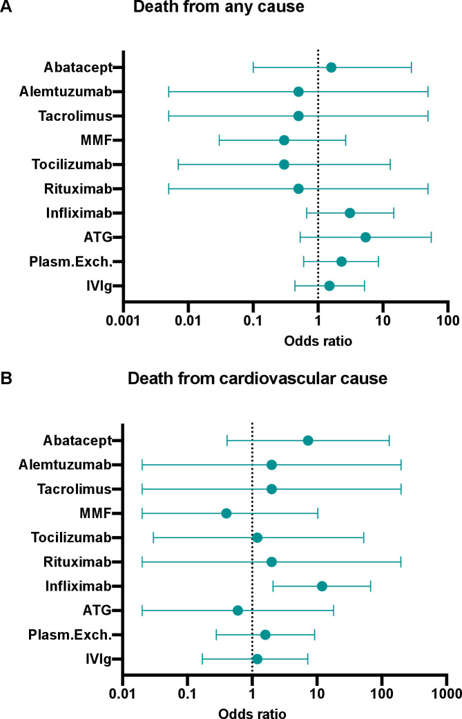
Twenty-three patients (38%) died from any cause, and eight (13%) died from cardiovascular causes. Patients who required IIST were more likely to die from any cause (50% vs 21%, p=0.02) (table 2). According to the type of IIST, patients who received infliximab were more likely to die from cardiovascular causes (OR 12.0; 95% CI 2.1 to 67.1; p=0.005) (figure 4) (table 3). Indications for infliximab as a single or combination therapy were hemodynamic (n=4), biological (n=1) and others (n=3).
Figure 4.
Forest plot of the risk of death from any cause (A) and from a cardiovascular cause according to the type of IIST. ATG, antithymocyte globulin; IIST, intensified immunosuppressive therapy; IV, intravenous; MMF, mycophenolate mofetil; Plasm.Exch., plasma exchange therapy.
Table 3.
All-cause and cardiovascular mortality according to intensified immunosuppressive therapy
| All-cause mortality | OR (95% CI) | P value | Cardiovascular mortality | OR (95% CI) | P value | |
| No IIST | 5/24 (21) | 0.3 (0.06 to 0.96) | 0.02 | 1/24 (4) | 0.2 (0.004 to 1.60) | 0.13 |
| IVIg | 9/20 (45) | 1.5 (0.44 to 5.18) | 0.57 | 3/20 (15) | 1.2 (0.17 to 7.22) | 1.0 |
| Plasm.Exch. | 6/11 (55) | 2.3 (0.60 to 8.50) | 0.23 | 2/11 (18) | 1.6 (0.28 to 9.20) | 0.60 |
| ATG | 3/4 (75) | 5.4 (0.53 to 55.4) | 0.16 | 0/4 (0) | 0.6 (0.02 to 18.04) | 0.79 |
| Infliximab | 5/8 (63) | 3.1 (0.67 to 14.7) | 0.15 | 4/8 (50) | 12.0 (2.1 to 67.1) | 0.005 |
| Rituximab | 0/1 (0) | 0.5 (0.005 to 49.3) | 0.78 | 0/1 (0) | 2.0 (0.02 to 197.1) | 0.76 |
| Tocilizumab | 0/2 (0) | 0.3 (0.007 to 12.92) | 0.52 | 0/2 (0) | 1.2 (0.03 to 52.62) | 0.93 |
| MMF | 1/6 (17) | 0.3 (0.03 to 2.66) | 0.27 | 0/6 (0) | 0.4 (0.02 to 10.3) | 0.59 |
| Tacrolimus | 0/1 (0) | 0.5 (0.005 to 49.3) | 0.78 | 0/1 (0) | 2.0 (0.02 to 197.9) | 0.76 |
| Alemtuzumab | 0/1 (0) | 0.5 (0.005 to 49.3) | 0.78 | 0/1 (0) | 2.0 (0.02 to 197.9) | 0.76 |
| Abatacept | 1/2 (50) | 1.6 (0.10 to 27.5) | 0.73 | 1/2 (50) | 7.3 (0.41 to 130.1) | 0.18 |
Values are n (%).
ATG, antithymocyte glogulin; IIST, intensified immunosuppressive therapy; IV, intravenous; MMF, mycophenolate mofetil; Plasm.Exch., plasma exchange therapy.
Discussion
The management of irAEs during ICI therapy is challenging because some of them can lead to life-threatening complications.2 6–9 21 Despite many limitations, our study is the first to investigate the treatment of ICI-induced myocarditis by immunosuppressive therapy other than corticosteroids alone. In our opinion, it provides two important results. The first is to have clarified the profile of patients who required the intensification of their immunosuppressive treatment. Compared with patients not treated with IIST, patients who required IIST were more likely to receive a combination of ICIs and to experience myocarditis earlier after the first ICI administration. Moreover, the episode of myocarditis was more frequently complicated by hemodynamic or electrical instability and associated with neuromuscular irAEs. The second important finding was that infliximab was associated with a greater risk of cardiovascular death in patients requiring IIST. To the best of our knowledge, this is the first study that raises concern over infliximab use as second-line therapy.
Although any organ can be involved in ICI therapy, some irAEs may be very serious and lethal, such as myocarditis. This adverse event is infrequent but is associated with a high case fatality rate.13 14 It most commonly occurs with combined ICI therapy (especially PD-1i/PD-L1i+CTLA-4i) within the first months after the initiation of cancer treatment.8 11 The high risk of death justifies monitoring and management strategies for which strong data are lacking to provide recommendations with a high level of evidence. However, in the recent American and European guidelines, experts highlight the importance of very early diagnosis and management.13 14 As soon as a diagnosis of ICI-induced myocarditis is suspected, ICI treatment should be interrupted, the patient should be admitted to a cardiology unit (ideally in the intensive care unit), and corticosteroid treatment should be promptly initiated.22 Therefore, it is of crucial importance to detect patients with more severe myocarditis for whom corticosteroid therapy will be insufficient and IIST will be needed. From this perspective, our study provides insights into the profile of patients requiring IIST who will, therefore, be closely monitored. These were those presenting the first signs of myocarditis very early after the start of ICI therapy. While previous studies have shown that the median time was approximately 30 days for all patients with ICI-induced myocarditis,11 in our work, it was only 18 days for patients who were going to require IIST vs 60 days for others. Therefore, early onset of myocarditis symptoms/signs after the start of ICI treatment should encourage clinicians to be more attentive to the evolution of these patients, especially if they had received a combination of ICIs. The analysis of ICI-induced myocarditis cases based on the need for IIST allows us to confirm that the occurrence of cardiogenic shock, ventricular arrhythmia, atrioventricular block or concomitant neuromuscular adverse events are poor prognostic factors that may lead to IIST.9 11 Thus, in our study, the two main indications for IIST were the presence of other irAEs and an unfavorable hemodynamic outcome. Nevertheless, the risk of sudden death related to ventricular arrhythmia should also be considered because this complication can occur even in the presence of a stable hemodynamic state.6 A recent work has shown that the persistence of elevated troponin T (≥1.5 ng/mL) at hospital discharge was associated with a fourfold increased risk of major adverse cardiac events.11 In our work, the persistence of elevated troponin or its increase was also a frequent indication of IIST.
As soon as the evolution is unfavorable under corticosteroids, the guidelines recommend IIST, but this is an empirical strategy due to lack of evidence.13 14 The pathophysiology of ICI-induced myocarditis remains poorly understood.23 Since the histological lesions observed are similar to those observed during acute cardiac transplant cell rejection, experts logically recommended drugs indicated in this situation. These include high doses of methylprednisolone (1 g per day for 3 days) as well as ATG, MMF or tacrolimus. However, an in vitro study showed that B cells also widely express both PD-1 and PD-L1. Thus, the blocking of PD-1/PD-L1 can restore and enhance B-cell proliferation, interleukin-6 production and antibodies to self-antigens, such as acetylcholine receptor, striated muscle or Ro proteins.24 This may justify other therapies, such as plasma exchange, intravenous Ig, infliximab (antitumor necrosis factor-alpha (TNF-α)), rituximab (anti-CD20), tocilizumab (anti-interleukin-6), alemtuzumab (anti-CD52) and abatacept (CTLA-4 agonist).
In our study, these strategies were used either alone or in combination. Although our work was not designed to determine which of them is the most effective, it does suggest a deleterious effect of infliximab, which was associated with a significant increase in the risk of cardiovascular death. This monoclonal antibody is indicated in the treatment of several chronic inflammatory diseases because the neutralization of TNF-α regulates the inflammatory response by reducing the release of interleukin-1, interleukin-6 and TNF-β. Although TNF-α is increased in the serum, macrophages and myocardial cells of patients with heart failure syndrome,25 cardiac toxicity has been well reported with infliximab, and it has been shown to adversely affect the clinical condition of patients with moderate to severe heart failure.26 27 In addition, cases of myocarditis have also been described under this treatment.28 These data, combined with those from our study, suggest that infliximab should not be used as first-line therapy after corticosteroids in patients with ICI-induced myocarditis.
Our work has many limitations that we acknowledge. This study was retrospective in design, and we pooled patients from our center with patients in previously published case reports. Thus, the quality of the data collected in case reports was not controlled. However, we chose to limit the number of case reports by including only those where patients received IIST. Moreover, data from patients from our center were prospectively collected into a controlled and protected database immediately on admission. We acknowledge that this methodology is questionable, but it made it possible to increase the number of patients treated with IIST for this infrequent serious disease. Due to the design, we were unable to determine the exact rate of patients requiring IIST in the whole population. Nevertheless, in the subgroup of patients admitted to our center, this rate was 14%. Several relevant covariates could not be analyzed, such as the evolution of troponin and natriuretic peptide levels, doses of drugs administered, and the evolution of cancer after the ICI-induced myocarditis episode. Mortality rates should be interpreted with caution, as follow-up in case reports was most often limited to a very short period following the adverse event. Finally, results on the potential deleterious effect of infliximab should be interpreted with caution in regard to the small sample size and the lack of controlled design. These are only exploratory data, but they raise concern over infliximab use as second-line therapy.
Conclusion
The need for IIST was more common in patients who developed myocarditis very early after the start of ICI therapy, especially combination of ICIs, as well as when hemodynamic/electrical instability or neuromuscular irAEs occurred. In patients receiving IIST therapy, treatment with infliximab was associated with a significantly increased risk of cardiovascular death. Thus, this study identifies patients at high risk of adverse events and provides opportunities for further work to determine the most effective immunosuppressive therapeutic strategy for ICI-induced myocarditis.
jitc-2020-001887supp002.pdf (613.7KB, pdf)
Footnotes
Twitter: @franckthuny
JC and SZ contributed equally.
Contributors: JC and SZ contributed equally to this work and drafted the manuscript with guidance from FT. All authors participated in the conceptualization, writing, review and revision of this manuscript. In addition, all authors have read and approved the final version of this manuscript.
Funding: This study received support from Assistance Publique Hôpitaux de Marseille, France. FT has been supported by The French Federation of Cardiology and The French League against Cancer.
Competing interests: FT received modest fees for lectures outside the submitted work from Novartis, Merck Sharp and Dohme, Bristol-Myers Squibb, Roche, AstraZeneca. JC received modest lecture fees outside the submitted work from Merck Sharp and Dohme, Novartis, AstraZeneca. FB received modest consultant fees outside the submitted work from AstraZeneca, Bayer, Bristol-Myers Squibb, Boehringer–Ingelheim, Eli Lilly Oncology, F. Hoffmann–La Roche, Novartis, Merck, MSD, Pierre Fabre, Pfizer and Takeda.
Patient consent for publication: Not required.
Ethics approval: Comité de protection des personnes "Est IV" IDRCB2017-A003362-51. The protocol was approved by our institutional review board.
Provenance and peer review: Not commissioned; externally peer reviewed.
Data availability statement: Data are available on reasonable request. Data are available on reasonable request to the corresponding author.
Supplemental material: This content has been supplied by the author(s). It has not been vetted by BMJ Publishing Group Limited (BMJ) and may not have been peer-reviewed. Any opinions or recommendations discussed are solely those of the author(s) and are not endorsed by BMJ. BMJ disclaims all liability and responsibility arising from any reliance placed on the content. Where the content includes any translated material, BMJ does not warrant the accuracy and reliability of the translations (including but not limited to local regulations, clinical guidelines, terminology, drug names and drug dosages), and is not responsible for any error and/or omissions arising from translation and adaptation or otherwise.
References
- 1.Ribas A, Wolchok JD. Cancer immunotherapy using checkpoint blockade. Science 2018;359:1350–5. 10.1126/science.aar4060 [DOI] [PMC free article] [PubMed] [Google Scholar]
- 2.Champiat S, Lambotte O, Barreau E, et al. . Management of immune checkpoint blockade dysimmune toxicities: a collaborative position paper. Ann Oncol 2016;27:559–74. 10.1093/annonc/mdv623 [DOI] [PubMed] [Google Scholar]
- 3.Cautela J, Lalevee N, Thuny F. Potential of Oncocardiology. JAMA Cardiol 2017;2:817–8. 10.1001/jamacardio.2017.0119 [DOI] [PubMed] [Google Scholar]
- 4.Cautela J, Rouby F, Salem J-E, et al. . Acute coronary syndrome with immune checkpoint inhibitors: a proof-of-concept case and pharmacovigilance analysis of a life-threatening adverse event. Can J Cardiol 2020;36:476–81. 10.1016/j.cjca.2019.11.035 [DOI] [PubMed] [Google Scholar]
- 5.Salem J-E, Manouchehri A, Moey M, et al. . Cardiovascular toxicities associated with immune checkpoint inhibitors: an observational, retrospective, pharmacovigilance study. Lancet Oncol 2018;19:1579–89. 10.1016/S1470-2045(18)30608-9 [DOI] [PMC free article] [PubMed] [Google Scholar]
- 6.Johnson DB, Balko JM, Compton ML, et al. . Fulminant myocarditis with combination immune checkpoint blockade. N Engl J Med 2016;375:1749–55. 10.1056/NEJMoa1609214 [DOI] [PMC free article] [PubMed] [Google Scholar]
- 7.Lyon AR, Yousaf N, Battisti NML, et al. . Immune checkpoint inhibitors and cardiovascular toxicity. Lancet Oncol 2018;19:e447–58. 10.1016/S1470-2045(18)30457-1 [DOI] [PubMed] [Google Scholar]
- 8.Escudier M, Cautela J, Malissen N, et al. . Clinical features, management, and outcomes of immune checkpoint Inhibitor-Related cardiotoxicity. Circulation 2017;136:2085–7. 10.1161/CIRCULATIONAHA.117.030571 [DOI] [PubMed] [Google Scholar]
- 9.Wang DY, Salem J-E, Cohen JV, et al. . Fatal toxic effects associated with immune checkpoint inhibitors: a systematic review and meta-analysis. JAMA Oncol 2018;4:1721–8. 10.1001/jamaoncol.2018.3923 [DOI] [PMC free article] [PubMed] [Google Scholar]
- 10.Jain V, Mohebtash M, Rodrigo ME, et al. . Autoimmune myocarditis caused by immune checkpoint inhibitors treated with antithymocyte globulin. J Immunother 2018;41:332–5. 10.1097/CJI.0000000000000239 [DOI] [PubMed] [Google Scholar]
- 11.Mahmood SS, Fradley MG, Cohen JV, et al. . Myocarditis in patients treated with immune checkpoint inhibitors. J Am Coll Cardiol 2018;71:1755–64. 10.1016/j.jacc.2018.02.037 [DOI] [PMC free article] [PubMed] [Google Scholar]
- 12.Hu J-R, Florido R, Lipson EJ, et al. . Cardiovascular toxicities associated with immune checkpoint inhibitors. Cardiovasc Res 2019;115:854–68. 10.1093/cvr/cvz026 [DOI] [PMC free article] [PubMed] [Google Scholar]
- 13.Brahmer JR, Lacchetti C, Schneider BJ, et al. . Management of immune-related adverse events in patients treated with immune checkpoint inhibitor therapy: American Society of clinical oncology clinical practice guideline. J Clin Oncol 2018;36:1714–68. 10.1200/JCO.2017.77.6385 [DOI] [PMC free article] [PubMed] [Google Scholar]
- 14.Haanen JBAG, Carbonnel F, Robert C, et al. . Management of toxicities from immunotherapy: ESMO clinical practice guidelines for diagnosis, treatment and follow-up. Ann Oncol 2017;28:iv119–42. 10.1093/annonc/mdx225 [DOI] [PubMed] [Google Scholar]
- 15.Alexandre J, Cautela J, Ederhy S, et al. . Cardiovascular toxicity related to cancer treatment: a pragmatic approach to the American and European Cardio-Oncology guidelines. J Am Heart Assoc 2020;9:e018403. 10.1161/JAHA.120.018403 [DOI] [PMC free article] [PubMed] [Google Scholar]
- 16.Salem J-E, Allenbach Y, Vozy A, et al. . Abatacept for severe immune checkpoint inhibitor-associated myocarditis. N Engl J Med 2019;380:2377–9. 10.1056/NEJMc1901677 [DOI] [PubMed] [Google Scholar]
- 17.Esfahani K, Buhlaiga N, Thébault P, et al. . Alemtuzumab for immune-related myocarditis due to PD-1 therapy. N Engl J Med 2019;380:2375–6. 10.1056/NEJMc1903064 [DOI] [PubMed] [Google Scholar]
- 18.Wang H, Tian R, Gao P, et al. . Tocilizumab for fulminant programmed death 1 inhibitor-associated myocarditis. J Thorac Oncol 2020;15:e31–2. 10.1016/j.jtho.2019.09.080 [DOI] [PubMed] [Google Scholar]
- 19.Bonaca MP, Olenchock BA, Salem J-E, et al. . Myocarditis in the setting of cancer therapeutics: proposed case definitions for emerging clinical syndromes in Cardio-Oncology. Circulation 2019;140:80–91. 10.1161/CIRCULATIONAHA.118.034497 [DOI] [PMC free article] [PubMed] [Google Scholar]
- 20.Ferreira VM, Schulz-Menger J, Holmvang G, et al. . Cardiovascular Magnetic Resonance in Nonischemic Myocardial Inflammation: Expert Recommendations. J Am Coll Cardiol 2018;72:3158–76. 10.1016/j.jacc.2018.09.072 [DOI] [PubMed] [Google Scholar]
- 21.Moslehi JJ, Salem J-E, Sosman JA, et al. . Increased reporting of fatal immune checkpoint inhibitor-associated myocarditis. Lancet 2018;391:933. 10.1016/S0140-6736(18)30533-6 [DOI] [PMC free article] [PubMed] [Google Scholar]
- 22.Zhang L, Zlotoff DA, Awadalla M, et al. . Major adverse cardiovascular events and the timing and dose of corticosteroids in immune checkpoint Inhibitor–Associated myocarditis. Circulation 2020;141:2031–4. 10.1161/CIRCULATIONAHA.119.044703 [DOI] [PMC free article] [PubMed] [Google Scholar]
- 23.Mangan BL, McAlister RK, Balko JM, et al. . Evolving insights into the mechanisms of toxicity associated with immune checkpoint inhibitor therapy. Br J Clin Pharmacol 2020:1778–89. 10.1111/bcp.14433 [DOI] [PMC free article] [PubMed] [Google Scholar]
- 24.Thibult M-L, Mamessier E, Gertner-Dardenne J, et al. . Pd-1 is a novel regulator of human B-cell activation. Int Immunol 2013;25:129–37. 10.1093/intimm/dxs098 [DOI] [PubMed] [Google Scholar]
- 25.Tsutamoto T, Wada A, Matsumoto T, et al. . Relationship between tumor necrosis factor-alpha production and oxidative stress in the failing hearts of patients with dilated cardiomyopathy. J Am Coll Cardiol 2001;37:2086–92. 10.1016/S0735-1097(01)01299-2 [DOI] [PubMed] [Google Scholar]
- 26.de' Clari F, Salani I, Safwan E, et al. . Sudden death in a patient without heart failure after a single infusion of 200 Mg infliximab: does TNF-alpha have protective effects on the failing heart, or does infliximab have direct harmful cardiovascular effects? Circulation 2002;105:e183. 10.1161/01.CIR.0000017216.41471.DF [DOI] [PubMed] [Google Scholar]
- 27.Kwon HJ, Coté TR, Cuffe MS, et al. . Case reports of heart failure after therapy with a tumor necrosis factor antagonist. Ann Intern Med 2003;138:807–11. 10.7326/0003-4819-138-10-200305200-00008 [DOI] [PubMed] [Google Scholar]
- 28.Slattery E, Ismail N, Sheridan J, et al. . Myocarditis associated with infliximab: a case report and review of the literature. Inflamm Bowel Dis 2011;17:1633–4. 10.1002/ibd.21546 [DOI] [PubMed] [Google Scholar]
Associated Data
This section collects any data citations, data availability statements, or supplementary materials included in this article.
Supplementary Materials
jitc-2020-001887supp001.pdf (35.5KB, pdf)
jitc-2020-001887supp002.pdf (613.7KB, pdf)


