. 2020 Nov 25;9:e59489. doi: 10.7554/eLife.59489

Figure 3. Wnt3 source and target regions at 48 hpf.

3D dorsal projection of Wnt3 (A) source regions at 48 hpf and (B) target regions at 48 hpf (top view). 3D ventral projection of Wnt3 (C) source regions at 48 hpf and (D) target regions at 48 hpf (bottom view). (E) 3D projection of Wnt3 source and target regions at 48 hpf (lateral view). See Video 4 for a detailed view. dCe, dorsal regions of cerebellum; Epi, epithalamus; FP, floor plate; MHB, midbrain–hindbrain boundary; mRP, midbrain roof plate; OT, optic tectum; vCe, ventral regions of cerebellum. Images orientation: anterior to the left. Scale bar 40 μm.

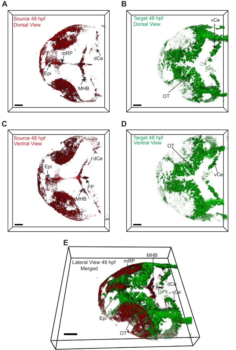
Figure 3.

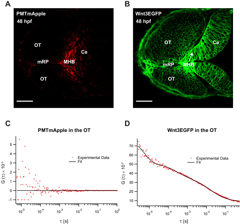
Figure 3—figure supplement 1. Fluorescence correlation spectroscopy (FCS) measurements of Wnt3EGFP and PMTmApple in the Wnt3 target regions at 48 hpf.

Figure 3—figure supplement 1.

Expression of (A) PMTmApple and (B) Wnt3EGFP in Tg(−4.0wnt3:Wnt3EGFP) × Tg(−4.0wnt3:PMTmApple) double transgenics at 48 hpf. Representative autocorrelation function (ACF) for (C) PMTmApple and (D) Wnt3EGFP in the OT at 48 hpf. No ACFs were obtained for PMTmAPple in the target regions indicating an absence of mApple signal in these regions. Ce, cerebellum; MHB, midbrain–hindbrain boundary; OT, optic tectum. Images orientation: anterior to the left. Scale bar 30 μm.
Figure 3—figure supplement 2. Expression of EGFP in Tg(−4.0wnt3:EGFP) at 48 hpf.

Figure 3—figure supplement 2.

The expression of EGFP in Tg(-4.0wnt3:EGFP) closely mimics the expression of PMTmApple in Tg(−4.0wnt3:PMTmApple). Ce, cerebellum; MHB, midbrain–hindbrain boundary; OT, optic tectum. Images orientation: anterior to the left. Scale bar 30 μm.
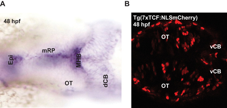
Figure 3—figure supplement 3. Expression of wnt3 transcripts and downstream wnt signaling transcription factors at 48 hpf.

Figure 3—figure supplement 3.

(A) Whole mount in situ hybridization (dorsal view) of wnt3 at 48 hpf. The expression pattern is similar to previously published results in Clements et al., 2009Duncan et al., 2015, and Teh et al., 2015. (B) Expression of NLS-mCherry in wnt reporter line Tg(7XTcf-Xla.Siam:NLS-Cherry) at 48 hpf. dCB, cerebellum; Epi, epithalamus; FP, floor plate; MHB, midbrain–hindbrain boundary; mRP, midbrain roof plate; OT, optic tectum. Orientation: anterior to left.