Abstract
Background:
Decelerating and cutting are two common movements during which non-contact anterior cruciate ligament (ACL) injuries occur in soccer players. Retrospective video analysis of ACL injuries has demonstrated that players are often in knee valgus at the time of injury.
Purpose:
To determine whether prospectively measured components of valgus collapse during a deceleration and 90 ° cut can differentiate between collegiate women's soccer players who go on to non-contact ACL injury.
Design:
Secondary analysis of prospectively collected data.
Methods:
51 NCAA women's soccer players completed motion analysis of a deceleration and 90 ° before the competitive season. Players were classified as Injured (noncontact ACL injury during the season) or Uninjured at the end of the season. Differences between groups for peak hip adduction, internal rotation, and knee abduction angles, and knee valgus collapse were analyzed with a MANOVA.
Results:
Four non-contact ACL injuries were reported at the end of the season. There was a significant difference between groups for hip adduction angle during the 90 ° cut (p = 0.02) and deceleration (p = 0.03). Players who went on to ACL injury were in more hip adduction.
Conclusions:
Hip adduction angle is larger in players who go on to ACL injury than those who do not during two sport-specific tasks. The components of knee injury prevention programs that address proximal control and strength are likely crucial for preventing ACL injuries.
Level of Evidence:
2b
Keywords: injury, knee, movement system, soccer, training
INTRODUCTION
Women soccer players incur anterior cruciate ligament (ACL) injuries at a rate disproportionately higher than their male peers.1 The majority of ACL injuries among women occur without contact from another player.2 These non-contact injuries may result from poor on-field biomechanics and lack of neuromuscular control. Neuromuscular control is a modifiable risk factor for ACL injury, and therefore non-contact injuries may be preventable.3 Several injury prevention programs address presumed risk factors. These risk factors have been traditionally studied in the laboratory setting using tasks not specific to soccer (e.g. drop jump).3 To improve existing injury prevention programs and to design new ones, ACL injury risk factors should be studied in sport-specific tasks.
Cutting and decelerating are two sport-specific movements that soccer players frequently execute. Planting and cutting or a sudden deceleration with a cut have also been identified as two of the most common mechanisms for non-contact ACL injuries.2,4,5 Retrospective video analysis of team handball players demonstrated that the majority of non-contact ACL injuries occurred during a plant and cut movement,6 and retrospective video analysis of women's soccer players demonstrated that the second most common mechanism of ACL injuries was cutting.7 Epidemiological work in the German women's national soccer league also reported that seven out of eleven non-contact ACL injuries recorded in a single season resulted from a sudden change of direction.8 Through these observational and video analysis studies, several biomechanical patterns have been associated with non-contact deceleration and cutting related ACL injuries.
Sudden cutting and pivoting maneuvers in soccer are frequently preceded by a deceleration. Decelerating with the knee in close to full extension and valgus have been observed as antecedents to non-contact ACL injury.2,4,6 Cadaveric studies support this by demonstrating that the ACL is under greater strain at lower flexion angles, especially when combined with a valgus moment and internal tibial torque.9 But, there is a paucity of controlled laboratory studies examining how the biomechanics of deceleration affect injury risk.
One of the most commonly cited biomechanical risk factors for ACL injury is valgus collapse.3 Valgus collapse combines three motions: hip internal rotation, hip adduction, and knee abduction.3 In the laboratory setting, valgus collapse has been studied using a bilateral drop jump, with athletes at higher risk for injury demonstrating greater levels of valgus collapse and higher peak knee abduction moments.3 However, it has been estimated that only 25% of ACL injuries among women's soccer players occur during jump landings.10 Observational studies have indicated that during deceleration and cutting related injuries the knee is frequently collapsed medially.6,7,11 Several studies have attempted to bridge this gap by examining whether there is a relationship between valgus collapse measured during landing tasks and cutting tasks.12-14 Although moderate to strong correlations between knee abduction angles measured during a cut and bilateral and unilateral drop jump landings respectively have been found,12,13 other comparisons between bilateral drop jumps and pre-planned cutting have demonstrated weak relationships.12,15 No research yet has used injury data to directly examine whether valgus collapse measured prospectively during a cut differentiates between players who go on to non-contact ACL injury and those who do not go on to injury.
Without establishing whether there is a direct association between the biomechanics of the sport-specific tasks of decelerating and cutting and ACL injury risk, it is difficult to design effective injury prevention programs to make these tasks safer. It is also challenging to interpret the impact of injury prevention programs on a player's on-field movement strategies if there is not a direct, quantifiable relationship between the biomechanics of sport-specific tasks and injury. Therefore the purposes of this exploratory study were to identify whether prospectively measured components of valgus collapse: peak hip adduction, hip internal rotation, and knee abduction angles during a declaration and a 90 ° cut differentiate soccer players who go on to ACL injury from those who do not, and whether these components of valgus collapse are associated with future ACL injury in collegiate women's soccer players. The hypothesis was that participants who went on to ACL injury would demonstrate greater hip adduction, hip internal rotation, and knee abduction angles, and more knee valgus collapse, on both tasks than participants who did not go on to ACL injury.
METHODS
Three NCAA Division I and II women's soccer teams were invited to participate in this research as part of a larger study. Sixty-eight participants were enrolled. This study was approved by the institution's internal review board and all participants provided informed consent before participation. Participant demographics and injury histories were collected during motion analysis testing. Preseason motion analysis of two tasks was completed: a deceleration and a 90 ° cut. Six marker clusters and twenty-two retroreflective markers were placed on bony prominences by a single investigator (AA). Individual markers were placed bilaterally on the: acromion, iliac crest, greater trochanter, medial and lateral femoral condyle, medial and lateral malleolus, base of the fifth metatarsal, base of the first metatarsal, and the posterior calcaneus, and the rigid marker clusters were placed on the: trunk, pelvis, thighs, and shanks. For the deceleration, participants were given a demonstration of the task and instructed to: “Accelerate over a 10-meter distance to full speed and decelerate as the lower extremity strikes the center of the force plate. Ensure that your foot lands in the center of the plate and your heel does not touch the perimeter of the plate”. For the 90 ° cut, participants were also given a demonstration of the task and instructed to, “Quickly run forward, plant your entire foot straight forward on the force plate, turn your hip 90 °, and continue your run. Ensure that your entire foot lands on the center of the plate.” Participants completed three trials of each task on the dominant and non-dominant limbs. Limb dominance was defined by which foot a participant would kick a ball with. Kinematic and kinetic data were recorded simultaneously using an eight-camera motion system (Vicon, Nexus) and an embedded force plate (Bertec Worthington, OH) sampling at 240Hz and 1080Hz respectively.
During the soccer season, each team's athletic trainer recorded injuries for their respective team. Injury data were shared with study investigators postseason, and four non-contact, grade 3 sprain ACL injuries were reported. Two ACL injuries on dominant limbs were reported and two ACL injuries on non-dominant limbs were reported.
Post-processing of motion capture data for the 90 ° cut and for the deceleration was completed in Vicon Nexus (v 1.85 VICON, Oxford Metrics Ltd, London, England) and Visual 3D (C-Motion Inc., Germantown. MD). Makers were labelled in Nexus and gaps in the markers less than five frames were filled using Nexus's spline-based algorithm. If gaps were greater than five frames for either task, participants were excluded from the analysis. Kinematic and kinetic data were low pass filtered at 6Hz and 40Hz respectively. Inverse dynamics and rigid body analysis were then completed in Visual 3D using custom written scripts. Peak hip and knee kinematics and kinetics were exported from Visual 3D. Only the second and third trials of both the 90 ° cut and deceleration were analyzed secondary to higher reliability (Appendix 1).
STATISTICAL METHODS
An innovative measure was used to analyze changes in lower extremity valgus, which was termed “knee valgus collapse”. Since valgus collapse is a composite of hip adduction, hip internal rotation, and knee abduction, it was assessed at peak knee flexion as a global measure. The equation used for this was Knee Valgus Collapse = hip adduction angle + hip internal rotation angle + (knee abduction angle x -1), with (-) values indicating less knee valgus collapse and (+) values indicating more knee valgus collapse.16
After accounting for marker dropout during both the 90 ° cut and the deceleration, 51 participants had complete data sets. Participants were divided into two groups: ACL injury, “Injured” (N = 4) and no ACL injury, “Uninjured” (N = 47). Independent t-tests were used to determine whether there were differences between Injured and Uninjured participants for height, weight, and age. Paired samples t-tests were used to determine whether there were significant differences between the dominant and non-dominant limbs of Uninjured participants for peak hip and knee kinematics. There were no significant differences between limbs, except for dominant limb knee valgus collapse (Table 1). So, peak hip and knee kinematics were averaged across limbs for Uninjured participants so each Uninjured participant contributed a single healthy limb to the statistical model, and each Injured participant contributed a single ACL injury limb. Participants with previous history of ACL reconstruction were excluded from this analysis.17 All data were analyzed in SPSS for Windows, version 25 (SPSS Inc. Chicago, IL). MANOVAs were used to determine whether there were significant differences between the Injured and Uninjured groups for peak hip internal rotation, hip adduction, and knee abduction angles and knee valgus collapse. Separate MANOVAs were used for the 90 ° cut and deceleration tasks.
Table 1.
Comparison between limbs for kinematic variables.
| Variable | Dominant Limb | Non-Dominant Limb | p-value |
|---|---|---|---|
| 90 ° Cut Hip Adduction Angle (°) | −5.26 ± 5.97 | −6.09 ± 04.97 | 0.31 |
| 90 ° Cut Hip Internal Rotation Angle (°) | 10.61 ± 6.28 | 9.29 ± 7.61 | 0.26 |
| 90 ° Cut Knee Abduction Angle (°) | −3.89 ± 3.11 | −3.75 ± 3.10 | 0.75 |
| 90 ° Cut Valgus Collapse (°) | −5.39 ± 9.34 | −10.48 ± 11.50 | 0.00 |
| Deceleration Hip Adduction Angle (°) | 2.24 ± 6.71 | 1.06 ± 6.90 | 0.22 |
| Deceleration Hip Internal Rotation Angle (°) | 8.04 ± 5.51 | 8.41 ± 6.73 | 0.76 |
| Deceleration Knee Abduction Angle (°) | −2.00 ± 2.54 | −1.32 ± 2.91 | 0.09 |
| Deceleration Valgus Collapse (°) | 3.02 ± 8.45 | 0.42 ± 10.13 | 0.11 |
RESULTS
There were no significant differences between Injured and Uninjured participants for height, weight, or age (Table 2).
Table 2.
Demographic variables for Injured and Uninjured participants.
| Variable | Injured | Uninjured | p-value |
|---|---|---|---|
| Height (m) | 1.65 ± 0.05 | 1.67 ± 0.05 | 0.45 |
| Weight (kg) | 66.1 ± 6.95 | 63.6 ± 6.55 | 0.46 |
| Age (years) | 19.8 ± 1.5 | 19.4 ± 1.2 | 0.57 |
90° Cut
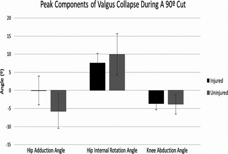
The MANOVA for the 90 ° cut was not significant (p = 0.15). There was a significant difference between groups for hip adduction angle (p = 0.02), with the Injured participants in greater hip adduction than the Uninjured participants (Injured mean: -0.02 ± 3.96 °, Uninjured mean: -5.75 ± 4.73 °) (Figure 1). There were no significant differences between groups for hip internal rotation angle (p = 0.43, Injured mean: 7.60 ± 2.58 °, Uninjured mean: 9.94 ± 5.75 °) knee abduction angle (p = 0.89, Injured mean: -3.66 ± 1.68 °, Uninjured mean: -3.84 ± 2.68 °), or for knee valgus collapse (p = 0.86, Injured mean: -9.02 ± 11.63 °, Uninjured mean: -8.20 ± 8.72 °).
Figure 1.
A visual comparison between Injured and Uninjured players of peak components of valgus collapse during a 90 degree cut.
Deceleration
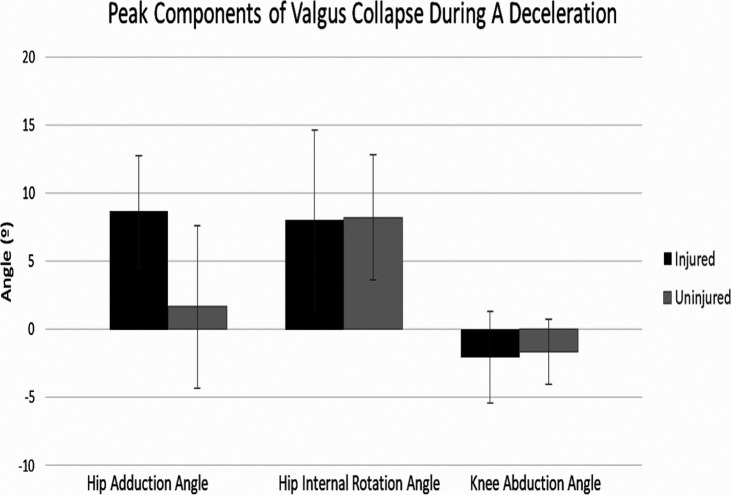
The MANOVA for the deceleration was not significant (p = 0.17). There was a significant difference between groups for hip adduction angle (p = 0.03), with the Injured participants in greater hip adduction than the Uninjured participants (Injured mean: 8.63 ± 4.12 °, Uninjured mean: 1.66 ± 5.98 °) (Figure 2). There was also a significant difference between groups for knee valgus collapse (p = 0.04), with Injured participants in more knee valgus collapse than the Uninjured participants (Injured mean: 8.57 ± 8.34 °, Uninjured mean: 0.65 ± 6.90 °). There were no significant differences between groups for hip internal rotation angle (p = 0.92, Injured mean: 7.98 ± 6.66 °, Uninjured mean: 8.22 ± 4.59 °) or knee abduction angle (p = 0.77, Injured mean: -2.03 ± 3.28 °, Uninjured mean: -1.66 ± 2.38 °).
Figure 2.
A visual comparison between Injured and Uninjured players of peak components of valgus collapse during a deceleration.
DISCUSSION
The primary finding of this study was that women soccer players who incurred an ACL injury were in less hip abduction during preseason testing of two sport-specific tasks than women who did not incur ACL injury. These results partially supported the hypothesis. Participants who went on to ACL injury were in more hip adduction than those who did not go on to injury, however, hip internal rotation and knee abduction angles did not differ between players who went on to injury and those who did not. And, participants who went on to ACL injury were in more knee valgus collapse during the deceleration task than participants who did not go on to ACL injury. However, this difference did not exceed conservative measures of the smallest detectable change (SDC) for knee valgus collapse (Appendix 1).
These results partly confirm that components of valgus collapse measured during the soccer-specific tasks of decelerating and cutting may distinguish between players who will go on to ACL injury. The statistically significant differences between groups for both tasks exceeded the SDC values for hip adduction (Appendix 1), indicating the changes exceed noise in the signal. Hip internal rotation, another component of valgus collapse, did not differentiate between players who went on to injury and those who did not. This follows previous research which has indicated a lack of relationship between hip internal rotation and knee abduction angles while cutting, and has also suggested that hip internal rotation may not be predictive of ACL injury.18 Another interesting finding of this analysis was that peak knee abduction angle during a 90 ° cut and deceleration also did not differentiate between Injured and Uninjured players. Knee abduction angle has been hypothesized as a risk factor for ACL injury during cutting and decelerating.2,4,3 Although knee abduction angle did not differentiate between groups in this prospective cohort, high knee abduction angles while cutting and decelerating are still likely a risk factor for ACL injury secondary to the increased load placed on the ACL.19,9 And, previous research has reported that knee abduction angle and hip adduction angle are highly correlated among women's soccer players during a 90 ° cut.18 Therefore, reduction of hip adduction and knee abduction angles while decelerating and cutting may lead to lower non-contact ACL injury risk. For this study, hip adduction may differentiate between players who go on to ACL injury and those who do not because there may be more room for physiologic variability in this component of valgus collapse during a deceleration or 90 ° cut when compared to hip internal rotation and knee abduction angles. Identification of hip adduction as a risk factor for non-contact ACL injury helps confirm that reducing hip adduction angle with neuromuscular training is a favorable biomechanical change. And, identification of this modifiable biomechanical variable as a discriminating factor for players who go on to ACL injury can lead to improvements in the design of injury prevention programs.
Although this is the first study to analyze whether prospectively measured hip adduction angles during two soccer-specific tasks can differentiate between players who go on to ACL injury and those who do not, several studies have examined gender differences in hip adduction angle during unilateral, athletic tasks. In both a randomly cued cutting task and a single leg landing task, women demonstrated a tendency toward greater hip adduction angles than men.20,21 Since women soccer players incur non-contact ACL injuries at a rate higher than men,22 these studied gender differences, along with this study's prospectively measured group differences, confirm the need for women's participation in neuromuscular training to address this modifiable risk factor. Hip adduction angle can be modified through exercises focused on proximal control.23-24 Inclusion of exercises addressing proximal control is a key factor in designing effective injury prevention programs.25 Descriptions of proximal control exercises to reduce knee valgus vary, but strengthening of the hip abductors and core and functional stability exercises with a focus on biomechanical alignment may be effective for reducing hip adduction angle during dynamic tasks.24,25 The results of this study affirm that increased hip adduction during decelerating and cutting may be a risk factor for ACL injury and should be addressed through participation in an injury prevention program that focuses on proximal neuromuscular control and on biomechanical technique.25-27 As a biomechanical variable, hip adduction angle during these tasks may be modifiable through strengthening of the hip abductor muscles and participation in neuromuscular training.28-30
Several studies have expressed the need for a sport-specific ACL injury risk screening task.31,18 In this sample of collegiate women's soccer players, biomechanical analysis of a deceleration task could differentiate between players who go on to ACL injury and those who don't. Therefore, this task may hold potential to be developed into a screening task for ACL injury risk, but more research is needed in a larger cohort and in a broader age-group. And, screening large volumes of soccer players with three-dimensional biomechanical analysis is not practical. This study does however guide clinicians and coaches for training safe biomechanical technique during soccer specific tasks, especially avoiding hip adduction.
A primary limitation of this study was analysis of a pre-planned cutting task. While analyzing a pre-planned task improved the reliability of the task between trials, previous literature has examined differences between anticipated and unanticipated cutting and reported significant differences between the biomechanics of these tasks.32,33 Unanticipated cutting may lead to greater valgus loads at the knee.32 Therefore, the differences observed between Injured and Uninjured participants in this study may not reflect the magnitude of biomechanical differences between players who go on to injury and those who do not in on-field situations. Another limitation of this study was the small number of ACL injuries recorded. The small number of Injured participants reduced statistical power of this analysis; however, significant differences between Injured and Uninjured participants were observed despite this.
CONCLUSION
Biomechanical risk factors for ACL injury have been studied using a bilateral drop jump, and knee valgus has been identified as a biomechanical risk factor for non-contact ACL injuries in women.3 This study found that peak hip adduction angle, a component of valgus collapse, measured prospectively during a 90 ° cut and deceleration can differentiate between collegiate women's soccer players who go on to ACL injury and those who do not. Injury prevention programs that contain proximal hip control exercises have been effective in reducing ACL injury rates.25,27 These results support the existing evidence that athletes should participate in neuromuscular training to reduce ACL injury risk. These results suggest there is potential for these tasks to be developed into tests to screen athletes for injury risk. Future work should examine these tasks in a larger cohort to further explore their screening properties.
APPENDIX 1.
Smallest Detectable Change (SDC) and Minimal Important Difference (MID) Calculation Methods
Smallest detectable change (SDC) and minimal clinical difference (MDC) values were calculated to determine whether biomechanical changes were clinically meaningful. Interclass correlation coefficients were calculated to assess between trial reliability. Only the second and third trials of the cutting task were analyzed secondary to higher reliability between trials1,2. Interclass correlation coefficients (ICCs) demonstrated good to excellent reliability (>0.75-0.91) for all variables except dominant knee valgus collapse and non-dominant hip adduction angle, which demonstrated moderate reliability (>0.5-0.75)3. ICCs were used to calculate the standard error of the mean (SEM), which was then used to calculated the SDC for each variable using the equation: SDC = SEM*1.96*1,4.
Minimal important differences (MID) were also calculated. Injury logs across the soccer season were provided for one soccer team (N = 19*) by the team's athletic trainer. Study investigators verified that the soccer team was not participating in a structured injury prevention program, as this hypothetically would have altered injury risk. MANOVAs were used to determine whether there were pre-season differences between the players who went on to sustain a non-contact, time loss, lower extremity injury during the season 5. One MANOVA was calculated for the dominant limb and one MANOVA was calculated for the non-dominant limb for each task. Neither the MANOVA for the dominant (p = 0.52) or non-dominant (p = 0.24) limb for the 90 ° cut was statistically significant. And, neither the MANOVA for the dominant (p = 0.86) or non-dominant (p = 0.69) limb for the deceleration was statistically significant. If the mean difference between injured and uninjured players exceeded the SDC, this was considered to be a MID6. MID = (Injured player mean- uninjured player mean) > SDC. *19 participants had usable 90 ° cut data, and 15 participants had usable deceleration data.
Smallest Detectable Change and Minimal Important Difference Values.
| Variable | Side | SDC | MID |
|---|---|---|---|
| Cut Hip Adduction Angle (°) | Dominant | 3.58 | |
| Non-Dominant | 5.44 | ||
| Cut Hip Internal Rotation Angle (°) | Dominant | 2.79 | |
| Non-Dominant | 1.81 | ||
| Cut Knee Abduction Angle (°) | Dominant | 1.04 | 2.00 |
| Non-Dominant | 0.91 | 2.38 | |
| Cut Valgus Collapse (°) | Dominant | 8.59 | |
| Non-Dominant | 6.40 | ||
| Deceleration Hip Adduction Angle (°) | Dominant | 2.27 | |
| Non-Dominant | 2.65 | ||
| Deceleration Hip Internal Rotation Angle (°) | Dominant | 1.11 | 2.72 |
| Non-Dominant | 1.22 | ||
| Deceleration Knee Abduction Angle (°) | Dominant | 0.83 | 1.37 |
| Non-Dominant | 1.02 | ||
| Deceleration Valgus Collapse (°) | Dominant | 4.46 | |
| Non-Dominant | 7.07 |
REFERENCES
- 1.Prodromos CC Han Y Rogowski J, et al. A Meta-analysis of the Incidence of anterior cruciate ligament tears as a function of gender, sport, and a knee injury-reduction regimen. Arthrosc - J Arthrosc Relat Surg. 2007;23(12):1320-1325. [DOI] [PubMed] [Google Scholar]
- 2.Alentorn-Geli E Myer GD Silvers HJ et al. Prevention of non-contact anterior cruciate ligament injuries in soccer players. Part 1: Mechanisms of injury and underlying risk factors. Knee Surgery, Sport Traumatol Arthrosc. 2009;17(7):705-729. [DOI] [PubMed] [Google Scholar]
- 3.Hewett TE Myer GD Ford KR, et al. Biomechanical measures of neuromuscular control and valgus loading of the knee predict anterior cruciate ligament injury risk in female athletes: A prospective study. Am J Sports Med. 2005;33(4):492-501. [DOI] [PubMed] [Google Scholar]
- 4.Boden BP Dean GS Feagin JAJ Garrett WEJ. Mechanisms of anterior cruciate ligament injury. Orthopedics. 2000;23(6):573-578. [DOI] [PubMed] [Google Scholar]
- 5.Waldén M Krosshaug T Bjørneboe J Andersen TE Faul O Hägglund M. Three distinct mechanisms predominate in non- contact anterior cruciate ligament injuries in male professional football players?: a systematic video analysis of 39 cases. Br J Sports Med. 2015;(49):1452-1460. [DOI] [PMC free article] [PubMed] [Google Scholar]
- 6.Olsen O-E Myklebust G Engebretsen L Bahr R. Injury mechanisms for anterior cruciate ligament injuries in team handball. Am J Sports Med. 2004;32(4):1002-1012. [DOI] [PubMed] [Google Scholar]
- 7.Brophy RH Stepan JG Silvers HJ Mandelbaum BR. Defending puts the anterior cruciate ligament at risk during soccer: A gender-based analysis. Sports Health. 2015;7(3):244- [DOI] [PMC free article] [PubMed] [Google Scholar]
- 8.Faude O Junge A Kindermann W Dvorak J. Injuries in female soccer players: A prospective study in the German national league. Am J Sports Med. 2005;33(11):1694-1700. [DOI] [PubMed] [Google Scholar]
- 9.Markolf KL Burchfield DM Shapiro MM, et al. Combined knee loading states that generate high anterior cruciate ligament forces. J Orthop Res. 1995;(13):930-935. [DOI] [PubMed] [Google Scholar]
- 10.Piasecki D.P. Spindler K.P. Warren T.A. et al. Intraarticular injuries associated with anterior cruciate ligament tear: findings at ligament reconstruction in high school and recreational athletes. Am J Sports Med. 2003;31(4):601–605. [DOI] [PubMed] [Google Scholar]
- 11.Krosshaug T Nakamae A Boden BP, et al. Mechanisms of anterior cruciate ligament injury in basketball: Video analysis of 39 cases. Am J Sports Med. 2007;35(3):359-367. [DOI] [PubMed] [Google Scholar]
- 12.Kristianslund E Krosshaug T. Comparison of drop jumps and sport-specific sidestep cutting: Implications for anterior cruciate ligament injury risk screening. Am J Sports Med. 2013;41(3):684-688. d [DOI] [PubMed] [Google Scholar]
- 13.Jones PA Herrington LC Munro AG Graham-Smith P. Is there a relationship between landing, cutting, and pivoting tasks in terms of the characteristics of dynamic valgus? Am J Sports Med. 2014;42(9):2095-2102. [DOI] [PubMed] [Google Scholar]
- 14.O’Connor KM Monteiro SK Hoelker IA. Comparison of selected lateral cutting activities used to assess ACL injury risk. J Appl Biomech. 2009;25(1):9-21. [DOI] [PubMed] [Google Scholar]
- 15.Havens KL Sigward SM. Cutting mechanics: Relation to performance and anterior cruciate ligament injury risk. Med Sci Sports Exerc. 2015;47(4):818-824. [DOI] [PubMed] [Google Scholar]
- 16.Arundale AJH Silvers-Granelli HJ Marmon A, et al. Changes in biomechanical knee injury risk factors across two collegiate soccer seasons using the 11 + prevention program. Scand J Med Sci Sports. 2018:1-12. [DOI] [PMC free article] [PubMed] [Google Scholar]
- 17.Stearns KM Pollard CD. Abnormal frontal plane knee mechanics during sidestep cutting in female soccer athletes after anterior cruciate ligament reconstruction and return to sport. Am J Sports Med. 2013;41(4):918-923. [DOI] [PubMed] [Google Scholar]
- 18.Imwalle LE Myer GD Ford KR Hewett TE. Relationship between hip and knee kinematics in athletic women during cutting maneuvers: A possible link to noncontact anterior cruciate ligament injury and prevention. J Strength Cond Res. 2013;23(8):2223-2230. [DOI] [PMC free article] [PubMed] [Google Scholar]
- 19.Besier TF Lloyd DG Cochrane JL Ackland TR. External loading of the knee joint during running and cutting maneuvers. Med Sci Sports Exerc. 2001;33(7):1168-1175. [DOI] [PubMed] [Google Scholar]
- 20.Hewett TE Ford KR Myer GD, et al. Gender differences in hip adduction motion and torque during a single leg agility maneuver. J Orthop Res. 2006;(3):416-421. [DOI] [PubMed] [Google Scholar]
- 21.Pollard CD Davis IMC Hamill J. Influence of gender on hip and knee mechanics during a randomly cued cutting maneuver. Clin Biomech. 2004;19(10):1022-1031. [DOI] [PubMed] [Google Scholar]
- 22.Agel J Arendt EA Bershadsky B. Anterior cruciate ligament injury in National Collegiate Athletic Association basketball and soccer: A 13-year review. Am J Sports Med. 2005;33(4):524-530. [DOI] [PubMed] [Google Scholar]
- 23.Hewett TE Myer GD Ford KR. Anterior cruciate ligament injuries in female athletes: Part 1, mechanisms and risk factors. Am J Sports Med. 2006;34(2):299-311. [DOI] [PubMed] [Google Scholar]
- 24.Baldon RDM Lobato DFM Carvalho LP, et al. Effect of functional stabilization training on lower limb biomechanics in women. Med Sci Sports Exerc. 2012;44(1):135-145. [DOI] [PubMed] [Google Scholar]
- 25.Sugimoto D Myer GD Foss KDB Hewett TE. Specific exercise effects of preventive neuromuscular training intervention on anterior cruciate ligament injury risk reduction in young females: Meta-analysis and subgroup analysis. Br J Sports Med. 2015;49(5):282-289. [DOI] [PubMed] [Google Scholar]
- 26.Hewett TE Ford KR Myer GD. Anterior cruciate ligament injuries in female athletes: Part 2, a meta-analysis of neuromuscular interventions aimed at injury prevention. Am J Sports Med. 2006;34(3):490-498. [DOI] [PubMed] [Google Scholar]
- 27.Arundale AJH Bizzini M Giordano A, et al. Exercise-based knee and anterior cruciate ligament injury prevention. J Orthop Sports Phys Ther. 2018;48(9):A1-A42. [DOI] [PubMed] [Google Scholar]
- 28.Jacobs CA Uhl TL Mattacola CG, et al. Hip abductor function and lower extremity landing kinematics: Sex differences. J Athl Train. 2007;42(1):76-83. [PMC free article] [PubMed] [Google Scholar]
- 29.Pollard CD Sigward SM Ota S, et al. The influence of in-season injury prevention training on lower-extremity kinematics during landing in female soccer players. Clin J Sport Med. 2006;16(3):223-227. [DOI] [PubMed] [Google Scholar]
- 30.Myer GD Ford KR Palumbo JP Hewett TE. Neuromuscular training improves performance and lower-extremity biomechanics in female athletes. J Strength Cond Res. 2005;19(1):51-60. [DOI] [PubMed] [Google Scholar]
- 31.Krosshaug T Steffen K Kristianslund E, et al. The vertical drop jump is a poor screening test for ACL injuries in female elite soccer and handball players. Am J Sports Med. 2015;44(4):874-883. [DOI] [PubMed] [Google Scholar]
- 32.Besier TF Lloyd DG Ackland TR Cochrane JL. Anticipatory effects on knee joint loading during running and cutting maneuvers. Med Sci Sports Exerc. 2001;33(7):1176-1181. [DOI] [PubMed] [Google Scholar]
- 33.Neptune RR Wright IC Van Den Bogert AJ. Muscle coordination and function during cutting movements. Med Sci Sports Exerc. 1999;31(2):294-302. [DOI] [PubMed] [Google Scholar]


