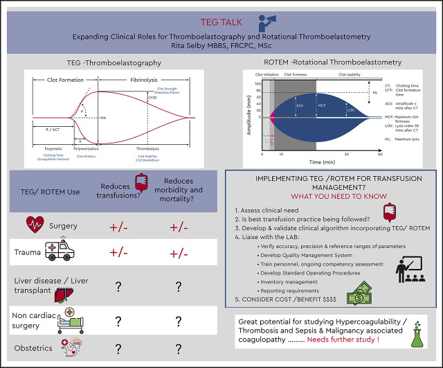
Visual Abstract
Abstract
Viscoelastic assays (VEAs) that include thromboelastography and rotational thromboelastometry add value to the investigation of coagulopathies and goal-directed management of bleeding by providing a complete picture of clot formation, strength, and lysis in whole blood that includes the contribution of platelets, fibrinogen, and coagulation factors. Conventional coagulation assays have several limitations, such as their lack of correlation with bleeding and hypercoagulability; their inability to reflect the contribution of platelets, factor XIII, and plasmin during clot formation and lysis; and their slow turnaround times. VEA-guided transfusion algorithms may reduce allogeneic blood exposure during and after cardiac surgery and in the emergency management of trauma-induced coagulopathy and hemorrhage. However, the popularity of VEAs for other indications is driven largely by extrapolation of evidence from cardiac surgery, by the drawbacks of conventional coagulation assays, and by institution-specific preferences. Robust diagnostic studies validating and standardizing diagnostic cutoffs for VEA parameters and randomized trials comparing VEA-guided algorithms with standard care on clinical outcomes are urgently needed. Lack of such studies represents the biggest barrier to defining the role and impact of VEA in clinical care.
Learning Objectives
Describe the principle of viscoelastic testing and the technologies of thromboelastography and rotational thromboelastometry
Appraise the evidence supporting viscoelastic assays in assessing coagulopathies and managing bleeding for various indications
Recognize important considerations prior to implementing thromboelastography or rotational thromboelastometry at your institution
What are viscoelastic assays, and how does one interpret them?
Viscoelastic assays (VEAs) are global tests of coagulation performed on whole blood at the point of care. The thromboelastography (TEG) assay was first described in 1948 by Dr. Helmut Hartert at the University of Heidelberg, Germany. As blood clots, the fluid becomes less viscous and more elastic in nature. TEG and rotational thromboelastometry (ROTEM) are VEAs that assess clot formation, strength, and dissolution by measuring the effect of a continuously applied rotational force on whole blood that is transmitted to an electromechanical transduction system (TEG) or optical detection system (ROTEM), with results displayed as a graph. This allows the quantitative and qualitative measurement of the function of almost all components of clot formation and lysis, including platelets, other blood cellular components, fibrinogen, microvesicles, and soluble factors.
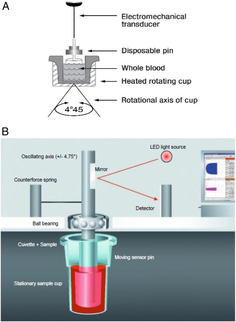
The TEG device has a pin suspended from a torsion wire immersed in a cup of whole blood (Figure 1A). The cup is held in a heating block and continually oscillates. Changes in viscoelastic clot strength are directly transmitted to the torsion wire and detected by an electromechanical transducer. The ROTEM device has a cup, which remains fixed in a heating block with whole blood while a pin suspended on a ball bearing mechanism oscillates (Figure 1B). The subsequent rotation of the pin is inversely related to the viscoelastic clot strength and is detected optically.
Figure 1.
(A) Thromboelastography principle. (B) Rotational thromboelastometry principle.
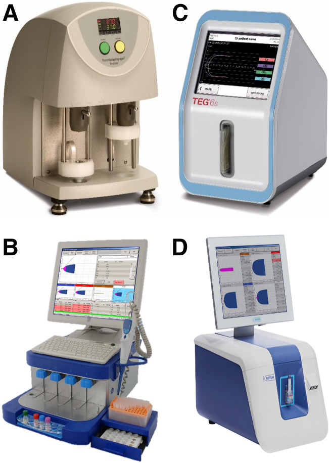
The 2 common devices in use currently are the TEG 5000 (Haemonetics, Braintree, MA) and ROTEM Delta (Instrumentation Laboratory, Bedford, MA), shown in Figures 2A and 2B. More recent versions of these analyzers, the TEG 6s and the ROTEM Sigma, use cartridge-based systems with dry reagents designed to improve both usability at point of care by non–laboratory-trained personnel and interoperator reproducibility (Figures 2C and 2D). Although all TEG and ROTEM devices measure the same viscoelastic property (shear modulus), the newer ROTEM Sigma device is still based on the rotating pin described above, whereas the TEG 6s has changed to a microfluidic assay that measures clot resonance frequency.
Figure 2.
(A) TEG 5000. (B) ROTEM Delta. (C) TEG 6S. (D) ROTEM Sigma.
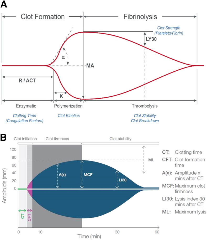
Although both TEG and ROTEM assess clot kinetics, strength, and lysis, the results are not interchangeable, owing to differences in both the operating characteristics and nomenclature of the parameters defined in the 2 systems. Figures 3A and 3B and Table 1 provide detailed descriptions of the parameters and their interpretation. A number of different assays can be performed on both TEG and ROTEM analyzers using various activators or inhibitors. In both systems, contact activation using ellagic acid or kaolin can provide information similar to the activated partial thromboplastin time. Tissue factor activation can provide information similar to the prothrombin time. Neutralization of heparin using lyophilized heparinase, blocking platelet contribution to clot formation by using platelet inhibitors, or inhibiting fibrinolysis with antifibrinolytics can help assess various aspects of coagulation. A comparison of the various assays available on the TEG 5000 and ROTEM Delta analyzers is summarized in Table 2. These assays can be customized for use in algorithms depending on the information needed to guide decision-making.
Figure 3.
(A) Thromboelastography output demonstrating clot initiation, propagation, strength, and lysis. (B) Rotational thromboelastometry output demonstrating clot initiation, propagation, strength, and lysis.
Table 1.
Comparison of TEG 5000 and ROTEM Delta parameters and their interpretation and physiological correlation to the phase of hemostasis
| Parameters | Interpretation | Physiological correlation to phase of hemostasis | |
|---|---|---|---|
| TEG 5000 (units) | ROTEM Delta (units) | ||
| Reaction rate (R) (min) | Clotting time (CT) (s) | Time for the trace to reach an amplitude of 2 mm | Activation of coagulation, thrombin generation, time to initial clot formation, and influence of anticoagulants |
| Kinetics time (K) (min) | Clot formation time (CFT) (s) | Time for the clot amplitude to reach from 2 mm to 20 mm | Fibrin activation and polymerization (ie, speed of clot propagation) |
| Angle (α) (degrees) | Angle (α) (degrees) | Angle created by drawing a tangent line from the point of clot initiation (R or CT) to the slope of the developing curve | Fibrin activation and polymerization (ie, speed of clot propagation) |
| N/A | A10 (mm) | Amplitude reached 10 min after CT | Fibrinogen and platelet contribution to the strength of the clot |
| Maximum amplitude (MA) (mm) | Maximum clot firmness (MCF) (mm) | Peak amplitude or strength of the clot | Fibrinogen and platelet contribution to the strength of the clot |
| Lysis 30 (LY 30) (%) | Lysis index 30 (LI 30) (%) | TEG: Percentage reduction in the area under the TEG curve (assuming MA remains constant) that occurs 30 min after MA is reachedROTEM: Percentage clot remaining (compared with MCF) when amplitude is measured 30 min after CT is detected | Fibrinolysis |
| Lysis 60 (LY 60) (%) | N/A | Percentage reduction in the area under the TEG curve (assuming MA remains constant) that occurs 60 min after MA is reached | Fibrinolysis |
| N/A | Maximum lysis (ML) (%) | Degree of fibrinolysis relative to MCF achieved during the measurement (percentage clot firmness lost). It is not calculated at a fixed time. | Fibrinolysis |
N/A, corresponding parameter not available on the analyzer; ROTEM, rotational thromboelastometry; TEG, thromboelastography.
Table 2.
Comparison of available assays on the TEG 5000 and ROTEM Delta analyzers
| Assays | Interpretation/clinical indications | |
|---|---|---|
| TEG 5000* | ROTEM Delta† | |
|
|
These assays assess the contact activation pathway of coagulation and provide information similar to the activated partial thromboplastin time (APTT). |
| N/A |
|
This assay assesses the tissue factor–initiated pathway of coagulation and provides information similar to the prothrombin time (PT). |
|
N/A | This assay assesses both the tissue factor–initiated and contact pathway–initiated coagulation and provides information similar to the PT and APTT. |
|
|
These assays can assess heparin’s effect when used in conjunction with the kaolin reagent (Standard TEG) and compared with the kaolin analysis on the TEG analyzer or when used in conjunction with the INTEM assay and compared with the INTEM analysis on the ROTEM analyzer. |
|
|
These assays can qualitatively assess the contribution of fibrinogen to clot strength independent of platelets when used in conjunction with the kaolin reagent (Standard TEG) and compared with the kaolin analysis on the TEG analyzer or when used in conjunction with the EXTEM assay and compared with the EXTEM analysis on the ROTEM analyzer. |
| N/A |
|
This assay is designed to allow discrimination between fibrinolysis and platelet-mediated clot retraction. |
|
N/A | This assay is designed to assess platelet function and the effect of antiplatelet agents. |
|
|
These assays are impractical for clinical use, given their long reaction rate (R) on the TEG analyzer and clot formation time (CFT) on the ROTEM analyzer, respectively. They can be used to run custom hemostasis tests. |
GP, glycoprotein; N/A, corresponding assay is not available on analyzer; ROTEM, rotational thromboelastometry; TEG, thromboelastography.
TEG 6S analyzer: Standard TEG (kaolin) is CK channel; rapid TEG (rTEG) is CRT channel; heparinase TEG (hTEG) is CKH channel; and functional fibrinogen TEG (FLEV-TEG) is CFF channel.
ROTEM Sigma analyzer: Assays have the same designation as the ROTEM Delta analyzer, except with a suffix C at the end (eg, EXTEM C).
It is essential to note that diagnostic cutoffs or thresholds for the various parameters are assay, institution, or algorithm specific and have not been standardized for many clinical indications. Evidence of correlation between VEA parameters and various conventional coagulation assays (CCAs) is also limited.
Case 1
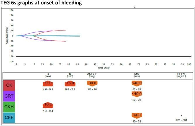
A 75-year-old man underwent a complicated aortic and mitral valve replacement with tricuspid repair on cardiopulmonary bypass (CPB). Due to the extent of the surgery, the CPB run time was long (2.5 hours), which is generally associated with increased bleeding. He received a prophylactic loading dose of 2 g of tranexamic acid (TXA) intravenously over the course of 10 minutes at sternotomy, followed by an infusion of 16 mg/kg/hour and heparin 32 000 U during CPB. As he was being weaned off CPB, during the rewarming phase, he started to bleed severely. Protamine was administered at 0.7 mg/100 U of an initial heparin dose with normalization of the activated clotting time. The TEG assay performed on the TEG 6s analyzer (4 simultaneous channels) in the operating room (OR) showed the results in Figure 4.
Figure 4.
TEG 6s graphs at onset of bleeding.
TEG 6s graphs at onset of bleeding
The kaolin-activated TEG channel (CK) demonstrated a prolonged R time, indicating coagulation factor deficiency. The CK channel also showed a prolonged K time and reduced angle with reduced maximum amplitude (MA) on the functional fibrinogen (CFF) channel, indicating decreased fibrinogen contribution to clot formation. The MA on the CK channel was reduced, indicating decreased platelet function. There was no significant residual heparin effect, as evidenced by the minimal difference in R time between the kaolin sample in the CK channel and the kaolin sample with heparinase (CKH channel).
Is there a role for VEAs in the management of post–cardiac surgery bleeding?
Excessive bleeding after CPB occurs in approximately 20% of patients and is severe or massive in 10% to 15%, with up to 5% of patients requiring emergency surgical reexploration.1 It accounts for almost 80% of blood transfusions after cardiac surgery and is an independent predictor of morbidity and mortality.1-5 Bleeding after CPB is multifactorial and is related to surgical damage to blood vessels, technically complicated procedures, redo operations, and an acquired acute coagulopathy. Contributors to this coagulopathy include qualitative and quantitative platelet function defects; hemodilution causing reduction of procoagulants; use of intraoperative anticoagulants (heparin and potentially protamine); and activation of coagulation, inflammation, complement, and fibrinolysis.1-7 Despite continuous efforts and clinical practice guidelines, blood product use after cardiac surgery has not shown significant declines over the past decade, remaining at ≥50% in high-risk patients.1,2 Interventions to reduce bleeding and allogeneic blood exposure include preoperative hemoglobin optimization, heparin reversal after CPB, minimizing hemodilution, cell salvage, restrictive red blood cell (RBC) transfusion strategies, and monitoring of coagulation and platelet function to guide blood product replacement.1,7
Over the past 2 decades, 16 randomized controlled trials (RCTs) and many observational studies have assessed the role of VEA-guided transfusion algorithms after cardiac surgery, and 5 recent meta-analyses have analyzed these data.8-12 In addition, an updated Cochrane systematic review and a health technology assessment (HTA) from the United Kingdom were published in 2015 and 2016, respectively, assessing the role of VEA in managing patients with bleeding. The majority of RCTs included in these 2 reviews were in adults undergoing elective CPB.13,14 Six RCTs used TEG and 10 used ROTEM, comparing them to either CCA-guided algorithms or algorithms using clinician judgment. The majority of RCTs were small (between 22 and 224 patients), conducted in a single center, of poor methodological quality, and with significant heterogeneity and bias, thereby limiting the reproducibility of VEA parameters and the generalizability of their conclusions. One large, multicenter study by Karkouti et al15 randomized 7402 patients at 12 Canadian hospitals in a stepped-wedge, clustered, pragmatic, randomized trial evaluating a combination of ROTEM and a point-of-care platelet function assay used in the OR in the context of a locally developed and validated transfusion algorithm.16-18 In the preintervention phase of this trial, hospitals were instructed to continue their usual management of post–cardiac surgery bleeding as per their institutional guidelines or standard of care.15 The cumulative assessment of all these RCTs and meta-analyses in summary is that VEA-based management algorithms may be beneficial in reducing allogeneic transfusion of RBCs, frozen plasma, and platelets, as well as postoperative blood loss 12 and 24 hours after surgery, with a trend toward lowering mortality. Other clinical outcomes, such as surgical reexploration and intensive care unit (ICU) or hospital length of stay, are not significantly reduced. However, these conclusions are based primarily on poor-quality RCTs with a high risk of bias.8-14 It is also possible that some of the benefit seen in these studies may be a result of VEA implementation often being paired with a renewed effort to standardize transfusion practice.
Case 1 follow-up
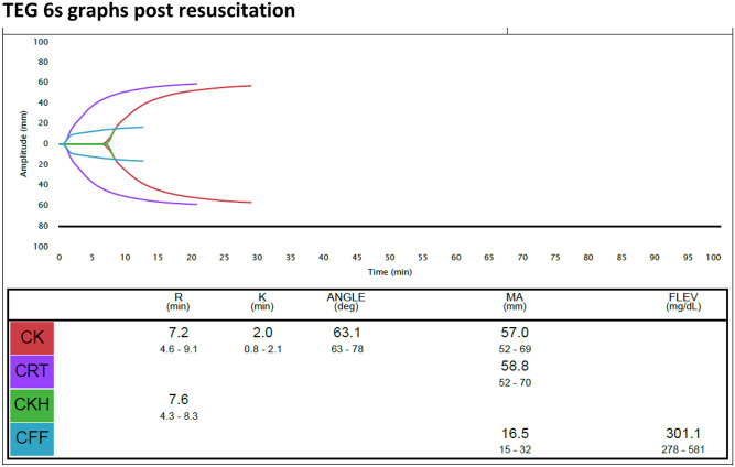
The initial patterns of TEG 6s graphs at the onset of bleeding indicated deficiency of coagulation factors, fibrinogen, and platelets. The following blood products were transfused: 4 U of frozen plasma, 4 g of fibrinogen concentrate, and 1 pool of platelets. TXA infusion was continued. Subsequent TEG 6s data at 80 minutes after transfusion are shown in Figure 5 and described below.
Figure 5.
TEG 6s graphs postresuscitation.
TEG 6s graphs after resuscitation
The postresuscitation TEG showed normal coagulation factors (CK R time, 7.2 minutes), fibrinogen (K time, 2 minutes; angle 63.1 degrees; CFF channel MA, 16.5 mm), and platelet function (CK MA, 57.0 mm). The bleeding slowed significantly, and the patient was transferred to the cardiovascular ICU in hemodynamically stable condition.
Case 2
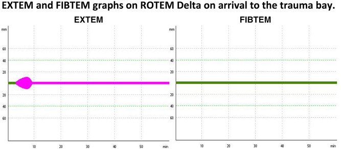
While jogging, a 48-year-old man was struck by a car traveling at 50 miles per hour. At the scene, he was alert and oriented but in severe pain in the lower abdomen, with no overt bleeding but with a systolic blood pressure of 85 mm Hg and a heart rate of 110 beats per minute. He was airlifted to a major trauma center. Initial resuscitation included IV fluids and a 1-g loading dose of TXA over the course of 10 minutes followed by 1 g of TXA infused over the course of 8 hours (per protocol). Trauma CT scans revealed a pelvic fracture with a large retroperitoneal hematoma. EXTEM and FIBTEM assays performed on a ROTEM Delta analyzer in the trauma bay showed the results in Figure 6.
Figure 6.
ROTEM Delta EXTEM and FIBTEM graphs on arrival at the trauma bay.
EXTEM MCF was 10 mm (reference interval [RI], 50 to 72 mm) with LI 30 at 0%. FIBTEM MCF showed no clot. This indicated severe coagulation factor and fibrinogen deficiency with severe hyperfibrinolysis. An initial complete blood count obtained from the laboratory within 15 minutes showed a hemoglobin level of 11.1 g/dL (RI, 12 to 15.5 g/dL) and a platelet count of 355 000 × 109/L (RI, 150 000 × 109 to 400 000 × 109/L).
Is there a role for VEA in the management of hemorrhage or coagulopathy in patients with major trauma?
Over the last 2 decades, more than 60 studies have evaluated either TEG or ROTEM assays in diagnosing early trauma coagulopathy and have assessed the role of VEA-guided transfusion algorithms in managing trauma-associated hemorrhage and in reducing mortality.19-28 Both trauma-induced coagulopathy and hemorrhage are strong predictors of inpatient morbidity (massive transfusion, increase in complications, multiorgan failure, length of ICU stay, and perioperative cardiac arrest) and mortality.19-21,29-31 Most of these studies are observational, single-center cohort studies; only one recent RCT has been published.23 Reliable estimates of diagnostic accuracy of either TEG or ROTEM compared with CCA are not available, because very few studies have reported this information.19,21 Almost half of the studies reported a trend toward improvement in blood product use and a mortality benefit when VEA-guided resuscitation algorithms were used.19-28 Recently, Gonzalez et al23 randomized 111 trauma patients who all met the criteria for activation of a massive transfusion protocol (MTP) to either a TEG-guided MTP algorithm or one guided by CCA, with a primary outcome of 28-day survival. The TEG-guided MTP algorithm significantly improved survival at 28 days compared with the CCA algorithm (36.4% deaths vs 19.6%; P = .049) and reduced deaths within the first 6 hours of arrival (7.1% in TEG group vs 21.8% in CCA group; P = .032). Patients randomized to TEG also received significantly fewer frozen plasma and platelet units than the CCA group. Although this is encouraging, large multicenter RCTs are needed to definitively determine the clinical effectiveness of VEAs in goal-directed resuscitation after major trauma.
One exception in which VEAs may uniquely contribute is in early hyperfibrinolysis due to hemorrhagic shock, seen in 2% to 5% of patients with major trauma but associated with up to 80% early mortality.30,32-35 VEAs can detect hyperfibrinolysis early when CCAs cannot, thereby allowing specific interventions such as antifibrinolytic therapy to prevent death.32-35 However, the lack of a reliable gold standard to diagnose hyperfibrinolysis makes it difficult to assess if VEA identification of hyperfibrinolysis is sensitive or specific.
Although recent international guidelines have recommended that VEAs be used early in trauma resuscitation to assess coagulation status and guide transfusion of blood products and antifibrinolytics, experts were unable to recommend the exact thresholds to be used as transfusion triggers due to lack of data.35,36 A recent large, prospective, multicenter study with 2287 patients from 6 European adult trauma centers attempted to systematically validate diagnostic cutoffs for both ROTEM and TEG parameters for the detection of coagulopathy, thrombocytopenia, hypofibrinogenemia, and hyperfibrinolysis and specific transfusion therapies based on these triggers.24 Algorithms based on these VEA thresholds are now being compared with CCAs in an ongoing RCT and will require external validation in subsequent studies.
Case 2 follow-up
Initial EXTEM and FIBTEM graphs upon arrival indicated severe coagulation factor and fibrinogen deficiency with severe hyperfibrinolysis. On this basis, resuscitation was begun within 10 minutes with 8 U of frozen plasma and 4 g of fibrinogen concentrate. TXA infusion was continued. Eight units of RBC were administered. Coagulation laboratory results obtained 65 minutes after arrival confirmed the coagulopathy and hypofibrinogenemia: the patient’s international normalized ratio (INR) was 2.9 (RI, 0.9 to 1.2), and his fibrinogen level was 0.29 g/L (RI, 1.7 to 4 g/L).
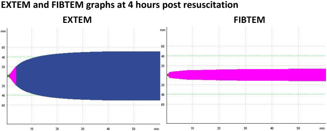
EXTEM and FIBTEM graphs at 4 hours after resuscitation (Figure 7)
Figure 7.
EXTEM and FIBTEM graphs at 4 hours postresuscitation.
The patient’s EXTEM MCF corrected to 51 mm (RI, 50 to 72 mm), with LI 30 improving to 100%. His FIBTEM MCF was now normal at 11 mm (RI, 9 to 25 mm). His INR and fibrinogen were 1.35 (RI, 0.9 to 1.2) and 1.6 g/L (RI, 1.7 to 4 g/L), respectively, at the same time point. The patient was taken to the operating room for internal fixation of his pelvic fracture in stable hemodynamic condition.
Other clinical indications for VEAs
VEAs have been used in the management of bleeding in liver transplant (LT) surgery37 and other noncardiac surgeries,38 as well as in the assessment of the coagulopathy of end-stage liver disease (ESLD)37,39 and sepsis.40,41 In ESLD and LT surgery, the evidence base is limited to small, single-center, observational studies and only one RCT with 28 LT patients that compared a TEG-based algorithm with CCA.37 These studies have shown inconsistent trends toward reduced bleeding or transfusions with the use of VEA. A recent meta-analysis of VEA in noncardiac surgical settings (trauma, burns, and ESLD and LT surgery) included only 4 small RCTs (a total of 229 participants) and concluded that data were insufficient to determine any reduction in transfusions in these settings.38 Despite this, VEAs are used in the majority of LT surgeries, likely extrapolating the evidence from cardiac surgery studies. This is concerning because the coagulopathic states of ESLD and during LT surgery are markedly different from those seen in cardiac surgery.37,39 Using VEAs for these indications must be accompanied by a clear understanding that VEA parameters suggesting coagulation factor, fibrinogen, or platelet deficiency or hyperfibrinolysis have not been validated in ESLD or during LT surgery and, if being used in the context of a locally validated VEA-based algorithm, should be used only to manage bleeding (not prophylactically).
In the evaluation of sepsis-induced coagulopathy (SIC), limited evidence suggests that, compared with CCAs, VEAs can assess both hypo- and hypercoagulability (associated with disseminated intravascular coagulation) and impaired fibrinolysis (associated with systemic inflammatory response syndrome).40,41 The evidence thus far is limited to the characterization of SIC and early associations with clinical outcomes such as mortality.
VEAs have also been used in assessing hypercoagulability following surgery and its relationship to venous thromboembolism (VTE); characterization of malignancy-associated coagulopathy; and, most recently, the hypercoagulopathy seen in patients with severe coronavirus disease 2019.42-44 Similar to studies in SIC, these studies are mostly observational and describe VEA parameters most indicative of a hypercoagulable state, the temporal course of this hypercoagulability, and associations with VTE. They are limited by varying definitions of VEA parameters that suggest hypercoagulability, varying populations of patients, and small sample sizes.42,43 Several such studies have shown that hypercoagulable parameters in VEA testing are associated with increased thromboembolic complications and may be used to guide thromboprophylaxis.42,43 Because CCAs cannot specifically identify hypercoagulable states or predict VTE, VEAs present an exciting potential opportunity for future research in this area.
VEAs have been evaluated extensively in pregnancy and puerperium in both the management of postpartum hemorrhage (PPH) and assessment of hypercoagulability. Normal reference ranges for both TEG and ROTEM have been developed for all trimesters of pregnancy, during labor, postpartum, and pre- and postoperatively for women undergoing cesarean delivery, and they correlate well with CCAs.45 PPH is the commonest obstetrical scenario in which VEAs have been used; the ROTEM FIBTEM assay has been shown to correlate with fibrinogen, predict progression of PPH, and guide transfusion needs.45,46 The Women’s Health Scientific and Standardization Committee of the International Society on Thrombosis and Haemostasis has identified the lack of large RCTs comparing VEA-guided care with usual care with evaluation of important clinical outcomes as the biggest barrier to the adoption of VEAs in obstetrics.45,46
Evolving roles for VEAs include the management of hemophilia and the complex anticoagulation management of patients receiving extracorporeal membrane oxygenation (ECMO) and left ventricular assist devices. In hemophilia A, VEAs have been studied in determining clinical bleeding phenotype correlated with factor VIII (FVIII) levels, monitoring of routine FVIII prophylaxis, assessing the response to bypassing agents, and as a means to determine the optimal dose of factor replacement in the perioperative period for patients undergoing surgery.47 Although VEAs have been incorporated into institution-specific algorithms to assess the complexities of concurrent bleeding and thrombosis and to monitor anticoagulants in patients receiving ECMO and left ventricular assist devices, widespread use has been hampered by a lack of robust evidence.48,49
How do I implement VEAs at my institution?
VEAs are being used in clinical algorithms despite lack of evidence demonstrating benefit for many indications. This is driven largely by the inherent limitations associated with the CCAs, including their poor correlation with bleeding; their inability to reflect the contribution of platelets, factor XIII, and plasmin during coagulation; their lack of immediate turnaround times; and the need for rapid, serial monitoring for guiding blood products during severe hemorrhage. Given transport to the laboratory and centrifugation requirements, CCAs using citrated plasma will (at a minimum) have a turnaround time of 30 to 60 minutes. VEAs help to address this gap by providing coagulation information at the point of care within 10 to 20 minutes. However, until rigorous evidence establishing and externally validating diagnostic cutoffs for various parameters, their interoperator reproducibility, and their independent predictive value for clinical outcomes is available, the following considerations will help to ensure that VEAs are being used in a beneficial and safe manner. This requires close collaboration between clinicians and the laboratory.
Clinicians must clearly define their goals for introducing VEAs (eg, ensuring appropriate transfusion during MTPs to reduce allogeneic blood exposure).
Current practice in the area must be evaluated first to ensure that best transfusion practices are being adhered to.
If slow turnaround time of laboratory results is the major reason for needing to institute VEAs, assess if laboratory turnaround times for CCAs can be improved through institution of “stat” protocols that operationalize clinical, laboratory, and transport teams to achieve this goal.
Clinicians must develop a detailed clinical algorithm using the chosen VEA and must locally validate diagnostic thresholds for the specific clinical indication.16-18
-
The laboratory must be involved in decision-making about selection of both VEA devices and assays, their ideal location (laboratory vs point of care) based on device type and clinical goals, and assisting with the following implementation steps before the device can be put into clinical use:
Evaluating and validating the selected VEA for accuracy and precision (ie, verifying manufacturer claims) and assessing interoperator reproducibility;
Developing local reference ranges for various parameters or verifying manufacturer-provided reference ranges;
Setting up a quality management program for the VEA, including the type and frequency of quality controls, enrolling in an external quality assessment program, training of individuals who will be performing the VEA (often nonlaboratory professionals), and assessing their competency at regular intervals;
Developing and maintaining standard operating procedures;
Assisting with managing inventory of reagents and consumables and service contracts with vendors, as well as troubleshooting device malfunctions and downtimes; and
Assisting with reporting of VEA results in accordance with clinical needs and compliant with local laboratory accreditation standards.
Are VEAs cost-effective?
An HTA on VEAs conducted on behalf of the National Institute for Health and Care Excellence in the United Kingdom in 2015 concluded that VEAs are effective and cost-effective compared with CCAs in cardiac surgery, primarily on the basis of projected reduction in transfusions.14 Depending on the assay combinations used, the costs may vary between TEG and ROTEM. For the trauma indication, both this HTA and a Canadian HTA conducted in 2017 concluded that VEAs may potentially be more cost-effective than CCAs in patients with trauma, given the larger volumes of blood transfusions than in cardiac surgery patients; however, the lack of high-quality evidence of clinical effectiveness limits the validity of their conclusions.50 In the absence of robust cost-effectiveness data, institutions adopting VEAs must calculate annual costs of analyzer service contracts, test reagents, consumables, and quality control materials, as well as the costs of acquiring and maintaining a backup analyzer, training of personnel, and participation in an external quality assessment program, against the potential cost savings associated with reduced transfusions and associated complications.
Conclusions
VEAs have the potential to provide a complete picture of all elements of coagulation, clot strength, and lysis in assessing various coagulopathies. Although CCAs provide informative data on individual elements of coagulation, the presentation of clot formation and lysis visually and at the point of care have likely made VEAs an attractive alternative to CCAs.
Current evidence shows that using VEA-guided transfusion algorithms in cardiac surgery and major trauma may reduce allogeneic blood exposure and blood loss, which can translate to reduction in morbidity and mortality and can be cost-effective. There are also exciting opportunities to advance understanding of hypercoagulability and thrombosis prediction using VEAs, which is a gap currently not addressed by CCAs.
However, there is an urgent need for appropriately designed diagnostic studies correlating VEAs to CCAs, determining and externally validating diagnostic cutoffs for VEA parameters, and large multicenter RCTs establishing the independent contribution of VEA-guided algorithms in improving clinical outcomes compared with standard practice. Until such rigorous evidence is available, clinicians and institutions wishing to use TEG or ROTEM must do so appreciating the uncertainty regarding effectiveness, paying attention to monitoring the quality of results, and evaluating their benefits and costs locally.
Acknowledgments
Oksana Volod and Jeannie Callum provided images of TEG and ROTEM outputs for cases 1 and 2, respectively. Haemonetics and Instrumentation Laboratory (Werfen) provided the images for Figures 1 to 3. Carolyne Elbaz provided creative and formatting assistance for the visual abstract.
References
- 1.Raphael J, Mazer CD, Subramani S, et al. Society of Cardiovascular Anesthesiologists clinical practice improvement advisory for management of perioperative bleeding and hemostasis in cardiac surgery patients. Anesth Analg. 2019;129(5):1209-1221. [DOI] [PubMed] [Google Scholar]
- 2.Ferraris VA, Brown JR, Despotis GJ, et al. ; International Consortium for Evidence Based Perfusion. 2011 Update to the Society of Thoracic Surgeons and the Society of Cardiovascular Anesthesiologists blood conservation clinical practice guidelines. Ann Thorac Surg. 2011;91(3):944-982. [DOI] [PubMed] [Google Scholar]
- 3.Ranucci M. Outcome measures and quality markers for perioperative blood loss and transfusion in cardiac surgery. Can J Anaesth. 2016;63(2):169-175. [DOI] [PubMed] [Google Scholar]
- 4.Murphy GJ, Reeves BC, Rogers CA, Rizvi SI, Culliford L, Angelini GD. Increased mortality, postoperative morbidity, and cost after red blood cell transfusion in patients having cardiac surgery. Circulation. 2007;116(22):2544-2552. [DOI] [PubMed] [Google Scholar]
- 5.Karkouti K, Wijeysundera DN, Yau TM, et al. The independent association of massive blood loss with mortality in cardiac surgery. Transfusion. 2004;44(10):1453-1462. [DOI] [PubMed] [Google Scholar]
- 6.Sarkar M, Prabhu V. Basics of cardiopulmonary bypass. Indian J Anaesth. 2017;61(9):760-767. [DOI] [PMC free article] [PubMed] [Google Scholar]
- 7.Sniecinski RM, Chandler WL. Activation of the hemostatic system during cardiopulmonary bypass. Anesth Analg. 2011;113(6):1319-1333. [DOI] [PubMed] [Google Scholar]
- 8.Meco M, Montisci A, Giustiniano E, et al. Viscoelastic blood tests use in adult cardiac surgery: meta-analysis, meta-regression, and trial sequential analysis. J Cardiothorac Vasc Anesth. 2020;34(1):119-127. [DOI] [PubMed] [Google Scholar]
- 9.Li C, Zhao Q, Yang K, Jiang L, Yu J. Thromboelastography or rotational thromboelastometry for bleeding management in adults undergoing cardiac surgery: a systematic review with meta-analysis and trial sequential analysis. J Thorac Dis. 2019;11(4):1170-1181. [DOI] [PMC free article] [PubMed] [Google Scholar]
- 10.Lodewyks C, Heinrichs J, Grocott HP, et al. Point-of-care viscoelastic hemostatic testing in cardiac surgery patients: a systematic review and meta-analysis. Can J Anaesth. 2018;65(12):1333-1347. [DOI] [PubMed] [Google Scholar]
- 11.Serraino GF, Murphy GJ. Routine use of viscoelastic blood tests for diagnosis and treatment of coagulopathic bleeding in cardiac surgery: updated systematic review and meta-analysis. Br J Anaesth. 2017;118(6):823-833. [DOI] [PubMed] [Google Scholar]
- 12.Deppe AC, Weber C, Zimmermann J, et al. Point-of-care thromboelastography/thromboelastometry-based coagulation management in cardiac surgery: a meta-analysis of 8332 patients. J Surg Res. 2016;203(2):424-433. [DOI] [PubMed] [Google Scholar]
- 13.Wikkelsø A, Wetterslev J, Møller AM, Afshari A. Thromboelastography (TEG) or thromboelastometry (ROTEM) to monitor haemostatic treatment versus usual care in adults or children with bleeding. Cochrane Database Syst Rev. 2016;(8):CD007871. [DOI] [PMC free article] [PubMed] [Google Scholar]
- 14.Whiting P, Al M, Westwood M, et al. Viscoelastic point-of-care testing to assist with the diagnosis, management and monitoring of haemostasis: a systematic review and cost-effectiveness analysis. Health Technol Assess. 2015;19(58). [DOI] [PMC free article] [PubMed] [Google Scholar]
- 15.Karkouti K, Callum J, Wijeysundera DN, et al. ; TACS Investigators. Point-of-care hemostatic testing in cardiac surgery: a stepped-wedge clustered randomized controlled trial. Circulation. 2016;134(16):1152-1162. [DOI] [PubMed] [Google Scholar]
- 16.Karkouti K, McCluskey SA, Callum J, et al. Evaluation of a novel transfusion algorithm employing point-of-care coagulation assays in cardiac surgery: a retrospective cohort study with interrupted time-series analysis. Anesthesiology. 2015;122(3):560-570. [DOI] [PubMed] [Google Scholar]
- 17.Orlov D, McCluskey SA, Selby R, Yip P, Pendergrast J, Karkouti K. Platelet dysfunction as measured by a point-of-care monitor is an independent predictor of high blood loss in cardiac surgery. Anesth Analg. 2014;118(2):257-263. [DOI] [PubMed] [Google Scholar]
- 18.Mace H, Lightfoot N, McCluskey S, et al. Validity of thromboelastometry for rapid assessment of fibrinogen levels in heparinized samples during cardiac surgery: a retrospective, single-center, observational study. J Cardiothorac Vasc Anesth. 2016;30(1):90-95. [DOI] [PubMed] [Google Scholar]
- 19.Da Luz LT, Nascimento B, Shankarakutty AK, Rizoli S, Adhikari NK. Effect of thromboelastography (TEG) and rotational thromboelastometry (ROTEM) on diagnosis of coagulopathy, transfusion guidance and mortality in trauma: descriptive systematic review. Crit Care. 2014;18(5):518. [DOI] [PMC free article] [PubMed] [Google Scholar]
- 20.Müller MC, Balvers K, Binnekade JM, et al. Thromboelastometry and organ failure in trauma patients: a prospective cohort study. Crit Care. 2014;18(6):687. [DOI] [PMC free article] [PubMed] [Google Scholar]
- 21.Hunt H, Stanworth S, Curry N, et al. Thromboelastography (TEG) and rotational thromboelastometry (ROTEM) for trauma induced coagulopathy in adult trauma patients with bleeding. Cochrane Database Syst Rev. 2015;(2):CD010438. [DOI] [PMC free article] [PubMed] [Google Scholar]
- 22.Veigas PV, Callum J, Rizoli S, Nascimento B, da Luz LT. A systematic review on the rotational thrombelastometry (ROTEM) values for the diagnosis of coagulopathy, prediction and guidance of blood transfusion and prediction of mortality in trauma patients. Scand J Trauma Resusc Emerg Med. 2016;24(1):114. [DOI] [PMC free article] [PubMed] [Google Scholar]
- 23.Gonzalez E, Moore EE, Moore HB, et al. Goal-directed hemostatic resuscitation of trauma-induced coagulopathy: a pragmatic randomized clinical trial comparing a viscoelastic assay to conventional coagulation assays. Ann Surg. 2016;263(6):1051-1059. [DOI] [PMC free article] [PubMed] [Google Scholar]
- 24.Baksaas-Aasen K, Van Dieren S, Balvers K, et al. ; TACTIC/INTRN collaborators. Data-driven development of ROTEM and TEG algorithms for the management of trauma hemorrhage: a prospective observational multicenter study. Ann Surg. 2019;270(6):1178-1185. [DOI] [PubMed] [Google Scholar]
- 25.Juffermans NP, Wirtz MR, Balvers K, et al. ; TACTIC partners. Towards patient-specific management of trauma hemorrhage: the effect of resuscitation therapy on parameters of thromboelastometry. J Thromb Haemost. 2019;17(3):441-448. [DOI] [PMC free article] [PubMed] [Google Scholar]
- 26.Peng HT, Nascimento B, Tien H, et al. A comparative study of viscoelastic hemostatic assays and conventional coagulation tests in trauma patients receiving fibrinogen concentrate. Clin Chim Acta. 2019;495:253-262. [DOI] [PubMed] [Google Scholar]
- 27.Smith AR, Karim SA, Reif RR, et al. ROTEM as a predictor of mortality in patients with severe trauma. J Surg Res. 2020;251:107-111. [DOI] [PubMed] [Google Scholar]
- 28.Cohen J, Scorer T, Wright Z, et al. A prospective evaluation of thromboelastometry (ROTEM) to identify acute traumatic coagulopathy and predict massive transfusion in military trauma patients in Afghanistan. Transfusion. 2019;59(S2):1601-1607. [DOI] [PubMed] [Google Scholar]
- 29.Braz LG, Carlucci MTO, Braz JRC, Módolo NSP, do Nascimento P Jr, Braz MG. Perioperative cardiac arrest and mortality in trauma patients: a systematic review of observational studies. J Clin Anesth. 2020;64:109813. [DOI] [PubMed] [Google Scholar]
- 30.Moore HB, Moore EE. Temporal changes in fibrinolysis following injury. Semin Thromb Hemost. 2020;46(2):189-198. [DOI] [PubMed] [Google Scholar]
- 31.Spinella PC, Holcomb JB. Resuscitation and transfusion principles for traumatic hemorrhagic shock. Blood Rev. 2009;23(6):231-240. [DOI] [PMC free article] [PubMed] [Google Scholar]
- 32.Cotton BA, Harvin JA, Kostousouv V, et al. Hyperfibrinolysis at admission is an uncommon but highly lethal event associated with shock and prehospital fluid administration. J Trauma Acute Care Surg. 2012;73(2):365-370, discussion 370. [DOI] [PubMed] [Google Scholar]
- 33.David JS, Lambert A, Bouzat P, et al. Fibrinolytic shutdown diagnosed with rotational thromboelastometry represents a moderate form of coagulopathy associated with transfusion requirement and mortality: a retrospective analysis. Eur J Anaesthesiol. 2020;37(3):170-179. [DOI] [PubMed] [Google Scholar]
- 34.Wang IJ, Park SW, Bae BK, et al. FIBTEM improves the sensitivity of hyperfibrinolysis detection in severe trauma patients: a retrospective study using thromboelastometry. Sci Rep. 2020;10(1):6980. [DOI] [PMC free article] [PubMed] [Google Scholar]
- 35.Inaba K, Rizoli S, Veigas PV, et al. ; Viscoelastic Testing in Trauma Consensus Panel. 2014 Consensus conference on viscoelastic test-based transfusion guidelines for early trauma resuscitation: report of the panel. J Trauma Acute Care Surg. 2015;78(6):1220-1229. [DOI] [PubMed] [Google Scholar]
- 36.Spahn DR, Bouillon B, Cerny V, et al. The European guideline on management of major bleeding and coagulopathy following trauma: fifth edition. Crit Care. 2019;23(1):98. [DOI] [PMC free article] [PubMed] [Google Scholar]
- 37.Blaine KP, Sakai T. Viscoelastic monitoring to guide hemostatic resuscitation in liver transplantation surgery. Semin Cardiothorac Vasc Anesth. 2018;22(2):150-163. [DOI] [PubMed] [Google Scholar]
- 38.Franchini M, Mengoli C, Cruciani M, et al. The use of viscoelastic haemostatic assays in non-cardiac surgical settings: a systematic review and meta-analysis. Blood Transfus. 2018;16(3):235-243. [DOI] [PMC free article] [PubMed] [Google Scholar]
- 39.Abeysundara L, Mallett SV, Clevenger B. Point-of-care testing in liver disease and liver surgery. Semin Thromb Hemost. 2017;43(4):407-415. [DOI] [PubMed] [Google Scholar]
- 40.Müller MC, Meijers JC, Vroom MB, Juffermans NP. Utility of thromboelastography and/or thromboelastometry in adults with sepsis: a systematic review. Crit Care. 2014;18(1):R30. [DOI] [PMC free article] [PubMed] [Google Scholar]
- 41.Davies GR, Lawrence M, Pillai S, et al. The effect of sepsis and septic shock on the viscoelastic properties of clot quality and mass using rotational thromboelastometry: a prospective observational study. J Crit Care. 2018;44:7-11. [DOI] [PubMed] [Google Scholar]
- 42.Brown W, Lunati M, Maceroli M, et al. Ability of thromboelastography to detect hypercoagulability: a systematic review and meta-analysis. J Orthop Trauma. 2020;34(6):278-286. [DOI] [PubMed] [Google Scholar]
- 43.Walsh M, Moore EE, Moore H, et al. Use of viscoelastography in malignancy-associated coagulopathy and thrombosis: a review. Semin Thromb Hemost. 2019;45(4):354-372. [DOI] [PMC free article] [PubMed] [Google Scholar]
- 44.Panigada M, Bottino N, Tagliabue P, et al. Hypercoagulability of COVID-19 patients in intensive care unit: a report of thromboelastography findings and other parameters of hemostasis. J Thromb Haemost. 2020;18(7):1738-1742. [DOI] [PMC free article] [PubMed] [Google Scholar]
- 45.Othman M, Han K, Elbatarny M, Abdul-Kadir R. The use of viscoelastic hemostatic tests in pregnancy and puerperium: review of the current evidence - communication from the Women’s Health SSC of the ISTH. J Thromb Haemost. 2019;17(7):1184-1189. [DOI] [PubMed] [Google Scholar]
- 46.Amgalan A, Allen T, Othman M, Ahmadzia HK. Systematic review of viscoelastic testing (TEG/ROTEM) in obstetrics and recommendations from the women’s SSC of the ISTH. J Thromb Haemost. 2020;18(8):1813-1838. [DOI] [PubMed] [Google Scholar]
- 47.Ramiz S, Hartmann J, Young G, Escobar MA, Chitlur M. Clinical utility of viscoelastic testing (TEG and ROTEM analyzers) in the management of old and new therapies for hemophilia. Am J Hematol. 2019;94(2):249-256. [DOI] [PubMed] [Google Scholar]
- 48.Bercovitz RS. An introduction to point-of-care testing in extracorporeal circulation and LVADs. Hematology (Am Soc Hematol Educ Program). 2018;2018(1):516-521. [DOI] [PMC free article] [PubMed] [Google Scholar]
- 49.Koster A, Ljajikj E, Faraoni D. Traditional and non-traditional anticoagulation management during extracorporeal membrane oxygenation. Ann Cardiothorac Surg. 2019;8(1):129-136. [DOI] [PMC free article] [PubMed] [Google Scholar]
- 50.MacDonald E, Severn M. Thromboelastography or rotational thromboelastography for trauma: a review of the clinical and cost-effectiveness and guidelines. Ottawa, ON, Canada: Canadian Agency for Drugs and Technologies in Health; 2017. [PubMed] [Google Scholar]


