

A 30-year-old male presented with diminished vision in the left eye of 3 days duration, following trauma sustained to after a fall from motor bike. His visual acuity was 20/20 in the right eye and 20/200 in the left eye. His intraocular pressure with non-contact tonometry was 11 and 13 mm of Hg respectively in the right eye and the left eye. Ocular examination showed peri-orbital edema and ecchymosis on the left side with no evidence of orbital fracture. Ocular movements were full and painless. Pupillary light reflex was normal. Slit-lamp examination showed subconjuctival hemorrhage in the temporal conjunctiva. Indirect ophthalmoscopy showed a normal optic disc, inferior vitreous hemorrhage, severe commotio retinae and sub foveal hemorrhage with choroidal rupture involving the macula. Extensive pre retinal hemorrhages were seen over the posterior pole and temporal retina. There was retinal edema with a large radial, wedge shaped, horizontal retinal tear in temporal periphery, extending till the equatorial region [Fig. 1], with no retinal detachment. The patient was advised for orbital imaging, B-scan ultrasonography and barrage laser photocoagulation. This case is unique due to the shape and orientation of the tear and such radial tears being rarely described in the literature.
Figure 1.
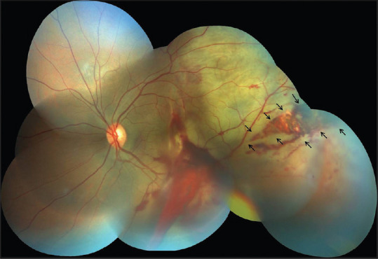
Left eye: Fundus photograph showing preretinal and subretinal hemorrhage at the posterior pole, choroidal rupture involving the fovea, extensive Berlin's edema, and an atypical wedge-shaped horizontal irregular retinal tear with underlying bare choroid (black arrows) secondary to closed globe injury
Discussion
Posterior segment injuries and their sequelae can cause severe and permanent visual morbidity. Thirty-one percent of all serious injuries have retinal involvement and among these, 34% are closed globe injuries.[1] Blunt objects are the most common source of closed globe injuries ranging around 30%.[1] In closed globe injuries, the resulting damage can be explained by four mechanisms: compression, decompression, overfitting, and oscillations.[1]
Peripheral retinal tears can occur secondary to blunt trauma and can be divided into typical or horseshoe and atypical or irregular shaped. A type of an atypical tear is a stretch tear located anterior to equator, radial, irregular, and wedge-shaped configuration, postulated to occur due to rapid horizontal expansion of eyeball due to coup injury.[2]
The most common retinal defects in closed globe injury are retinal dialysis and peripheral retinal tear. A rare mechanism described as a coup injury can result from a direct concussive force, leading to full-thickness necrosis of the overlying retina, resulting in a large retinal tear at the scleral impact site [Fig. 1].[2,3,4] In conclusion, atypical retinal tears following closed globe injuries can be a result of retinal necrosis and edema at the scleral impact site, where the primary management is laser photocoagulation and requires close monitoring due to high risk of retinal detachment in ensuing weeks.
Financial support and sponsorship
Nil.
Conflicts of interest
There are no conflicts of interest.
References
- 1.Kuhn F, Morris R, Witherspoon D, Heimann K, Jeffers JB, Treister G. A standardized classification of ocular trauma. Ophthalmology. 1996;103:240–3. doi: 10.1016/s0161-6420(96)30710-0. [DOI] [PubMed] [Google Scholar]
- 2.Cox MS. Retinal breaks caused by blunt non-perforating trauma at the point of impact. Trans Am Ophthalmol Soc. 1980;78:414–66. [PMC free article] [PubMed] [Google Scholar]
- 3.Weidenthal DT, Schepens CL. Peripheral fundus changes associated with ocular contusion. Am J Ophthalmol. 1966;62:465–77. doi: 10.1016/0002-9394(66)91326-2. [DOI] [PubMed] [Google Scholar]
- 4.Chaitanya K, Daggula DB, Dean Hart JC. Structural changes in the outer retinal layers following blunt mechanical non-perforating trauma to the globe: An experimental study. Br J Ophthalmol. 1977;61:573–87. doi: 10.1136/bjo.61.9.573. [DOI] [PMC free article] [PubMed] [Google Scholar]