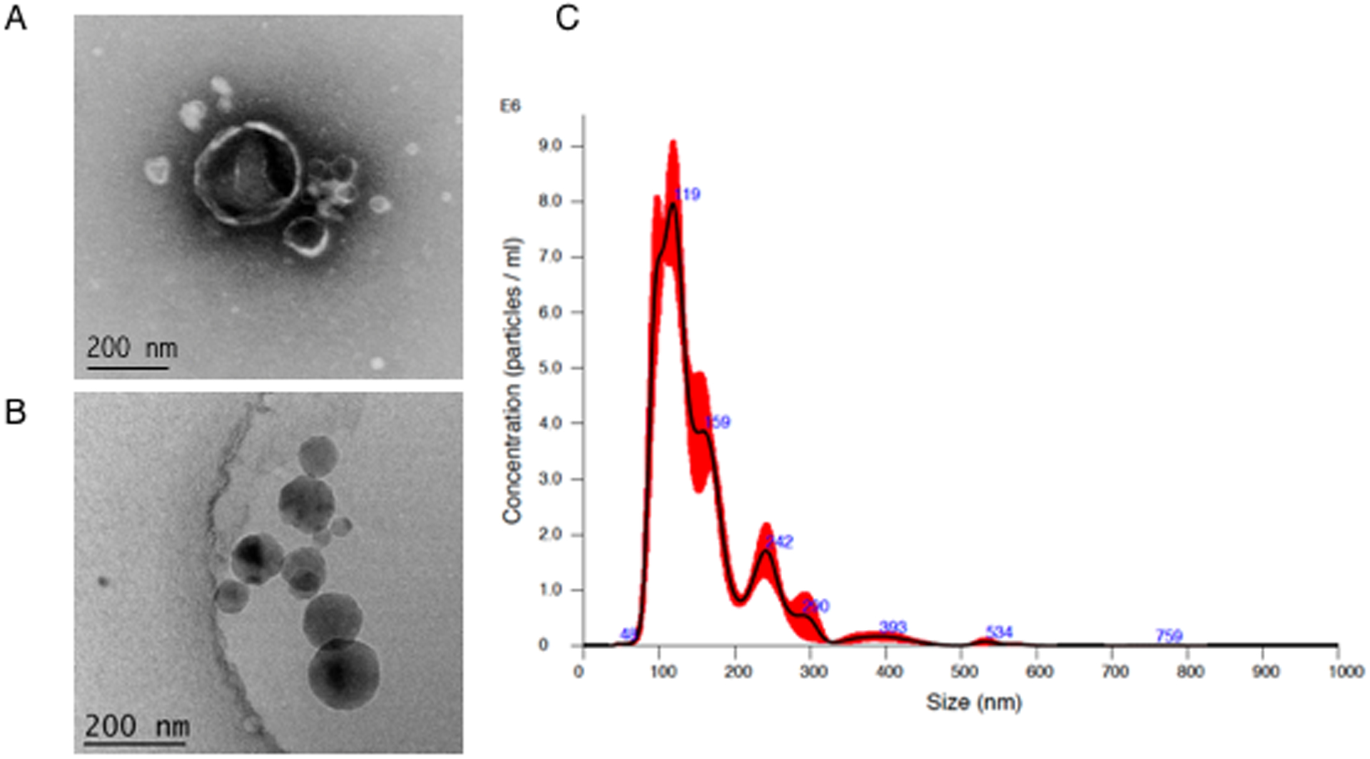
Fig 1. Characterization of extracellular vesicles from Pseudomonas aeruginosa (P.a.).

EVs from P.a. were isolated as described in methods. For visualization, EVs were negatively stained for TEM (A) or flash frozen for cryo-electron microscopy (B). Nanoparticle tracking analysis via NanoSight NS300 was used for EV particle counting and sizing (C).
