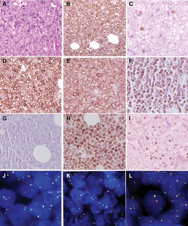
Figure 1.

Breast mass, Case 1. (A), Biopsy shows an infiltrate of atypical lymphoid cells, medium to large in size with vesicular nuclei and basophilic nucleoli (400×). The tumor cells are CD20 positive (B), CD5 negative (C), CD10 positive (D), BCL2 positive (E), cyclin D1 positive (F), SOX11 negative (G), MYC positive (H), and focally positive for TDT (I). FISH studies of the breast mass revealed breaks in BCL2 (J), MYC (K), and CCND1 (L) in approximately 80% of nuclei. BCL6 gene rearrangement was negative. FISH = fluorescence in situ hybridization.
