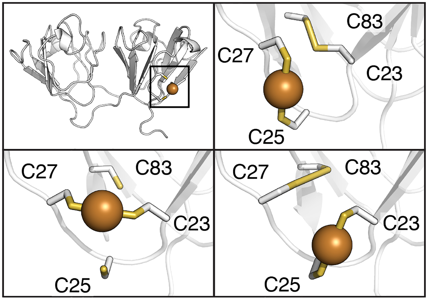
Figure 11:

Potential intramolecular disulfide bond and copper binding site interactions for γS-crystallin. The copper binding sites shown here involve ligands 180° apart with bond distances less than 2.4 Å. (Top left) The bonding interactions are localized to the cysteine loop area of the NTD. (Top right) Potential C23-C83 intramolecular disulfide bond and C25/C27 copper binding site modeled in γS-crystallin using PDBID 6MYG.106 (Bottom right) Alternative possible intramolecular disulfide between C27-C83 and copper binding sites in γS-crystallin modeled using PDBID 2M3T.64 (Bottom left) Copper binding site in S-crystallin between C23 and C27 modeled using PDBID 2M3T.64
