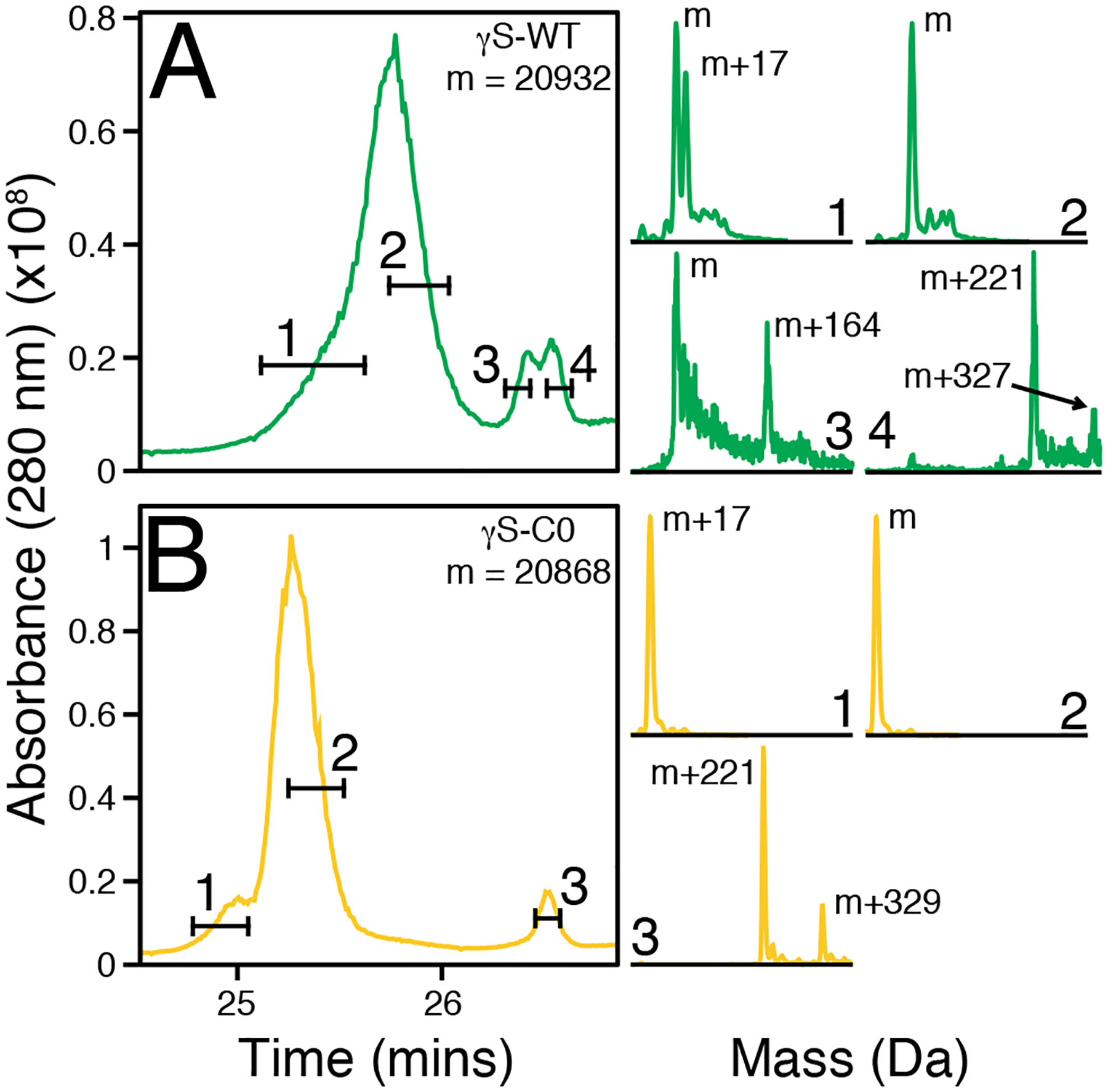
Figure 6:

The dimer peaks from chromatographic separation (analytical SEC) of γS-WT (A) and γS-C0 (B) after EDTA resolubilization were separated on a C4 column prior to mass spectrometry. Two distinct peaks are visible for both proteins, with shouldering or splitting present for most peaks. The selected time frames drawn across the chromatographic traces correspond to the data used for the corresponding mass reconstructions. The fourth selected time frame for γS-WT and third selected time frame of γS-C0 correspond to the same chromatographic retention time.
