Abstract
We present an atypical case of histopathology suggesting hemangioendothelioma and immunohistochemistry-proven Ewing’s sarcoma in a 39-year-old lady who presented with multiple stony hard swellings involving the occipital region of the scalp, right cervical lymph node, right scapular region, left infraclavicular region of the chest, right anterior abdominal wall swelling, and inner aspect of right thigh. She underwent left-sided below-knee amputation for parosteal osteosarcoma in the left distal tibia 3 years back. Palliative radiotherapy with dose of 30 Gy in 10 fractions over 2 weeks was administered to the right neck and right upper back following which she attained moderate pain relief but no reduction in swellings as was expected had it been a case of hemangioendothelioma or Ewing's sarcoma..
Keywords: Hemangioendothelioma, Ewing’s sarcoma, Immunohistochemistry
Case Report
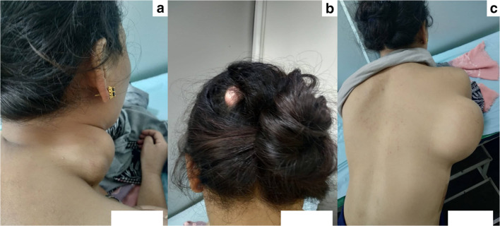
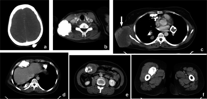
A 39-year-old gentle lady with no comorbidities presented with right-sided chest wall and neck pain and swellings at the scalp, right neck, right chest wall, upper back, abdomen, and right thigh for 1 year. She underwent left-sided below-knee amputation for parosteal osteosarcoma in the left distal tibia 3 years back. Present-day examination reveals multiple stony hard swellings involving the occipital region of the scalp (2 × 2cm2), right cervical lymph node (4 × 4 cm2), right scapular region (10 × 8 cm2), left infraclavicular region of chest (3 × 3 cm2), right anterior abdominal wall swelling (5 × 5cm2), and inner aspect of right thigh (2 × 2cm2), left knee stump being healthy (Fig. 1). Contrast-enhanced computed tomography (CECT) scan of the brain, neck, thorax, and abdomen showed well-defined calcified lesions in the left side of the occipital scalp, right posterior triangle of neck, bilateral hilar region, bilateral pleura based and intraparenchymal lung nodules, right upper thigh, and adjacent to segment IV B of the liver. Large heterogeneously enhancing nodular lesions were noted in the right lateral chest wall without any intrathoracic extension and right side of anterior abdominal wall with eccentric calcifications without intraperitoneal extension (Fig. 2). Trucut biopsy from right upper back swelling showed spindle cell neoplasm suggestive of hemangioendothelioma (Fig. 3). Further immunohistochemistry (IHC) findings were consistent with Ewing’s sarcoma as tumour cells were strongly positive for CD99, vimentin, CD56, FLI 1, and Ki-67 = 35% and negative for CK, chromogranin A, synaptophysin, EMA, desmin, S 100, SMA, WT1, h-Caldesmon, CD 57, CD 31, and CD 34 (Fig. 3). Palliative radiotherapy of a dose of 30 Gy in 10 fractions over 2 weeks was administered to the right neck and right upper back as she was having severe pain (Fig. 4). She attained 50–60% improvement in pain at the end of 3 weeks of treatment completion, but to our dismay, no reduction in size happened. She was planned for systemic chemotherapy henceforth with alternating cycles of VAC (vincristine, Adriamycin, and cyclophosphamide) regimen and IE (ifosfamide and etoposide) regimen alternatively, but due to financial constraints, she refused for further management. She succumbed to her illness after 6 months.
Fig. 1.
Showing (a) right cervical swelling, (b) scalp swelling, and (c) swelling at the right scapular region
Fig. 2.
CECT scan showing lesions in the (a) occipital scalp, (b) neck, (c) hilar region, pleura based and intraparenchymal lung nodules, right chest wall (arrow), (d) adjacent to liver, (e) anterior abdominal wall, and (f) right upper thigh
Fig. 3.
(a) Showing haematoxylin and eosin stained histopathologic section suggesting hemangioendothelioma and IHC showing positivity for (b) CD 99, (c) vimentin, (d) Ki 67, (e) FLI1, and (f) CD 56
Fig. 4.
Three-dimensional conformal treatment plan for palliative radiotherapy to (a) right lateral chest wall lesion and (b) right cervical lesion
Discussion
Ewing’s sarcoma (ES) is basically a neoplasm found in the paediatric population involving bone predominantly [1], on the contrary of which presented case was an adult with widespread and bizarre soft tissue involvement. In spite of such extensive involvement, her symptoms were quite minimal and long standing which is usually seen in benign conditions. The metastatic lesions were mainly calcified, which is again not keeping in with Ewing’s sarcoma [2]. Successful treatment of ES includes a multidisciplinary approach with surgery, radiotherapy, and chemotherapy [3]. As index case presented with disseminated disease, surgical removal was unfeasible. Radiation therapy alone results in a local control rate of 65–75% [4]. The tumour has a distinctive property of being highly sensitive to radiation, sometimes acknowledged by the phrase “melting like snow” [5]; however, this was not true in our case. The recommended chemotherapy regimen is alternating cycles of vincristine-doxorubicin-cyclophosphamide and ifosfamide-etoposide [6].
Also considering hemangioendothelioma as a potential differential diagnosis as per histopathologic examination, the presented case did not behave according to its natural course. Hemangioendothelioma is a vascular tumour with an intermediate-to-low-grade malignant potential, which commonly occurs in the superficial or deep soft tissue of the extremities, lungs, liver, bone, and lymph nodes [7]. It is characterized by a slow growing pattern with potential for local recurrence and even metastasis [7]. It has conventionally been treated by excision in localized cases with favourable outcome [7]. Chemotherapy and radiotherapy have also been employed in metastatic setting with promising responses [7, 8]. Response rate and long-term effectiveness of radiotherapy in adjuvant and metastatic setting have been found to be commendable [7, 8]. However, similar outcome has not been observed in our case.
Extensive literature search does not reveal a similar case in the past. Hence, index case might shed light on dilemma that hemangioendothelioma/Ewing’s sarcoma may also portend an atypical clinical behaviour or it might be an all new total malignancy for which present-day science is unaware about.
Footnotes
Publisher’s Note
Springer Nature remains neutral with regard to jurisdictional claims in published maps and institutional affiliations.
Contributor Information
Anirban Halder, Email: dranirban666@gmail.com.
Rituparna Biswas, Email: mail4r_biswas@yahoo.co.in.
Mimi Gangopadhyay, Email: mimi_ray@rediffmail.com.
Dipanwita Biswas, Email: drdipanwita.biswas94@gmail.com.
Rajni Parmar, Email: rajni.parmar@oncquest.net.
References
- 1.Pappo AS, Navid F, Brennan RC, Krasin MJ, Davidoff AM, Furman WL. Solid tumors of childhood. In: DeVita VT Jr, Lawrence TS, Rosenberg SA, editors. Devita, Hellman, and Rosenberg’s cancer: principles & practice of oncology. 10. Philadelphia: Wolters Kluwer; 2015. pp. 1484–1488. [Google Scholar]
- 2.Koreich OM, Zaytonia R, Sidki K (1989) Ewing’s sarcoma presenting as calcified metastases. Published online 1 Mar 1989. Ann Saudi Med 9(2). 10.5144/0256-4947.1989.216a
- 3.Yeshvanth SK, Ninan K, Bhandary SK, Lakshinarayana KPH, Shetty JK, Makannavar JH. Rare case of extraskeletal Ewings sarcoma of the sinonasal tract. J Cancer Res Ther. 2012;8(1):142–144. doi: 10.4103/0973-1482.95197. [DOI] [PubMed] [Google Scholar]
- 4.Adisesh AM, Neeraja M, Prasad T. Metastatic Ewing sarcoma - a case report. Indian J Orthop. 2005;39:127–129. doi: 10.4103/0019-5413.36793. [DOI] [Google Scholar]
- 5.Ebnezar J, John R. Textbook of Orthopedics. 5. New Delhi: Jaypee Brothers medical publishers(P) limited; 2016. [Google Scholar]
- 6.Womer RB, West DC, Krailo MD, Dickman PS, Pawel BR, Grier HE, Marcus K, Sailer S, Healey JH, Dormans JP, Weiss AR. Randomized controlled trial of interval-compressed chemotherapy for the treatment of localized Ewing sarcoma: a report from the children's oncology group. J Clin Oncol. 2012;30(33):4148–4154. doi: 10.1200/JCO.2011.41.5703. [DOI] [PMC free article] [PubMed] [Google Scholar]
- 7.Heera R, Cherian LM, Lav R, Ravikumar V. Hemangioendothelioma of palate: a case report with review of literature. J Oral Maxillofac Pathol. 2017;21(3):415–420. doi: 10.4103/jomfp.JOMFP_194_14. [DOI] [PMC free article] [PubMed] [Google Scholar]
- 8.Sardaro A, Bardoscia L, Petruzzelli MF, Nikolaou A, Detti B, Angelelli G. Pulmonary epithelioid hemangioendothelioma presenting with vertebral metastases: a case report. J Med Case Rep. 2014;8:201. doi: 10.1186/1752-1947-8-201. [DOI] [PMC free article] [PubMed] [Google Scholar]


