Abstract
Homeotherms maintain a stable internal body temperature despite changing environments. During energy deficiency, some species can cease to defend their body temperature and enter a hypothermic and hypometabolic state known as torpor. Recent advances have revealed the medial preoptic area (MPA) as a key site for the regulation of torpor in mice. The MPA is estrogen-sensitive and estrogens also have potent effects on both temperature and metabolism. Here, we demonstrate that estrogen-sensitive neurons in the MPA can coordinate hypothermia and hypometabolism in mice. Selectively activating estrogen-sensitive MPA neurons was sufficient to drive a coordinated depression of metabolic rate and body temperature similar to torpor, as measured by body temperature, physical activity, indirect calorimetry, heart rate, and brain activity. Inducing torpor with a prolonged fast revealed larger and more variable calcium transients from estrogen-sensitive MPA neurons during bouts of hypothermia. Finally, whereas selective ablation of estrogen-sensitive MPA neurons demonstrated that these neurons are required for the full expression of fasting-induced torpor in both female and male mice, their effects on thermoregulation and torpor bout initiation exhibit differences across sex. Together, these findings suggest a role for estrogen-sensitive MPA neurons in directing the thermoregulatory and metabolic responses to energy deficiency.
Subject terms: Neural circuits, Metabolism
Torpor is a state of reduced metabolism and body temperature that conserves energy when food is scarce. Here the authors show that estrogen-sensitive neurons in the hypothalamus regulate torpor in mice, maintaining torpor in both sexes but initiating torpor and regulating core temperature differentially across sex.
Introduction
Maintaining a stable internal body temperature requires complex responses that coordinately modulate facultative thermogenesis, heat dissipation, and basal metabolic rate. These thermoregulatory effectors are energetically demanding and may be downregulated when energy is scarce. Thus, when food is unavailable, some mammalian species can enter a regulated state called torpor, in which body temperature, physical activity, metabolism, and reproduction are dramatically reduced1–3. Pioneering studies have identified the preoptic area (POA) as a key regulator of temperature and energy balance4,5. There are neuronal populations within the POA that respond to local temperature changes and receive ascending temperature signals from the periphery6–8. Integrative temperature information in the POA is then transmitted to various brain sites that modulate physiological and behavioral thermal responses9,10. In addition to thermoregulation, the POA also monitors metabolic state and regulates food intake11. The dual functions of the POA in maintaining temperature and energy balance suggest the hypothesis that this region may coordinate the thermal and metabolic responses to energy deficiency. Elegant studies have begun to pinpoint neuron populations in the POA and associated neural circuits that control body temperature (reviewed in refs. 9,10,12); however, it is unclear if these or other neuron populations in the POA orchestrate the thermoregulatory and the metabolic changes observed in torpor.
The medial POA (MPA)13,14 is densely enriched for estrogen receptor alpha (ERα, gene Esr1). Whereas estrogen signaling in the POA has well-characterized effects on sexual, parental, and aggressive behaviors15–17, the role of ERα signaling in the POA on thermoregulation and metabolism is less clear. Estrogens are potent modulators of both temperature and metabolism18,19. Daily body temperature fluctuates during the menstrual cycle in women20,21 and the estrous cycle in mice22,23, and estradiol administration alters body temperature in humans24,25 and rodents26,27. Within the hypothalamus, ERα signaling modulates various metabolic processes, including glucose metabolism28,29, food intake30, thermogenesis31, physical activity32, and adiposity33,34. Single-cell RNA profiling of the POA has revealed that Esr1 transcripts are expressed within many of the neuronal clusters that control body temperature and metabolism14. Considering the overlapping functions of the POA and estrogens on thermoregulation and energy homeostasis, we questioned whether ERα positive (ERα+) neurons in the MPA also coordinate torpor. Specifically, we asked if ERα+ MPA neurons are sufficient to reduce body temperature and metabolism as observed in torpor, exhibit changes in endogenous activity during torpor, and are required for the expression of torpor in mice. Here, we find that chemogenetic activation of ERα+ MPA neurons is sufficient to induce hypothermia and hypometabolism, that the natural activity of ERα+ MPA neurons is elevated during fasting-induced torpor, and that ERα+ MPA neurons are required for the full expression of fasting-induced torpor in mice.
Results
ERα+ neurons in the MPA drive a rapid decrease in core body temperature
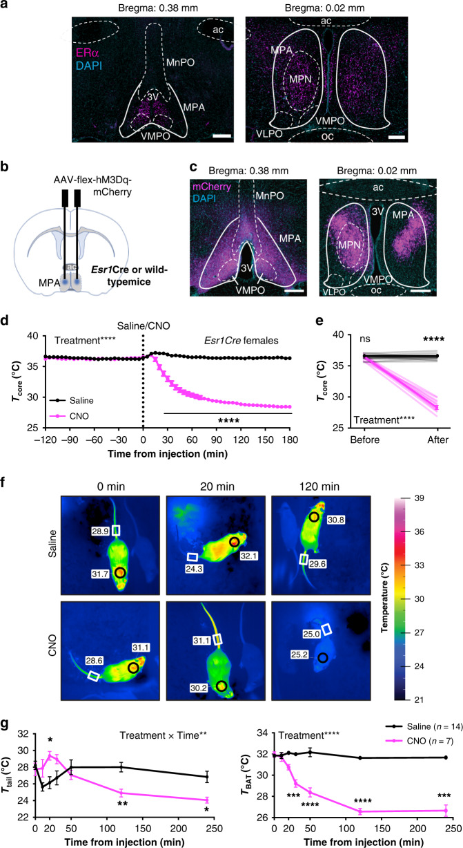
To visualize the anatomical distribution of ERα neurons in the MPA, we evaluated ERα immunoreactivity in brain sections ranging from anterior POA (Bregma 0.6 mm) to posterior POA (Bregma −0.4 mm) according to the mouse brain atlas35 in both male and female mice. We found enrichment of ERα+ cells within the anatomical boundaries of the MPA, particularly within the medial preoptic nucleus (MPN) and rostral sections of the MPA (Fig. 1a). Overall, ERα immunoreactivity in the MPA was higher in females than in males (Fig. 1a and Supplementary Fig. 1a, b). These patterns of expression are consistent with previous findings showing ERα immunoreactivity13 or Esr1 transcripts14 in the mouse MPA and sexually dimorphic ERα expression in this and associated hypothalamic regions36,37. Additionally, Esr1 is co-expressed with transcripts that mark warm-responsive14,38 or torpor-regulating neurons39,40 in the MPA.
Fig. 1. ERα+ neurons in the MPA drive a profound decrease in core body temperature.
a ERα immunoreactivity (magenta) in the MPA of adult female mice. Image representative of n = 4 mice. Scale bar: 200 µm. 3v third ventricle, ac anterior commissure, MnPO median preoptic nucleus, MPA medial preoptic area, MPN medial preoptic nucleus, oc optic chiasm, VLPO ventrolateral preoptic nucleus, VMPO ventromedial preoptic nucleus. b Schematic of the AAV encoding a Gq-coupled DREADD (AAV-flex-hM3Dq-mCherry) delivered to the MPA of Esr1Cre mice. c mCherry reporter expression in the MPA of Esr1Cre females. Image representative of n = 13 mice. 3 v third ventricle, ac anterior commissure, oc optic chiasm. Scale bar: 200 µm. d Core body temperature measured every 5 min before and after injection (dotted line at x = 0) of saline (black) or CNO (pink) in Esr1Cre females (n = 13). e Per-animal averages of core temperature before (−120 min to 0 min) and after (120 min to 180 min) saline or CNO injection (n = 13). f Representative infrared thermal images showing temperature above BAT (circles) and tail (rectangles) after saline or CNO injection. g Quantification of thermography images in regions above the BAT and tail before and after saline (n = 14) or CNO (n = 7) injection in Esr1Cre female mice. *p < 0.05; **p < 0.01; ***p < 0.001; ****p < 0.0001 for Sidak’s multiple comparison tests comparing saline and CNO following a significant effect of treatment or treatment time in a two-way RM ANOVA or Mixed-effects model. All error bars show SEM.
To test the role of ERα+ MPA neurons in torpor, we investigated the effect of selectively activating ERα+ MPA neurons using chemogenetic Designer Receptors Exclusively Activated by Designer Drugs (DREADDs), a powerful tool for modulating neuronal activity41. An adeno-associated virus (AAV) construct encoding the Cre-dependent, excitatory, Gq-coupled hM3Dq receptor and an mCherry reporter was stereotaxically injected into the MPA of mice that express Cre in Esr1-expressing cells (Esr1Cre) and wild-type littermates (Fig. 1b). mCherry was detected in the MPA of female and male Esr1Cre mice (Fig. 1c and Supplementary Fig. 1c) but not in wild-type controls that received the same AAV. Upon intraperitoneal administration of the exogenous ligand for the hM3Dq receptor, clozapine-N-oxide (CNO, 0.3 mg/kg), male and female Esr1Cre mice exhibited a rapid reduction in core body temperature (Fig. 1d, e and Supplementary Fig. 1d, e). Core temperature dipped below 30 °C within 1 h of CNO injection (Fig. 1e and Supplementary Fig. 1e) but was not altered in the same mice following saline injection on a different day (Fig. 1d, e and Supplementary Fig. 1d, e). The effect on body temperature cannot be attributed to CNO alone or its conversion to clozapine, as CNO did not alter body temperature in wild-type mice that received AAV (Supplementary Fig. 1f). In addition, administration of an alternative DREADD ligand, Compound 21 (1 mg/kg)42, elicited a reduction in body temperature similar to CNO (Supplementary Fig. 1g).
Thermal homeostasis is maintained by the balance of heat production and heat dissipation. In rodents, the brown adipose tissue (BAT) is critical in adaptive thermogenesis43,44, whereas a modulation of blood flow to the tail skin plays an active role in heat dissipation45,46. To further understand how ERα+ MPA neuronal activity affects this equilibrium, we monitored the temperature of the body core, BAT, and tail skin. Thermal probes were implanted intraperitoneally to measure core temperature, and thermal imaging of the interscapular region was used to monitor BAT thermogenesis. A temperature logger attached near the ventral vein of the tail, together with thermal imaging of the tail, were used to measure heat dissipation (Supplementary Fig. 1h). Infrared thermal images indicated a profound increase in tail skin temperature 20 min after CNO injection, whereas saline treatment was associated with a transient decrease in tail skin temperature, perhaps due to handling stress47 (Fig. 1f, g). Consistent with regulated hypothermia, the rapid drop in core temperature (Fig. 1d) was not accompanied by an increase in heat generation, as indicated by both infrared imaging at multiple time points and gene expression in BAT measured 90 min following saline or CNO treatment (Fig. 1f, g and Supplementary Fig. 1i). Using thermo-loggers attached to the tail (Supplementary Fig. 1h), we observed similar changes in tail skin temperature and used these to estimate heat dissipation by calculating the heat loss index (HLI): HLI = (Tskin − Tambient)/(Tcore − Tambient)48. HLI largely corrects for the effect of overall body cooling on tail skin temperature by comparing the temperature of the skin and core to the ambient temperature. Analysis of HLI revealed that CNO injection elicited a profound increase in heat loss that was coincident with the initial decrease in core temperature (Supplementary Fig. 1j, k). Consistent with previous studies showing that the POA mediates responses to warmth, including cutaneous vasomotor control10,49, these results indicate that activating ERα+ MPA neurons initiates a rapid heat loss and suppresses heat production to induce hypothermia.
Chemogenetic activation of ERα+ MPA neurons depresses temperature and metabolism
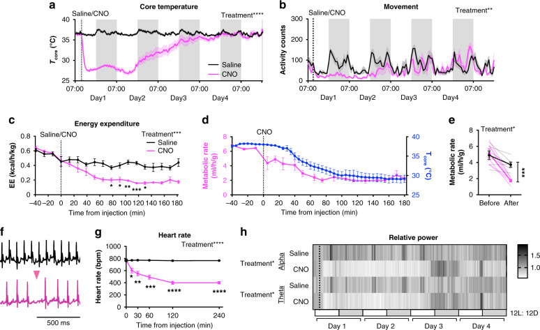
Analysis of the changes in body temperature revealed that a single CNO injection could induce a state of prolonged hypothermia. Of the 42 Esr1Cre females expressing hM3Dq and treated with CNO, 31 mice (74%) exhibited a core body temperature below 31 °C and 12 mice (29% of all subjects) maintained this level of hypothermia for over 20 h (Supplementary Fig. 2a, b). Indeed, core temperature returned to baseline (36.5 °C) up to 4 days after a single CNO injection (Fig. 2a). The reduction in core temperature also was accompanied by diminished physical activity (Fig. 2b). The expression of torpor has been previously associated with body weight50–52. We also find that body weight was significantly correlated with the minimum core temperature reached and with duration of the hypothermia following CNO administration (Supplementary Fig. 2b). Even in warmer conditions (ambient temperature of 30 °C) that might diminish thermogenesis45,53,54, activating ERα+ MPA neurons resulted in a rapid decrease in core temperature (Supplementary Fig. 2c), suggesting that reduced metabolic rate may also contribute to the reduction in body temperature. Indeed, indirect calorimetry revealed a reduction in metabolic rate and energy expenditure, as measured by oxygen consumption and calculated heat production, respectively. Both measures were reduced by >50% in Esr1Cre mice following CNO injection, compared to the effects of saline injection in the same mice and to baseline levels before injection (Fig. 2c–e and Supplementary Fig. 2d). We did not detect an effect of CNO on respiratory exchange ratio (RER), a measure of fuel preference, within 3 h of injection. However, RER seems to decline in both groups, consistent with measurement during a daytime fast (Supplementary Fig. 2e). As further indication of an overall reduction in metabolic rate, electrocardiogram recordings revealed a significant reduction in heart rate as early as 15 min after CNO injection, dropping from 776 ± 3.2 (mean ± SEM) bpm at baseline to 401 ± 18.8 bpm 4 h after CNO injection. Control saline injection did not affect heart rate (Fig. 2f, g and Supplementary Fig. 2g). The CNO-induced reduction in heart rate was frequently accompanied by pronounced sinus arrhythmia (Fig. 2f and Supplementary Fig. 2g), a known effect of enhanced cardiac vagal tone55 that was never observed after saline treatment. These results suggest that the cardiac autonomic nervous system may be involved in this coordinated response. Finally, electroencephalography (EEG) was used to measure overall brain activity during hypothermia and hypometabolism. CNO induced reductions of relative alpha and theta power in the EEG signal (Fig. 2h). Comparisons of relative delta power following saline or CNO injection revealed significant differences from non-rapid eye movement (NREM) or wake states (Extended Fig. 2h). Reductions such as these are common in experimental models of hibernation and torpor (see ref. 56 for review). The induction of this phenotype was repeatable, and mice seemed to recover without adverse impacts on their health (Supplementary Fig. 2i, j). Taken together, these findings indicate that activation of ERα+ MPA neurons induces a torpor-like hypothermic and hypometabolic state in mice.
Fig. 2. Chemogenetic activation of ERα+ MPA neurons induces a torpor-like state.
AAV-flex-hM3Dq-mCherry was delivered to the MPA of female Esr1Cre mice. a, b Four-day consecutive recordings of core temperature and movement after saline (black, n = 8) or CNO (pink, n = 6) injection. Lines show group means, shaded area shows SEM. Results of pairwise comparisons for a, b are listed in Supplementary Files. c Energy expenditure (EE or Heat) calculated from indirect calorimetry data and normalized to lean body mass following saline or CNO injection (n = 6). d Comparison of CNO-induced changes in metabolic rate (VO2 ml/h/g, pink) and Tcore (blue) in the same mice (n = 4 Esr1Cre females). e Metabolic rate (VO2 ml/h/g) before (−40 min to 0 min) and after (120 min to 180 min) saline or CNO injection calculated from indirect calorimetry data and normalized to lean body mass (n = 6 Esr1Cre females). f ECG traces in mice 4 h after saline or CNO injection. Traces representative of n = 8 mice. Pink arrowhead indicates a skipped heartbeat. g Heart rate (n = 8) before (0 min) to 4 h after saline or CNO injection. h Heatmap of relative alpha and theta power from four-day consecutive EEG recording after saline or CNO injection (dash line, n = 4). White and gray bars below represent light and dark periods, respectively. Group means ± SEM shown. *p < 0.05; **p < 0.01;***p < 0.001; ****p < 0.0001 for Sidak’s multiple comparison tests comparing saline and CNO following a significant effect of treatment a (p < 0.0001), b (p = 0.0020), c (p = 0.0006), e (p < 0.05), g (p < 0.0001), and h (Alpha p = 0.04 and theta p = 0.02) in a two-way RM ANOVA.
ERα+ neuronal activity increases during fasting-induced torpor
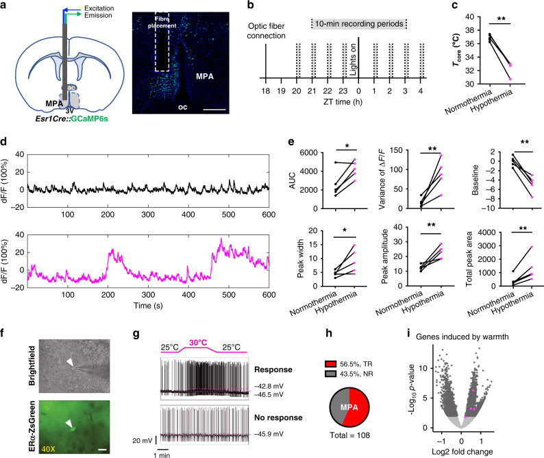
The state triggered by activating ERα+ neurons suggests the hypothesis that ERα+ neuron activity is modulated during natural hypothermic and hypometabolic states. Prolonged food deprivation has been shown to induce torpor in mice57. Here, a 48 h fast resulted in consistent torpor bouts (Supplementary Fig. 3a). To record neural activity from ERα+ MPA neurons in live animals during fasting-induced torpor, we targeted the high-sensitivity sixth-generation, slow-kinetic calcium reporter GCaMP6s58 to ERα+ neurons by delivering Cre-dependent AAV9-FLEX-Syn-GCaMP6s to the MPA of Esr1Cre mice (Fig. 3a). Following a 3-week recovery, mice were fasted for 48 h and fluorescence from ERα+ MPA neurons was measured during the last 8 h of fasting. Calcium transients were measured over eight 10-minute periods, 4 h before and 4 h after lights on, when torpor bouts are more likely to occur (Fig. 3b). Fasting-induced bouts of reduced core body temperature were consistent with fasting-induced torpor (Fig. 3c and Supplementary Fig. 3a). We detected larger and more variable calcium transients during bouts of hypothermia (core temperature < 33 °C) compared to times of normothermia (core temperature > 36 °C) following a fast (Fig. 3d, e). The decrease in baseline activity during hypothermia is in line with recently reported neuronal activity changes in torpor neurons39. Despite the reduced baseline, which may suggest a repression of tonic firing, the large peaks during hypothermia are consistent with an increase in synchronized burst firing. These results suggest a relationship between the natural activity of ERα+ MPA neurons and bouts of fasting-induced torpor.
Fig. 3. ERα+ neuronal activity during fasting-induced torpor in vivo and warmth exposure ex vivo.
a Schematic and coronal sections showing the optical fiber placement to record ERα+ neuronal activity in the MPA in vivo during fasting-induced torpor bouts. Image representative of n = 5 mice. oc optic chiasm. Scale bar 200 um. b Diagram showing the experimental design for photometry recordings during fasting. Mice were food deprived for 38 h at the time of optic fiber connection. c Core body temperature in mice showing normothermia and hypothermia (<33 °C) during the recording period following a fast. d Representative fluorescence traces of an individual mouse showing normothermia (black) and hypothermia (pink) during a 10 min recording period. e Mean area under the curve (AUC), variance, baseline, base width of peaks, peak amplitude, and total peak area of fluorescence detected from ERα+ MPA neurons in fasted mice showing normothermia (black) and hypothermia (pink) during a 10-min recording period (n = 5 mice, 4 females and 1 male). f Brightfield and fluorescence imaging of ERα-ZsGreen reporter expression in the MPA. Images representative of 179 cells from 4 mice. Scale bar 20 µm, white arrowhead denotes cell of interest. g Representative whole-cell current clamp recordings of ERα neurons in the MPA exposed to 25 °C and 30 °C. h Pie chart showing percentages of neurons showing a temperature response (TR, > 41.4% increase in firing rate) or no response (NR) in the MPA (MPN and rostral MPA). i Volcano plots comparing gene expression in TR (n = 4 samples, 2 neurons per sample) versus NR (n = 3 samples, 2 neurons per sample) ERα neurons in the MPN and rostral MPA regions combined. All genes shown as dots, with color denoting not significant in two-tailed differential expression testing (adjusted p > 0.05 using the Benjamini–Hochberg procedure, gray), significant (adjusted p < 0.05, dark gray), and genes induced by warmth66.
Recent studies have identified neurons and molecular markers in the POA that are responsive to changes in temperature and involved in thermoregulation38,59–62. To exclude the alternative hypothesis that ERα+ MPA neurons are responsive to warmth, primarily involved in driving cooling responses, and that the drop in metabolism is secondary to hypothermia, we measured temperature responsiveness using calcium imaging and electrophysiology. We did not detect changes in calcium transients in freely behaving mice exposed to warmth (40 °C) or cold (15 °C), suggesting that changes in ambient temperature do not alter the neural activity of the ERα+ MPA neuron population (Supplementary Fig. 3b–e). Although we were unable to detect a response to temperature at the population level, it is possible that temperature alters neural activity at the level of individual ERα+ MPA neurons. Thus, we recorded neural activity in brain slices from ERα-ZsGreen mice, in which ERα+ cells are identified by green fluorescence63 (Fig. 3f). Whole-cell current clamp recordings revealed heterogeneity in the temperature responsiveness of ERα+ neurons (Fig. 3g, h and Supplementary Fig. 3f, g). Using a 41.4% change in firing rate over a 5 °C temperature increase (Q10 > 2)64,65 as a cutoff for temperature responsiveness, approximately half (56.5%) of the 108 ZsGreen+ MPA neurons were considered temperature responsive. The ERα+ MPA neurons tested include ZsGreen+ neurons in the medial preoptic nucleus (MPN, 56.2% of 73 neurons) and rostral aspects of the MPA (57.1% of 35 neurons) (Fig. 3h and Supplementary Fig. 3g). In contrast, ZsGreen+ neurons in adjacent nuclei such as the VLPO and VMPO contained only 13.9% and 28.6% temperature-responsive ZsGreen+ neurons, respectively, suggesting heterogeneity of ERα+ neurons with respect to temperature responsiveness (Supplementary Fig. 3g).
Consistent with temperature responsiveness within some ERα+ MPA neurons, RNA sequencing analysis of cellular contents following electrophysiological recordings revealed higher expression of warmth-induced genes66 in temperature-responsive compared to non-responsive neurons (Fig. 3i). Global expression analysis of ERα+ neurons in the MPN after electrophysiology recordings revealed a strong separation of transcriptional signatures between temperature-responsive and non-responsive neurons (Supplementary Fig. 3h), indicating that these two ERα+ populations are transcriptionally distinguishable, possibly due to either their function in temperature responsiveness or changes induced by exposure to 30 °C. Whereas ERα+ cells in the MPA include glutamatergic and GABAergic populations14, temperature-responsive ERα+ neurons show enriched expression of GABAergic transcripts and reduced expression of glutamatergic transcripts when compared to non-responsive ERα+ neurons (Supplementary Fig. 3i). Additionally, temperature-responsive ERα neurons show enriched expression of the monoamine transporter Slc18a2, which is differentially expressed in a subpopulation of ERα neurons identified by high-resolution spatial RNA profiling14 (Supplementary Fig. 3i). Thus, temperature responsiveness was not detected in vivo and only detected in a GABAergic Slc18a2+ subpopulation of ERα+ neurons ex vivo. However, both temperature-responsive and non-responsive ERα+ neuron populations in the MPN express markers of neurons that are activated during torpor in mice39 (Supplementary Fig. 3i).
ERα+ MPA neurons are required for thermoregulatory homeostasis and the full expression of torpor
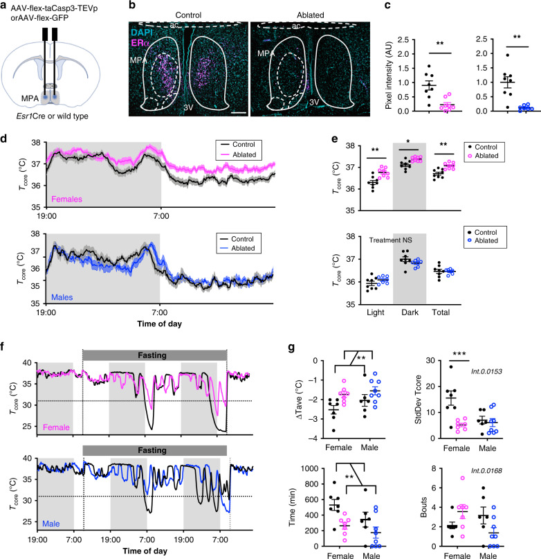
To determine if ERα+ MPA neurons are required for temperature homeostasis or fasting-induced torpor, we selectively ablated ERα+ neurons in adult mice using AAV that expresses a genetically modified caspase 3 (Fig. 4a). This approach has been shown to effectively delete cells in vivo by triggering cell-autonomous apoptosis67. AAV2-FLEX-Caspase3 was delivered to the MPA of Esr1Cre mice. Controls included wild-type mice receiving AAV encoding the Cre-dependent caspase 3 or Esr1Cre mice receiving AAV encoding a Cre-dependent GFP. Four weeks after AAV delivery, Esr1Cre mice showed a >40% reduction in ERα immunoreactivity in the MPA compared to controls (Fig. 4b, c). Ablating ERα+ cells in the MPA led to a significant increase in core temperature in female but not in male mice (Fig. 4d, e). In contrast, neuron ablation did not affect physical activity in either female or male mice, suggesting that the temperature increase was selective and not a consequence of changes in movement (Supplementary Fig. 4a–d). The effect on core temperature indicates that ERα+ MPA neurons are critical for maintaining normal thermal homeostasis in female but not in male mice.
Fig. 4. ERα+ cells in the MPA are required for thermoregulation and fasting-induced torpor.
a Schematic showing stereotaxic delivery of AAV encoding a Cre-dependent caspase to the MPA of Esr1Cre mice to ablate Esr1-expressing cells. b ERα immunoreactivity in the MPA of female mice following AAV-mediated ablation of Esr1-expressing cells (right, representative of n = 8 mice) or littermate controls (left, representative of n = 8 mice). 3v third ventricle, ac anterior commissure. Scale bar: 200 µm. c ERα immunoreactivity in the MPA of n = 8 control females (left plot, black circles), n = 8 ablated females (pink circles), n = 8 control males (right plot, black circles), and n = 7 ablated males. d Core body temperature (Tcore) over 24 h, measured every 5 min for 3 days. Group averages shown for control (black, n = 8) and ablated (pink, n = 8 female; blue, n = 7 male) mice. Shading along the curve denotes the SEM. e Average Tcore from mice shown in panel d highlighting per animal averages in light (7:00 to 19:00), dark (19:00 to 7:00), and total 24 h periods. f Representative Tcore measured every 5 min over 3 days during fasting-induced torpor in control (black) and ERα neuronal ablated (pink) female mice. Dashed line denotes Tcore = 31°C, the cutoff for hypothermia used here g Change in Tcore (ΔTcore = Lowest Tcore - Avg Tcore before the fast), variace of the Tcore, total time spent with Tcore ≤ 31°C, and number of bouts with Tcore ≤ 31 °C during the fasting period (n = 7 control females, 7 ablated females, 6 control males, 8 ablated males). Statistical significance denoted by NS, not significant; *p < 0.05; **p < 0.01; ***p < 0.001 for two tailed t-tests c, Sidak’s multiple comparison tests e, g following a significant effect of treatment, time or sex or interactions in a two-way (RM) ANOVA. All error bars show SEM.
We next investigated if ERα+ MPA neurons are required for fasting-induced torpor. Food deprivation induced bouts of hypothermia (Tcore < 31 °C) in mice with intact ERα+ MPA neurons (Fig. 4f). Ablating ERα+ neurons in mice of similar body weights (Extended Fig. 4e) greatly diminished the torpor response, affecting the changes in average core temperature, core temperature variability, total time with a core temperature below 31 °C, duration of the longest bout, and lowest core temperature reached in fasted mice (overall effect of treatment, Fig. 4g and Supplementary Fig. 4e). We did not detect a significant effect of neuron ablation on the number of bouts initiated, suggesting that ERα+ MPA neurons are required for maintaining rather than initiating torpor bouts. Comparisons by sex suggest that ERα+ MPA neurons may regulate torpor differently in females and males. We detected a significant effect of sex on core temperature variability, duration of the longest bout, and the lowest core temperature reached following a fast (Fig. 4g and Supplementary Fig. 4e). In pairwise comparisons within a sex, core temperature variability was significantly lower in females with neuron ablation but not in males (Fig. 4g). Additionally, the effect of neuron ablation on the number of bouts initiated varied by sex (interaction effect, Fig. 4g). While it appeared that neuronal ablation lead to more bouts initiated in females and fewer bouts initiated in males, pairwise comparisons did not reach statistical significance (Fig. 4g). Together, these findings suggest that ERα+ MPA neurons are necessary for the full expression of fasting-induced torpor. Specifically, ERα+ MPA neurons are critical for maintaining the duration of torpor in all sexes but may also have sex-dependent roles in the variability of body temperature during torpor and the initiation of torpor bouts.
ERα+ MPA neurons drive hypothermia through projections to the ARC and DMH
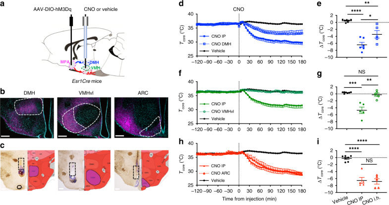
To visualize the downstream projections of ERα+ neurons from the MPA, we performed anterograde tracing using a Cre-dependent AAV expressing GFP (Supplementary Fig. 5a). We detected dense GFP+ fibers in several hypothalamic nuclei involved in thermoregulation, including the dorsomedial nucleus of the hypothalamus (DMH), ventromedial nucleus of the hypothalamus (VMH), and the arcuate nucleus of the hypothalamus (ARC) (Supplementary Fig. 5b, c). The DMH receives direct preoptic inputs38,62 and modulates the sympathetic outputs to BAT in response to cold and warm ambient temperature44. The VMH has been implicated in estrogenic regulation of BAT thermogenesis31 and many estrogenic effects on energy metabolism33,37. The ARC contains neurons implicated in the hormonal modulation of vasodilatation during hot flashes through projections to the POA68. To test the contribution of projections from ERα+ MPA neurons to these regions, we repeated the chemogenetic hM3Dq activation experiments but delivered the CNO locally to the DMH, VMH, or the ARC (Fig. 5a). This method exploits trafficking of hM3Dq to axon terminals to alter presynaptic activity and is effective both ex vivo and in vivo69,70. In line with the results of the anterograde tracing (Supplementary Fig. 5b, c) and previous studies69, we observed numerous mCherry positive fibers in the downstream target sites within the hypothalamus (Fig. 5b). We first confirmed that systemic administration of CNO by IP injection reduced core body temperature as in Fig. 2. When CNO was delivered to the DMH, core temperature was significantly reduced compared to delivery of vehicle but did not recapitulate the effects of systemic CNO administration (Fig. 5c–e). CNO delivery to the VMH had no effect on body temperature and was similar to vehicle injection (Fig. 5f, g). In contrast, local delivery of CNO to the ARC led to a reduction in body temperature that was indistinguishable from the effect of systemic CNO injection in the same mice (Fig. 5h, i). The effects of delivering CNO to distinct projection targets of ERα+ MPA neurons suggest that projections to the ARC, and to some extent DMH, could mediate the effects of ERα+ MPA neurons.
Fig. 5. Activation of estrogen-sensitive hypothalamic circuits drive decreases in core body temperature.
a Schematic strategy for testing the thermoregulatory hypothalamic circuits from the MPA by intrahypothalamic (i.h.) delivery of CNO or vehicle (artificial cerebrospinal fluid, aCSF) to the projected nuclei. b mCherry fibers indicating projections from MPA ERα+ neurons to DMH (represtentative of n = 6 mice), VMHvl (representative of n = 5 mice), and ARC (representative of n = 5 mice). ARC arcuate nucleus, DMH dorsal medial hypothalamus, MPA medial preoptic area, VMHvl ventral lateral subregion of ventromedial hypothalamus. Scare bars: 250 µm. c Brightfield coronal sections (left) and diagram coronal sections (right) showing the canular track and targeted nuclei for local CNO delivery. d, f, h Core temperature (Tcore) measured every 5 min before and after injection (dotted line at x = 0) of vehicle (saline and aCSF data combined, n = 7), CNO IP (0.3 mg/kg) or CNO i.h. (8.4 ng/mouse) in the DMH (blue, n = 6 mice shown in d and e), VMH (green, n = 5 mice shown in f and g), and ARC (red, n = 5 mice shown in h and i). e, g, i Change in core temperature (ΔTcore) after vehicle or CNO administration (120 min to 180 min after injection) compared to baseline (−120 min to 0 min before injection). Statistical significance denoted by NS, not significant; *p < 0.05; **p < 0.01; p < 0.01; ***p < 0.001, and ****p < 0.0001 for posthoc two-tailed Tukey’s multiple comparisons test following a significant effect of treatment (p < 0.0001) in a one-way ANOVA.
Discussion
Torpor has been investigated in numerous species, yet the neuronal mechanisms allowing endotherms to actively depress body temperature and metabolism in response to nutrient scarcity are only fragmentally understood1,3. Here we exploit the viral and genetic tools available in mice to show that selective activation of ERα+ MPA neurons induces a coordinated depression of metabolic rate, body temperature, physical activity, heart rate, respiratory rate, and brain activity. In the majority of cases, the induced torpid state lasted from 20 h to multiple days, far outlasting the pharmacokinetic duration of CNO (1–3 h71, although the duration could be longer if torpor also reduces CNO metabolism). The state induced by activating ERα+ MPA neurons resembles entry into torpor in several ways. As observed in several rodent species during entry into daily torpor72–74, we observe that the drop in metabolic rate declines faster than core temperature. We also observe a rapid drop in heart rate and characteristic skipped heart beats, as observed in multiple small mammalian species75,76, along with reduced theta power brain activity, as observed in ground squirrels entering torpor77. Additionally, we observe variation in the minimum core temperature, variation in the duration of hypothermia, and an association between the minimum body temperature and body weight, which have been reported in spontaneous torpor50. Indeed, individual variation in torpor expression has been well documented50,57,74 and associated with birth weight51 or body weight at the time of fast52 in mice. Overall, the state induced by activating ERα+ MPA neurons recapitulates several key facets of torpor, including hypometabolism, hypothermia, reduced physical activity, and reduced brain activity in fed mice.
The relationship between body weight and the hypothermic response to activating ERα+ MPA neurons suggests that nutritional experience or metabolic signals may alter the neural circuits that regulate torpor. In line with a proposed effect of nutritional signals, activating POA neurons that express the receptor for leptin, a signal of fat stores, also induces hypothermia61 and modulating leptin receptor signaling in the POA alters energy expenditure78. In addition, several molecules have been employed to induce torpor-like hypothermia, including nucleotides (adenosine, cyclohexyladenosine, AMP, and ADP), anesthetics, and 3-Iodothyronamine (T1AM)76,79–82. Central activation of the A1 adenosine receptor also produces a torpor-like state in rats82 but ablation of the adenosine A1 receptor does not affect entrance into torpor83. Although a detailed mechanism is unknown, the duration of DREADD-induced torpor suggests that ERα neurons may act as a “switch” to turn on torpor by triggering a neural circuit that mediates an inverted thermoregulatory state84.
Beyond the sufficiency of ERα+ MPA neurons to induce torpor, we examined the natural activity and the necessity of ERα+ MPA neurons in an endogenously regulated torpid state induced by fasting in mice57. Calcium imaging and neuron ablation studies suggest that ERα+ MPA neurons are activated during bouts of fasting-induced torpor and are important for the full expression of torpor in response to fasting. Our results suggest that ERα+ MPA neurons are involved in maintaining torpor in both female and male mice, and may also have a role in initiating torpor bouts that varies across the sexes. Ablating ERα+ MPA neurons appeared to increase the number of torpor bouts in females and decrease the number of torpor bouts in males. Although this effect was not significant in pairwise comparisons within a sex, there was a significant interaction between sex and neuron ablation. The trend in females could be explained by compensation, such that female mice with shorter torpor bouts might initiate more bouts to conserve energy. The trend in males is surprising, because they did not appear to initiate more bouts to compensate for shorter bout durations. This sex difference is consistent with evidence that torpor is regulated differently in males and females, and that females are generally more “thrifty” with their energy reserves than males85. Indeed, in several rodent species, females initiate hibernation earlier, have more or longer torpor bouts, spend more time in torpor, and terminate hibernation later than males86–90.
Ablating ERα+ MPA neurons also led to a female-specific effect on basal body temperature. This result is consistent with previous studies showing effects of estrogen-sensitive neurons on body temperature in female mice91 and with evidence that temperature-responsive neurons in nearby regions of the POA regulate blood flow to the skin7,92,93 and BAT activity12,38,62. Indeed, a GABAergic subset of ERα+ neurons in the MPN appear to be temperature responsive with respect to firing and gene expression changes. Although this responsiveness to temperature may not be strong enough to drive a response that is detected by calcium imaging from the whole population in vivo, it is consistent with previous findings of warm-responsive GABAergic neurons in the POA38,62 but different from warm-responsive glutamatergic POA neurons that decrease body temperature when activated in mice60,61,91,94. In total, about ~30% of neurons in the POA are temperature responsive64,95. Interestingly, approximately half of the warm-responsive neurons in the POA are responsive to estradiol, compared to only one-third of temperature-unresponsive neurons96. However, we do not have any evidence to implicate or exclude a role for ERα signaling in torpor. Future studies will determine if torpor neurons are modulated by changes in estrogen levels or estrogen receptor manipulations.
We also find that temperature-responsive ERα+ neurons differentially express Slc18a2, suggesting that an inhibitory cluster of ERα+ Slc18a2+ neurons in the MPN14 may be responsive to warmth and perhaps drive changes in body temperature. However, it is unclear if this neuron population mediates the effects on basal body temperature or the torpor response. Indeed, activating ERα+ MPA neurons induced rapid changes in tail skin vasodilation and inhibition of BAT thermogenesis. Because mice are highly dependent on somatic activity for thermoregulation, it is possible that the reduction in physical activity, inhibition of BAT thermogenesis, stimulation of heat dissipation at the tail, and bradycardia may together contribute to the rapid decrease in core body temperature observed in torpor. These effects may be mediated by modulation of multiple neural targets of the MPA or hormonal mediators such as thyroid hormone97. However, evidence that the drop in metabolic rate precedes hypothermia suggests that the multifaceted changes induced by activating ERα+ MPA neurons are not secondary to thermal changes (Q10)74. Because ERα+ neurons in the MPN appear to include temperature-responsive and non-responsive neurons as well as GABAergic and glutamatergic neurons, it will be important to dissect the contributions of the different subpopulations (i.e., ERα+ Slc18a2+ and ERα+Slc18a2− neurons) to thermoregulation and torpor.
Pioneering studies have identified hypothalamic nuclei and circuits that are critically involved in regulation of body temperature and metabolism9,12,98. Our tracing studies reveal projections from ERα+ neurons to several of these hypothalamic nuclei, including the DMH, VMH, and ARC. Local activation of ERα+ neurons that project to the DMH led to a partial decrease in core body temperature, whereas local activation of ERα+ neurons that project to the ARC recapitulated the effect of systemic CNO delivery on core body temperature. These findings are consistent with evidence that projections from the POA to the DMH regulate thermogenesis, heart rate, and energy expenditure38,62 and an increase in cFOS immunoreactivity within the DMH during fasting-induced torpor98. Similarly, the ARC is implicated in the regulation of heat dissipation68,99, thermogenesis100,101, and fasting-induced torpor98,102. It is possible that locally delivered CNO may diffuse to adjacent hypothalamic regions. For example, the partial effect of delivering CNO to the DMH could also be due to some diffusion to the ARC or elsewhere. However, CNO delivery to the VMH did not affect body temperature, excluding a role for the VMH in this circuit and suggesting that diffusion of CNO to adjacent regions may have been limited in these experiments. Additionally, CNO-induced activity may cause retrograde conduction and activate collateral projections. Although differential effects were observed when delivering CNO to the ARC, DMH, and VMH, we cannot exclude the possibility that ERα+ MPA neurons project to multiple regions and induce torpor via collateral projections. Considering the autonomic changes (cardiac function, BAT inhibition, and tail vasodilation) observed here, it is possible that ERα neurons also project to the raphe pallidus40,92,103 for the autonomic coordination of torpor. Although more selective circuit mapping and manipulations are required to fully understand the circuits that control torpor, these studies provide additional evidence of a potent role for ERα+ MPA neurons in the regulation of torpor.
In summary, these studies identify a neuron population that is critical for an adaptive response that minimizes energy costs and increases survival during food shortages in mice74. These findings are consistent with very recent studies showing that a hypothermic and hypometabolic state can be induced by activating POA neurons, specifically glutamatergic neurons that express pyroglutamylated RFamide peptide (Qrfp)40 or neurons expressing adenylate cyclase activating polypeptide 1 (Adcyap1)39. Correspondingly, we find expression of Adcyap1 and other gene markers of torpor neurons in ERα+ MPA neurons and general overlap in projection target regions. A major role for the ARC but not the VMH in the regulation of torpor is consistent with evidence that torpor neurons in the POA strongly innervate the ARC but not the VMH39. However, QRFP neurons appear to exert their effects primarily through projections to the DMH40, whereas our preliminary data suggest that ERα+ MPA neurons may exert their effects through projections to the ARC and partially through the DMH. Nonetheless, it is possible that the intersection of these three gene markers may define the neurons required for torpor or that multiple populations in this region together coordinate the torpor response.
Methods
Animals
Mice harboring the Esr1Cre knock in allele (Tm1.1(Cre)And)36 were maintained on a C57BL/6J genetic background and bred in approved facilities at University of California Los Angeles (UCLA). For all experiments using Esr1Cre mice, controls included Cre-negative (wild-type) littermates that received the same AAV as the experimental group and/or Esr1Cre mice receiving a control AAV, as described for each experiment. The ERα-ZsGreen transgenic mice63 used for electrophysiology were maintained on a C57BL/6J genetic background and bred in approved facilities at Baylor College of Medicine. All mice were maintained under a 12:12 h L/D schedule at room temperature (~22 °C), maintained at 30–70% relative humidity, and provided with food and water ad libitum unless otherwise indicated. Mice were at 8–10 weeks at the start of all the experiments.
Mouse procedures
All studies were carried out in accordance with the recommendations in the Guide for the Care and Use of Laboratory Animals of the National Institutes of Health. UCLA is AAALAC accredited and the UCLA Institutional Animal Care and Use Committee (IACUC) approved all animal procedures. Mice were anaesthetized with isoflurane and received combinatorial analgesics (buprenorphine and carprofen) pre- and post any surgeries.
Stereotactic surgery
The pAAV-flex-taCasp3-TEVp was a gift from Nirao Shah & Jim Wells (Addgene plasmid # 45580). The pAAV-Syn-FLEX-Mac-GFP was a gift from Edward Boyden (Addgene plasmid # 58852). The pAAV-hSyn-DIO-hM3D(Gq)-mCherry and pAAV-hSyn-DIO-mCherry were gifts from Bryan Roth (Addgene plasmid # 44361 and # 65417)104. The pAAV9-Syn-FLEX-GCaMP6s-WPRE-SV40 was a gift from Douglas Kim & GENIE Project (Addgene viral prep # 100843-AAV9; http://n2t.net/addgene:100843; RRID:Addgene_100843). AAVs (300 nl for GFP antegrade tracing, 150 nl for Caspase 3, Gq-coupled DREADDs and GCaMP6s) were injected bilaterally (or unilaterally for GCaMP6s) under stereotaxis to the MPA (coordinates: AP 0.2, ML ± 0.35, DV −5.3 from the surface of skull) of adult Esr1Cre or wild-type mice. Unilateral photometry fiber was implanted using the same coordinates for virus injections. For cannulation, bilateral stainless-steel guide cannulas 26 G (Plastics One) were implanted bilaterally targeted 2 mm above the DMH (AP −1.80, ML ± 0.45, DV −3.6), VMH (AP −1.5, ML ± 0.65, DV −3.9) or 3 mm above the ARC (AP −1.9, ML ± 0.25, DV −3.3). The guide cannulas or photometry fiber were mounted on top of the head using dental cement anchored with 2 screws fixed on skull. For the electroencephalogram (EEG) experiment, two anterior electrodes (frontal and ground, AP + 1.5, ML 1.5) and two posterior electrodes (parietal and common reference, AP −2.5, ML 1.5) were connected with a head mount (integrated 2 × 3 pin grid array) and secured to the skull with dental acrylic.
Temperature recording
A G2 eMitter (Starr Life Sciences) was implanted in the abdominal cavity and attached to the inside of the body wall. Mice were singly housed in cages placed on top of ER4000 Energizer/Receivers. Nesting material was held constant to normalize behavioral temperature regulation. Gross movement and core body temperature were measured every 5 min using VitalView software (version 5, Starr Life Sciences). Tail skin temperature was monitored every 5 min using a Nano-T temperature logger and analyzed with Mercury Software (version 5.7, Star-Oddi). The logger was attached to the ventral surface and 1 cm from the base of the tail in a 3D-printed polylactic acid collar modified from Krajewski-Hall et al27.
Chemogenetics
In DREADDs experiments, mice received i.p. injections of CNO (0.3 mg/kg, Sigma-Aldrich) or vehicle (saline, 0.15% DMSO) 3 h after the onset of the light phase. Saline and CNO were administered in the same mice in a randomized balanced design. Core and tail skin temperature were monitored continuously throughout the experiment. As a control, the DREADD ligand Compound 21 (1 mg/kg, Cayman Chemical Company) or vehicle (saline, 1% DMSO) was administered i.p. following the same experimental procedure as CNO injection. For intrahypothalamic injection, CNO was prepared at 2 mM in aCSF. Before injection, the mice were connected to a 33 G Stainless-Steel internal cannular (Plastic One) that was attached to 1 ul Hamilton Syringes through 40 cm non-compressive silicone tubing. The internal cannulae were cut 2 mm (for DMH and VMH) or 3 mm (for ARC) below the guide cannula. CNO or vehicle (aCSF 3.4% of DMSO) was injected at 50 nl/side in 1 min. The mice were able to move freely during injection.
Infrared thermal images were captured using an Industrial camera VarioCAM® HD head 800 (InfraTec infrared LLC) before (t0) CNO or vehicle injection, then 10, 20, 30, 50, 120, and 240 min after injection. The infrared images were analyzed using software IRBIS 3.1 (InfraTec infrared LLC). BAT skin temperature was the average temperature of a circular region above interscapular BAT and tail skin temperature was the average temperature of a 1 cm line along the tail starting at 1 cm from the base of the tail. Electrocardiogram (ECG) was recorded using ECGenie system (Mouse Specifics Inc.). Heart rate and other cardiological parameters were analyzed using EzCG Signal Analysis Software (version 7.0, Mouse Specifics Inc.). Indirect calorimetry was performed in Oxymax metabolic chambers and acquired by Oxymax software (version 3.3, Columbus Instruments). Body composition was determined using nuclear magnetic resonance (NMR) (Mouse Minispec, Brüker Corporation). EEG acquisition was performed by polysomnographic software (Sirenia Acquisition version 2.1, Pinnacle Technologies, Lawrence, KS). Signals were amplified (10x) and high-pass filtered (0.5 s−1) via a preamplifier. EEG signals were then further amplified, low-pass filtered with a 30 s−1 cutoff and collected continuously at a sampling rate of 200 s−1. For relative banded power, data were normalized to the total power and averaged as 5 min bins. For sleep definition, EEG/EMG waveforms were classified in 10-sec epochs as: (1) wake (low-voltage, high-frequency EEG; high-amplitude EMG); (2) NREM sleep (high-voltage, mixed-frequency EEG; low- amplitude EMG); or rapid-eye movement (REM) sleep (low-voltage EEG with a predominance of theta activity [6–10 s−1]; very low amplitude EMG) by a trained observer. EEG epochs determined to have artifact (interference caused by scratching, movement, eating, or drinking) were excluded from the analysis. Artifact comprised less than five percent of all recordings used for analysis. When mice were injected with CNO, their unscored band frequencies were compared to the band frequencies of wake and NREM of when the same mice were injected with saline. Fast Fourier Transform was completed on all recordings to determine the power analysis of each waveform.
Fasting-induced torpor
Mice were individually housed and core body temperature was monitored continuously as described above. Following baseline measurements (food and water ad libitum), mice were placed in new cages and food was removed from the cages at 10 am. After 48 h of fasting, the mice received their food back or were euthanized for perfusion. Mice are closely observed for symptoms of dehydration, sickness, and immobility during fasting. For torpor induction during ERα ablation experiment, torpor bouts were defined when the core body temperature was equal to or below 31 °C105. For photometry experiments, fasting-induced hypothermia was defined when core body temperature was below 33 °C.
Fiber photometry
Three weeks after AAV injection and temperature probe implantation, the mice were food deprived for 34 h and habituated 4 h before the start of recording. To record during periods of hypothermia and normothermia for each mouse, fluorescence was recorded for 10 min, hourly for the 4 h before and 4 h after lights on (Fig. 3b). Photometry was performed as described previously106. Briefly, we used a 405 nm LED and a 470 nm LED (Thorlabs, M405F1 and M470F1) for the Ca2+-dependent and Ca2+-independent isosbestic control measurements. The two LEDs were bandpass filtered (Thorlabs, FB410-10 and FB470-10) and then combined with a 425-nm longpass dichroic mirror (Thorlabs, DMLP425R) and coupled into the microscope using a 495-nm longpass dichroic mirror (Semrock, FF495-Di02-25 × 36). Mice were connected with a branched patchcord (400 μm, Doric Lenses, Quebec, Canada) using a zirconia sleeve to the optical system. The signal was captured at 20 s−1 (alternating 405 nm LED and 470 nm LED). To correct for signal artifacts of nonbiological origin (i.e., photobleaching and movement artifacts), custom Matlab (V9.5 R2008a) scripts leveraged the reference signal (405 nm), unaffected by calcium saturation, to isolate and remove these effects from the calcium signal (470 nm).
To test the effect of ambient temperature on ERα neuronal activity, the temperature of the mouse cage was manipulated using an iron plate and either a heat pad or ice block. Real-time temperature in the cage was monitored by digital thermometer with the extended sensor attached on the bottom of the cage. Changes in ambient temperature were recorded and depicted in Supplementary Fig. 3b. Photometry recordings were analyzed only after the ambient temperature reached 40 °C or 15 °C, not during the transitions.
Calcium signal was evaluated over 10 min recording traces for both normothermia (when Tcore > = 33 °C) or hypothermia (Tcore < 33 °C). A custom Matlab script was made to analyze the area under the curve (AUC), standard deviation of the ΔF/F (Variance of ΔF/F), average base width of the peaks (peak width), amplitude of the peaks measured from baseline (peak amplitude), and total peak area. A 1 min sliding window was applied to calculate the local baseline (10th percentile value) and standard deviation of the ΔF/F values39.
Electrophysiology
We used the ERα-ZsGreen reporter to identify ERα-expressing neurons in slice. This mouse line has been used to study electrophysiological properties of ERα+ neurons in multiple brain regions and shows higher ZsGreen reporter expression in the MPA of females than in males29,63. To identify ERα cells more broadly and provide a fuller picture of their responsiveness to temperature, we used only female ERα-ZsGreen mice. Mice (10–14 weeks old) were deeply anesthetized with isoflurane and transcardially perfused with a modified ice-cold sucrose-based cutting solution (pH 7.3) containing 10 mM NaCl, 25 mM NaHCO3, 195 mM sucrose, 5 mM glucose, 2.5 mM KCl, 1.25 mM NaH2PO4, 2 mM Na-Pyruvate, 0.5 mM CaCl2, and 7 mM MgCl2, bubbled continuously with 95% O2 and 5% CO2. Mice were decapitated and the entire brain was removed and immediately submerged in the cutting solution. Slices (250 µm) were cut with a Microm HM 650 V vibratome (Thermo Scientific). The brain slices containing the MPA region were obtained for each animal. The slices were recovered for 1 h at 34 °C and then maintained at room temperature (25 °C) in artificial cerebrospinal fluid (aCSF, pH 7.3) containing 126 mM NaCl, 2.5 mM KCl, 2.4 mM CaCl2, 1.2 mM NaH2PO4, 1.2 mM MgCl2, 5.0 mM glucose, and 21.4 mM NaHCO3, saturated with 95% O2 and 5% CO2 before recording. Slices were transferred to a recording chamber and allowed to equilibrate for at least 10 min before recording. ZsGreen-labeled neurons in the MPA were visualized using epifluorescence and IR-DIC imaging on an upright microscope (Eclipse FN-1, Nikon) equipped with a movable stage (MP-285, Sutter Instrument). Patch pipettes with resistances of 3–5 MΩ were filled with intracellular solution (pH 7.3) containing 128 mM K-Gluconate, 10 mM KCl, 10 mM HEPES, 0.1 mM EGTA, 2 mM MgCl2, 0.05 mM Na-GTP, and 0.05 mM Mg-ATP. Recordings were made using a MultiClamp 700B amplifier (Axon Instrument), sampled using Digidata 1440A, and analyzed offline with pClamp 10.3 software (Axon Instruments). Series resistance was monitored during the recording, and the values were generally <10 MΩ and were not compensated. The liquid junction potential was +12.5 mV, and was corrected after the experiment. Data were excluded if the series resistance increased dramatically during the experiment or without overshoot for action potential. Currents were amplified, filtered at 1000 s−1, and digitized at 10,000 s−1. Current clamp was engaged to test neural firing frequency and resting membrane potential at room temperature as reported107,108. For the temperature treatment, bath solution was heated by the in-line solution heater and maintained at 30 °C by the heating chamber (Warner). Temperature changes from 25 °C to 30 °C are sufficient to activate warm sensing neurons109,110. The values for firing frequency were averaged within 2-min bin at 25 °C or 30 °C. A neuron was considered warm responsive when there was a more than 50% increase in firing rate over a 5 °C temperature increase (Q10 > 2)64,65.
Patch-seq
After recording, the cellular component of each neuron was captured into the electrode pipet by delivering a gentle negative pressure, and then transferred into a PCR tube as in ref. 29. Two neurons from each cell type were pooled as one sample. RNA extraction was performed using SMART-seq V4 ultra-low input kit (Takara) and cDNA Library was built using Nextera XT DNA library preparation kit (Illumina). Samples were pooled and sequenced in a single lane of Illumina HiSeq 3000 (185 million reads over 7 samples for an average of 26.43 million reads per sample). Demultiplexed reads were aligned to the mouse genome (version mm10) using RNA STAR (Galaxy version 2.6.0b-1). PCR duplicates were removed using RmDup (Galaxy version 2.0.1). Gene level counts were determined from BAM files using htseq-count (Galaxy version 0.9.1). Sample distances and differentially expressed genes were determined using DESeq2 (Galaxy version 2.11.40.6). Highlighted volcano plots were created using a custom R function (Volcano_Plot_GS) available at https://github.com/jevanveen/zhang.
Immunohistochemistry
Mice were perfused transcardially with ice-cold DEPC treated PBS (pH = 7.4) followed by 4% paraformaldehyde (PFA). Brains were embedded in OCT and frozen in −80 °C after one overnight post fixation in 4% PFA and another overnight dehydration in 30% sucrose. Coronal sections were cut using a cryostat (Vibratome) into 8 equal series at 25 μm for the GFP tracing experiment and 18 μm for the rest of the experiments.
GFP, mCherry and GCaMP6s
Sections were washed 1× for 5 min in PBS and incubated with DAPI (1:1000, Thermo Fisher Scientific) for 5 min. Slides were then coverslipped with Fluoromount-G (Thermo Fisher Scientific) after a 5-min PBS wash.
ERα
Sections were first incubated for 40 min at 95 °C in 25 mM Tris–HCl (pH 8.5), 1 mM EDTA, and 0.05% SDS (Tris-EDTA-SDS) buffer for antigen retrieval and then blocked for 1 h in 10% BSA and 2% normal goat serum (NGS). Next, the sections were incubated overnight at 4 °C with primary antibody (ERα, 1:250, sc-8002, Santa Cruz). Following 3× 10 min washing in PBS, sections were incubated with Alexa fluor 488 conjugated goat anti-mouse secondary antibody (1:500, Thermo Fisher Scientific) for 2 h at room temperature. After 2× 10 min washing in PBS, sections were incubated with DAPI, washed, and coverslipped with Fluoromount-G.
The images were taken by DM1000 LED fluorescent microscope (Leica) or LSM780 confocal microscope (Zeiss). Confocal images that contain tiles and z-stacks were stitched and merged by maximum intensity projections using Zen Black (version 2.3, Zeiss). Cyan/magenta/yellow pseudo-colors were applied to all fluorescent images for accessibility. Image processing was performed using the Leica Application Suite (version 4.10, Leica), Zen Black, and ImageJ (version 2.0, NIH). Quantification was performed using CellProfiler software (version 3.1.8, Broad Institute).
RNA isolation and real-time PCR (qPCR)
Interscapular BAT was dissected 90 min after CNO or saline injections. BAT tissue was then snap frozen in liquid nitrogen and stored at −80 °C until analysis. Total RNA from BAT was isolated using the Zymo RNA isolation kit (ZYMO Research) and RNA yield was determined using the NanoDrop D1000 (Thermo Fisher Scientific). cDNA synthesis was performed with equal RNA input using the Transcriptor First Strand cDNA synthesis kit (Roche Molecular Biochemicals). Quantitative PCR was performed using C1000 Touch Thermal Cycler (BioRad) and SYBR mix (Bioline, GmbH, Germany). The primers used are listed in Supplementary Table 1.
Statistics
Data are represented as mean ± standard error of the mean (SEM). Data with normal distribution and similar variance were analyzed for statistical significance using two-tailed, unpaired Student’s t-tests. Paired data, such as within-subject comparisons, were analyzed by paired t-tests or ratio paired t-test. Comparisons for more than two groups were analyzed by one-way ANOVA followed by post-hoc Tukey’s analysis. Time course data and sex difference data were analyzed by two-way ANOVA or repeated measures two-way ANOVA for paired data or mixed model followed by Sidak’s multiple comparisons. Significance was defined at a level of P < 0.05. Plots were generated and statistical analyses were performed using GraphPad Prism version 8 or RStudio (using tidyverse version 1.3.0, nlme version 3.0, and R version 4.0).
Supplementary information
Acknowledgements
This research was supported by funds from the UCLA Division of Life Sciences and grant R01 AG066821 to S.M.C., a Pilot Award from the Iris Cantor-UCLA Women’s Health Center/UCLA National Center of Excellence in Women’s Health and NIH National Center for Advancing Translational Science (NCATS) UCLA CTSI Grant Number UL1TR001881 to Z.Z. and S.M.C., an American Heart Association Postdoctoral Fellowship to ZZ (18POST33960457), USDA/CRIS (51000-064-01S) to Y.X., the MCDB/BSCRC Microscopy Core, and the TCGB Technology Center for Genomics and Bioinformatics (supported by grant no. P30 CA016042). A.A. was supported by the National Institute for Mental Health (R00 MH106649 and R01 MH119089), the Brain and Behavior Research Foundation NARSAD Grant # 22663, and the Hellman Foundation. F.M.C.V.R. was supported with FAPESP grants #2015/23092-3 and #2017/08668-1 and a NARSAD grant #27654. We thank Dr. E. Gracheva and M. Massa for critical feedback on the manuscript and Dr. N. Rance for sharing methods to measure tail temperature in mice.
Source data
Author contributions
S.M.C. and Z.Z. conceived of and designed the studies, analyzed the data, and wrote the manuscript with input from all authors. F.M.C.V.R., S.P., Z.Z., and A.A. designed, performed, and analyzed the fiber photometry studies. Y.H. and Y.X. designed, performed, and analyzed the electrophysiology experiments. Y.H. and J.E.V. acquired and analyzed the patch-seq data. J.R.D. and N.S. analyzed the thermal imaging data. Z.Z. and O.A. acquired and analyzed the electrocardiography data. M.S., Z.Z., and M.L. designed, performed, and analyzed the indirect calorimetry experiments. I.N., S.A., Z.Z., J.E.V., and K.P. designed, performed, and analyzed electroencephalography data. Z.Z., J.W.P., J.R.D., and M.G.M. performed chemogenetic and ablation experiments and analyzed the data.
Data availability
The patch-seq data are deposited in the NCBI Gene Expression Omnibus (accession #GSE153350). All raw data generated during these studies (images, videos, phenotyping measures at finer time scales or during acclimations) are freely available from the corresponding author upon request. These will be provided as needed so that they can be delivered in the form, scale, and resolution that is requested. Source data are provided with this paper.
Code availability
Custom R and MATLAB scripts are available at https://github.com/jevanveen/zhang.
Competing interests
The authors declare no competing interests.
Footnotes
Peer review information Nature Communications thanks Shaun Morrison, Marc Reitman, and the other, anonymous, reviewer(s) for their contribution to the peer review of this work. Peer reviewer reports are available.
Publisher’s note Springer Nature remains neutral with regard to jurisdictional claims in published maps and institutional affiliations.
Supplementary information
Supplementary information is available for this paper at 10.1038/s41467-020-20050-1.
References
- 1.Jastroch, M. et al. Seasonal control of mammalian energy balance: recent advances in the understanding of daily torpor and hibernation. J. Neuroendocrinol.28, 10.1111/jne.12437 (2016). [DOI] [PubMed]
- 2.Carey HV, Andrews MT, Martin SL. Mammalian hibernation: cellular and molecular responses to depressed metabolism and low temperature. Physiol. Rev. 2003;83:1153–1181. doi: 10.1152/physrev.00008.2003. [DOI] [PubMed] [Google Scholar]
- 3.Melvin RG, Andrews MT. Torpor induction in mammals: recent discoveries fueling new ideas. Trends Endocrinol. Metab. 2009;20:490–498. doi: 10.1016/j.tem.2009.09.005. [DOI] [PMC free article] [PubMed] [Google Scholar]
- 4.Boulant JA. Hypothalamic mechanisms in thermoregulation. Fed. Proc. 1981;40:2843–2850. [PubMed] [Google Scholar]
- 5.Nakayama T. Thermosensitive neurons in the brain. Jpn. J. Physiol. 1985;35:375–389. doi: 10.2170/jjphysiol.35.375. [DOI] [PubMed] [Google Scholar]
- 6.Nakayama T, Eisenman JS, Hardy JD. Single unit activity of anterior hypothalamus during local heating. Science. 1961;134:560–561. doi: 10.1126/science.134.3478.560. [DOI] [PubMed] [Google Scholar]
- 7.Nakamura K, Morrison SF. A thermosensory pathway that controls body temperature. Nat. Neurosci. 2008;11:62–71. doi: 10.1038/nn2027. [DOI] [PMC free article] [PubMed] [Google Scholar]
- 8.Morrison SF, Nakamura K. Central neural pathways for thermoregulation. Front. Biosci. 2011;16:74–104. doi: 10.2741/3677. [DOI] [PMC free article] [PubMed] [Google Scholar]
- 9.Tan CL, Knight ZA. Regulation of body temperature by the nervous system. Neuron. 2018;98:31–48. doi: 10.1016/j.neuron.2018.02.022. [DOI] [PMC free article] [PubMed] [Google Scholar]
- 10.Madden CJ, Morrison SF. Central nervous system circuits that control body temperature. Neurosci. Lett. 2018;696:225–232. doi: 10.1016/j.neulet.2018.11.027. [DOI] [PMC free article] [PubMed] [Google Scholar]
- 11.Yu S, Francois M, Huesing C, Munzberg H. The hypothalamic preoptic area and body weight control. Neuroendocrinology. 2018;106:187–194. doi: 10.1159/000479875. [DOI] [PMC free article] [PubMed] [Google Scholar]
- 12.Siemens J, Kamm GB. Cellular populations and thermosensing mechanisms of the hypothalamic thermoregulatory center. Pflug. Arch. 2018;470:809–822. doi: 10.1007/s00424-017-2101-0. [DOI] [PubMed] [Google Scholar]
- 13.Merchenthaler I, Lane MV, Numan S, Dellovade TL. Distribution of estrogen receptor alpha and beta in the mouse central nervous system: in vivo autoradiographic and immunocytochemical analyses. J. Comp. Neurol. 2004;473:270–291. doi: 10.1002/cne.20128. [DOI] [PubMed] [Google Scholar]
- 14.Moffitt JR. Molecular, spatial, and functional single-cell profiling of the hypothalamic preoptic region. Science. 2018;362:eaau5324. doi: 10.1126/science.aau5324. [DOI] [PMC free article] [PubMed] [Google Scholar]
- 15.Paredes RG. Medial preoptic area/anterior hypothalamus and sexual motivation. Scand. J. Psychol. 2003;44:203–212. doi: 10.1111/1467-9450.00337. [DOI] [PubMed] [Google Scholar]
- 16.Ribeiro AC, et al. siRNA silencing of estrogen receptor-alpha expression specifically in medial preoptic area neurons abolishes maternal care in female mice. Proc. Natl Acad. Sci. USA. 2012;109:16324–16329. doi: 10.1073/pnas.1214094109. [DOI] [PMC free article] [PubMed] [Google Scholar]
- 17.Wei Y-C, et al. Medial preoptic area in mice is capable of mediating sexually dimorphic behaviors regardless of gender. Nat. Commun. 2018;9:279. doi: 10.1038/s41467-017-02648-0. [DOI] [PMC free article] [PubMed] [Google Scholar]
- 18.Mauvais-Jarvis F, Clegg DJ, Hevener AL. The role of estrogens in control of energy balance and glucose homeostasis. Endocr. Rev. 2013;34:309–338. doi: 10.1210/er.2012-1055. [DOI] [PMC free article] [PubMed] [Google Scholar]
- 19.Xu Y, Lopez M. Central regulation of energy metabolism by estrogens. Mol. Metab. 2018;15:104–115. doi: 10.1016/j.molmet.2018.05.012. [DOI] [PMC free article] [PubMed] [Google Scholar]
- 20.Matsuda-Nakamura M, Yasuhara S, Nagashima K. Effect of menstrual cycle on thermal perception and autonomic thermoregulatory responses during mild cold exposure. J. Physiol. Sci. 2015;65:339–347. doi: 10.1007/s12576-015-0371-x. [DOI] [PMC free article] [PubMed] [Google Scholar]
- 21.Charkoudian N, Stachenfeld NS. Reproductive hormone influences on thermoregulation in women. Compr. Physiol. 2014;4:793–804. doi: 10.1002/cphy.c130029. [DOI] [PubMed] [Google Scholar]
- 22.Weinert D, Waterhouse J, Nevill A. Changes of body temperature and thermoregulation in the course of the ovarian cycle in laboratory mice. Biol. Rhythm Res. 2004;35:171–185. doi: 10.1080/09291010412331335724. [DOI] [Google Scholar]
- 23.Sanchez-Alavez M, Alboni S, Conti B. Sex- and age-specific differences in core body temperature of C57Bl/6 mice. Age. 2011;33:89–99. doi: 10.1007/s11357-010-9164-6. [DOI] [PMC free article] [PubMed] [Google Scholar]
- 24.Brooks EM, et al. Chronic hormone replacement therapy alters thermoregulatory and vasomotor function in postmenopausal women. J. Appl. Physiol. 1997;83:477–484. doi: 10.1152/jappl.1997.83.2.477. [DOI] [PubMed] [Google Scholar]
- 25.Brooks-Asplund EM, Cannon JG, Kenney WL. Influence of hormone replacement therapy and aspirin on temperature regulation in postmenopausal women. Am. J. Physiol. Regul. Integr. Comp. Physiol. 2000;279:R839–R848. doi: 10.1152/ajpregu.2000.279.3.R839. [DOI] [PubMed] [Google Scholar]
- 26.Dacks PA, Rance NE. Effects of estradiol on the thermoneutral zone and core temperature in ovariectomized rats. Endocrinology. 2010;151:1187–1193. doi: 10.1210/en.2009-1112. [DOI] [PMC free article] [PubMed] [Google Scholar]
- 27.Krajewski-Hall SJ, Blackmore EM, McMinn JR, Rance NE. Estradiol alters body temperature regulation in the female mouse. Temperature. 2018;5:56–69. doi: 10.1080/23328940.2017.1384090. [DOI] [PMC free article] [PubMed] [Google Scholar]
- 28.Liu J, et al. Intrahypothalamic estradiol regulates glucose metabolism via the sympathetic nervous system in female rats. Diabetes. 2013;62:435–443. doi: 10.2337/db12-0488. [DOI] [PMC free article] [PubMed] [Google Scholar]
- 29.He Y, et al. Estrogen receptor-alpha expressing neurons in the ventrolateral VMH regulate glucose balance. Nat. Commun. 2020;11:2165. doi: 10.1038/s41467-020-15982-7. [DOI] [PMC free article] [PubMed] [Google Scholar]
- 30.Santollo J, Torregrossa AM, Eckel LA. Estradiol acts in the medial preoptic area, arcuate nucleus, and dorsal raphe nucleus to reduce food intake in ovariectomized rats. Hormones Behav. 2011;60:86–93. doi: 10.1016/j.yhbeh.2011.03.009. [DOI] [PMC free article] [PubMed] [Google Scholar]
- 31.Martinez de Morentin PB, et al. Estradiol regulates brown adipose tissue thermogenesis via hypothalamic AMPK. Cell Metab. 2014;20:41–53. doi: 10.1016/j.cmet.2014.03.031. [DOI] [PMC free article] [PubMed] [Google Scholar]
- 32.Correa SM, et al. An estrogen-responsive module in the ventromedial hypothalamus selectively drives sex-specific activity in females. Cell Rep. 2015;10:62–74. doi: 10.1016/j.celrep.2014.12.011. [DOI] [PMC free article] [PubMed] [Google Scholar]
- 33.Musatov S, et al. Silencing of estrogen receptor alpha in the ventromedial nucleus of hypothalamus leads to metabolic syndrome. Proc. Natl Acad. Sci. USA. 2007;104:2501–2506. doi: 10.1073/pnas.0610787104. [DOI] [PMC free article] [PubMed] [Google Scholar]
- 34.Xu Y, et al. Distinct hypothalamic neurons mediate estrogenic effects on energy homeostasis and reproduction. Cell Metab. 2011;14:453–465. doi: 10.1016/j.cmet.2011.08.009. [DOI] [PMC free article] [PubMed] [Google Scholar]
- 35.Franklin K. B. J., Paxinos G. The Mouse Brain in Stereotaxic Coordinates3rd edn (Academic Press, 2007).
- 36.Lee H, et al. Scalable control of mounting and attack by Esr1+ neurons in the ventromedial hypothalamus. Nature. 2014;509:627–632. doi: 10.1038/nature13169. [DOI] [PMC free article] [PubMed] [Google Scholar]
- 37.van Veen JE, et al. Hypothalamic oestrogen receptor alpha establishes a sexually dimorphic regulatory node of energy expenditure. Nat. Metab. 2020;2:351–363. doi: 10.1038/s42255-020-0189-6. [DOI] [PMC free article] [PubMed] [Google Scholar]
- 38.Tan CL, et al. Warm-sensitive neurons that control body temperature. Cell. 2016;167:47–59. doi: 10.1016/j.cell.2016.08.028. [DOI] [PMC free article] [PubMed] [Google Scholar]
- 39.Hrvatin S, et al. Neurons that regulate mouse torpor. Nature. 2020;583:115–121. doi: 10.1038/s41586-020-2387-5. [DOI] [PMC free article] [PubMed] [Google Scholar]
- 40.Takahashi TM, et al. A discrete neuronal circuit induces a hibernation-like state in rodents. Nature. 2020;583:109–114. doi: 10.1038/s41586-020-2163-6. [DOI] [PubMed] [Google Scholar]
- 41.Krashes MJ, et al. Rapid, reversible activation of AgRP neurons drives feeding behavior in mice. J. Clin. Investig. 2011;121:1424–1428. doi: 10.1172/JCI46229. [DOI] [PMC free article] [PubMed] [Google Scholar]
- 42.Thompson KJ, et al. DREADD agonist 21 is an effective agonist for muscarinic-based DREADDs in vitro and in vivo. ACS Pharmacol. Transl. Sci. 2018;1:61–72. doi: 10.1021/acsptsci.8b00012. [DOI] [PMC free article] [PubMed] [Google Scholar]
- 43.Cannon B, Nedergaard J. Brown adipose tissue: function and physiological significance. Physiol. Rev. 2004;84:277–359. doi: 10.1152/physrev.00015.2003. [DOI] [PubMed] [Google Scholar]
- 44.Morrison SF, Madden CJ. Central nervous system regulation of brown adipose tissue. Compr. Physiol. 2014;4:1677–1713. doi: 10.1002/cphy.c140013. [DOI] [PMC free article] [PubMed] [Google Scholar]
- 45.Gordon CJ. Thermal physiology of laboratory mice: defining thermoneutrality. J. Therm. Biol. 2012;37:654–685. doi: 10.1016/j.jtherbio.2012.08.004. [DOI] [Google Scholar]
- 46.Meyer CW, Ootsuka Y, Romanovsky AA. Body temperature measurements for metabolic phenotyping in mice. Front. Physiol. 2017;8:520. doi: 10.3389/fphys.2017.00520. [DOI] [PMC free article] [PubMed] [Google Scholar]
- 47.Vianna DML, Carrive P. Changes in cutaneous and body temperature during and after conditioned fear to context in the rat. Eur. J. Neurosci. 2005;21:2505–2512. doi: 10.1111/j.1460-9568.2005.04073.x. [DOI] [PubMed] [Google Scholar]
- 48.Romanovsky AA, Ivanov AI, Shimansky YP. Selected contribution: ambient temperature for experiments in rats: a new method for determining the zone of thermal neutrality. J. Appl Physiol. 2002;92:2667–2679. doi: 10.1152/japplphysiol.01173.2001. [DOI] [PubMed] [Google Scholar]
- 49.Tanaka M, McKinley MJ, McAllen RM. Roles of two preoptic cell groups in tonic and febrile control of rat tail sympathetic fibers. Am. J. Physiol. Regul. Integr. Comp. Physiol. 2009;296:R1248–R1257. doi: 10.1152/ajpregu.91010.2008. [DOI] [PubMed] [Google Scholar]
- 50.Ruf T, Geiser F. Daily torpor and hibernation in birds and mammals. Biol. Rev. Camb. Philos. Soc. 2015;90:891–926. doi: 10.1111/brv.12137. [DOI] [PMC free article] [PubMed] [Google Scholar]
- 51.Kato, G. A. et al. Individual differences in torpor expression in adult mice are related to relative birth mass. J. Exp. Biol. 221, jeb171983 (2018). [DOI] [PubMed]
- 52.Solymar M, Petervari E, Balasko M, Szelenyi Z. The onset of daily torpor is regulated by the same low body mass in lean mice and in mice with diet-induced obesity. Temperature. 2015;2:129–134. doi: 10.1080/23328940.2015.1014250. [DOI] [PMC free article] [PubMed] [Google Scholar]
- 53.Cannon B, Nedergaard J. Nonshivering thermogenesis and its adequate measurement in metabolic studies. J. Exp. Biol. 2011;214:242–253. doi: 10.1242/jeb.050989. [DOI] [PubMed] [Google Scholar]
- 54.Škop V, et al. Mouse thermoregulation: introducing the concept of the thermoneutral point. Cell Rep. 2020;31:107501. doi: 10.1016/j.celrep.2020.03.065. [DOI] [PMC free article] [PubMed] [Google Scholar]
- 55.Grossman P, Wilhelm FH, Spoerle M. Respiratory sinus arrhythmia, cardiac vagal control, and daily activity. Am. J. Physiol. Heart Circ. Physiol. 2004;287:H728–H734. doi: 10.1152/ajpheart.00825.2003. [DOI] [PubMed] [Google Scholar]
- 56.Deboer T. Brain temperature dependent changes in the electroencephalogram power spectrum of humans and animals. J. Sleep. Res. 1998;7:254–262. doi: 10.1046/j.1365-2869.1998.00125.x. [DOI] [PubMed] [Google Scholar]
- 57.Webb GP, Jagot SA, Jakobson ME. Fasting-induced torpor in Mus musculus and its implications in the use of murine models for human obesity studies. Comp. Biochem. Physiol. A Comp. Physiol. 1982;72:211–219. doi: 10.1016/0300-9629(82)90035-4. [DOI] [PubMed] [Google Scholar]
- 58.Chen TW, et al. Ultrasensitive fluorescent proteins for imaging neuronal activity. Nature. 2013;499:295–300. doi: 10.1038/nature12354. [DOI] [PMC free article] [PubMed] [Google Scholar]
- 59.Machado NLS, Bandaru SS, Abbott SBG, Saper CB. EP3R-expressing glutamatergic preoptic neurons mediate inflammatory fever. J. Neurosci. 2020;40:2573–2588. doi: 10.1523/JNEUROSCI.2887-19.2020. [DOI] [PMC free article] [PubMed] [Google Scholar]
- 60.Song K, et al. The TRPM2 channel is a hypothalamic heat sensor that limits fever and can drive hypothermia. Science. 2016;353:1393–1398. doi: 10.1126/science.aaf7537. [DOI] [PMC free article] [PubMed] [Google Scholar]
- 61.Yu S, et al. Glutamatergic preoptic area neurons that express leptin receptors drive temperature-dependent body weight homeostasis. J. Neurosci. 2016;36:5034–5046. doi: 10.1523/JNEUROSCI.0213-16.2016. [DOI] [PMC free article] [PubMed] [Google Scholar]
- 62.Zhao ZD, et al. A hypothalamic circuit that controls body temperature. Proc. Natl Acad. Sci. USA. 2017;114:2042–2047. doi: 10.1073/pnas.1616255114. [DOI] [PMC free article] [PubMed] [Google Scholar]
- 63.Saito K, et al. Visualizing estrogen receptor-alpha-expressing neurons using a new ERalpha-ZsGreen reporter mouse line. Metabolism. 2016;65:522–532. doi: 10.1016/j.metabol.2015.12.011. [DOI] [PMC free article] [PubMed] [Google Scholar]
- 64.Boulant JA, Dean JB. Temperature receptors in the central nervous system. Annu. Rev. Physiol. 1986;48:639–654. doi: 10.1146/annurev.ph.48.030186.003231. [DOI] [PubMed] [Google Scholar]
- 65.Boulant, J. A. Hypothalamic Neurons Regulating Body Temperature. In Comprehensive Physiology, (ed. Terjung, R.). 10.1002/cphy.cp040106 (2011).
- 66.Wang TA, et al. Thermoregulation via temperature-dependent PGD2 production in mouse preoptic area. Neuron. 2019;103:309–322. doi: 10.1016/j.neuron.2019.04.035. [DOI] [PMC free article] [PubMed] [Google Scholar]
- 67.Yang CF, et al. Sexually dimorphic neurons in the ventromedial hypothalamus govern mating in both sexes and aggression in males. Cell. 2013;153:896–909. doi: 10.1016/j.cell.2013.04.017. [DOI] [PMC free article] [PubMed] [Google Scholar]
- 68.Padilla SL, Johnson CW, Barker FD, Patterson MA, Palmiter RD. A neural circuit underlying the generation of hot flushes. Cell Rep. 2018;24:271–277. doi: 10.1016/j.celrep.2018.06.037. [DOI] [PMC free article] [PubMed] [Google Scholar]
- 69.Lichtenberg NT, et al. Basolateral amygdala to orbitofrontal cortex projections enable cue-triggered reward expectations. J. Neurosci. 2017;37:8374–8384. doi: 10.1523/JNEUROSCI.0486-17.2017. [DOI] [PMC free article] [PubMed] [Google Scholar]
- 70.Buchta W. C., Mahler S. V., Harlan B., Aston-Jones G. S., Riegel A. C. Dopamine terminals from the ventral tegmental area gate intrinsic inhibition in the prefrontal cortex. Physiol. Rep.5, e13198 (2017). [DOI] [PMC free article] [PubMed]
- 71.Roth BL. DREADDs for neuroscientists. Neuron. 2016;89:683–694. doi: 10.1016/j.neuron.2016.01.040. [DOI] [PMC free article] [PubMed] [Google Scholar]
- 72.Elvert R, Heldmaier G. Cardiorespiratory and metabolic reactions during entrance into torpor in dormice, glis glis. J. Exp. Biol. 2005;208:1373–1383. doi: 10.1242/jeb.01546. [DOI] [PubMed] [Google Scholar]
- 73.Song X, Kortner G, Geiser F. Reduction of metabolic rate and thermoregulation during daily torpor. J. Comp. Physiol. B. 1995;165:291–297. doi: 10.1007/BF00367312. [DOI] [PubMed] [Google Scholar]
- 74.Geiser F. Metabolic rate and body temperature reduction during hibernation and daily torpor. Annu. Rev. Physiol. 2004;66:239–274. doi: 10.1146/annurev.physiol.66.032102.115105. [DOI] [PubMed] [Google Scholar]
- 75.Milsom WK, Zimmer MB, Harris MB. Regulation of cardiac rhythm in hibernating mammals. Comp. Biochem. Physiol. A Mol. Integr. Physiol. 1999;124:383–391. doi: 10.1016/S1095-6433(99)00130-0. [DOI] [PubMed] [Google Scholar]
- 76.Vicent MA, Borre ED, Swoap SJ. Central activation of the A1 adenosine receptor in fed mice recapitulates only some of the attributes of daily torpor. J. Comp. Physiol. B. 2017;187:835–845. doi: 10.1007/s00360-017-1084-7. [DOI] [PMC free article] [PubMed] [Google Scholar]
- 77.Strijkstra AM, de Boer T, Daan S. Dynamics of cortical EEG power decrease rate during entry into natural hypothermia in European ground squirrels. Sleep.-Wake Res. Neth. 1999;10:157–162. [Google Scholar]
- 78.Yu S., et al. Preoptic leptin signaling modulates energy balance independent of body temperature regulation. Elife7, e33505 (2018). [DOI] [PMC free article] [PubMed]
- 79.Swoap SJ, Rathvon M, Gutilla M. AMP does not induce torpor. Am. J. Physiol. Regul. Integr. Comp. Physiol. 2007;293:R468–R473. doi: 10.1152/ajpregu.00888.2006. [DOI] [PubMed] [Google Scholar]
- 80.Shimaoka H, et al. Induction of hibernation-like hypothermia by central activation of the A1 adenosine receptor in a non-hibernator, the rat. J. Physiol. Sci. 2018;68:425–430. doi: 10.1007/s12576-017-0543-y. [DOI] [PMC free article] [PubMed] [Google Scholar]
- 81.Blackstone E, Morrison M, Roth MB. H2S induces a suspended animation-like state in mice. Science. 2005;308:518. doi: 10.1126/science.1108581. [DOI] [PubMed] [Google Scholar]
- 82.Tupone D, Madden CJ, Morrison SF. Central activation of the A1 adenosine receptor (A1AR) induces a hypothermic, torpor-like state in the rat. J. Neurosci. 2013;33:14512–14525. doi: 10.1523/JNEUROSCI.1980-13.2013. [DOI] [PMC free article] [PubMed] [Google Scholar]
- 83.Carlin JL, et al. Hypothermia in mouse is caused by adenosine A(1) and A(3) receptor agonists and AMP via three distinct mechanisms. Neuropharmacology. 2017;114:101–113. doi: 10.1016/j.neuropharm.2016.11.026. [DOI] [PMC free article] [PubMed] [Google Scholar]
- 84.Tupone D, Cano G, Morrison SF. Thermoregulatory inversion: a novel thermoregulatory paradigm. Am. J. Physiol. Regul. Integr. Comp. Physiol. 2017;312:R779–R786. doi: 10.1152/ajpregu.00022.2017. [DOI] [PMC free article] [PubMed] [Google Scholar]
- 85.Jonasson KA, Willis CKR. Changes in body condition of hibernating bats support the thrifty female hypothesis and predict consequences for populations with white-nose syndrome. PLOS ONE. 2011;6:e21061. doi: 10.1371/journal.pone.0021061. [DOI] [PMC free article] [PubMed] [Google Scholar]
- 86.Batavia M, Nguyen G, Harman K, Zucker I. Hibernation patterns of Turkish hamsters: influence of sex and ambient temperature. J. Comp. Physiol. B. 2013;183:269–277. doi: 10.1007/s00360-012-0706-3. [DOI] [PubMed] [Google Scholar]
- 87.Geiser F. Daily torpor and thermoregulation in antechinus (Marsupialia): influence of body mass, season, development, reproduction, and sex. Oecologia. 1988;77:395–399. doi: 10.1007/BF00378050. [DOI] [PubMed] [Google Scholar]
- 88.Healy JE, Burdett KA, Buck CL, Florant GL. Sex differences in torpor patterns during natural hibernation in golden-mantled ground squirrels (Callospermophilus lateralis) J. Mammal. 2012;93:751–758. doi: 10.1644/11-MAMM-A-120.1. [DOI] [Google Scholar]
- 89.Kart Gür M, Gür H. Age and sex differences in hibernation patterns in free-living Anatolian ground squirrels. Mamm. Biol. 2015;80:265–272. doi: 10.1016/j.mambio.2015.02.006. [DOI] [Google Scholar]
- 90.Michener GR. Sexual differences in over-winter torpor patterns of Richardson’s ground squirrels in natural hibernacula. Oecologia. 1992;89:397–406. doi: 10.1007/BF00317418. [DOI] [PubMed] [Google Scholar]
- 91.Krajewski-Hall SJ, Miranda Dos Santos F, McMullen NT, Blackmore EM, Rance NE. Glutamatergic neurokinin 3 receptor neurons in the median preoptic nucleus modulate heat-defense pathways in female mice. Endocrinology. 2019;160:803–816. doi: 10.1210/en.2018-00934. [DOI] [PMC free article] [PubMed] [Google Scholar]
- 92.Tanaka M, McKinley MJ, McAllen RM. Preoptic-raphe connections for thermoregulatory vasomotor control. J. Neurosci. 2011;31:5078–5088. doi: 10.1523/JNEUROSCI.6433-10.2011. [DOI] [PMC free article] [PubMed] [Google Scholar]
- 93.Dacks PA, Krajewski SJ, Rance NE. Activation of neurokinin 3 receptors in the median preoptic nucleus decreases core temperature in the rat. Endocrinology. 2011;152:4894–4905. doi: 10.1210/en.2011-1492. [DOI] [PMC free article] [PubMed] [Google Scholar]
- 94.Abbott SBG, Saper CB. Median preoptic glutamatergic neurons promote thermoregulatory heat loss and water consumption in mice. J. Physiol. 2017;595:6569–6583. doi: 10.1113/JP274667. [DOI] [PMC free article] [PubMed] [Google Scholar]
- 95.Boulant JA. Hypothalamic neurons. Mechanisms of sensitivity to temperature. Ann. N. Y Acad. Sci. 1998;856:108–115. doi: 10.1111/j.1749-6632.1998.tb08319.x. [DOI] [PubMed] [Google Scholar]
- 96.Silva NL, Boulant JA. Effects of testosterone, estradiol, and temperature on neurons in preoptic tissue slices. Am. J. Physiol. 1986;250:R625–R632. doi: 10.1152/ajpregu.1986.250.4.R625. [DOI] [PubMed] [Google Scholar]
- 97.Mullur R, Liu YY, Brent GA. Thyroid hormone regulation of metabolism. Physiol. Rev. 2014;94:355–382. doi: 10.1152/physrev.00030.2013. [DOI] [PMC free article] [PubMed] [Google Scholar]
- 98.Hitrec T, et al. Neural control of fasting-induced torpor in mice. Sci. Rep. 2019;9:15462. doi: 10.1038/s41598-019-51841-2. [DOI] [PMC free article] [PubMed] [Google Scholar]
- 99.Mittelman-Smith MA, Williams H, Krajewski-Hall SJ, McMullen NT, Rance NE. Role for kisspeptin/neurokinin B/dynorphin (KNDy) neurons in cutaneous vasodilatation and the estrogen modulation of body temperature. Proc. Natl Acad. Sci. USA. 2012;109:19846–19851. doi: 10.1073/pnas.1211517109. [DOI] [PMC free article] [PubMed] [Google Scholar]
- 100.Kong D, et al. GABAergic RIP-Cre neurons in the arcuate nucleus selectively regulate energy expenditure. Cell. 2012;151:645–657. doi: 10.1016/j.cell.2012.09.020. [DOI] [PMC free article] [PubMed] [Google Scholar]
- 101.Shi YC, et al. Arcuate NPY controls sympathetic output and BAT function via a relay of tyrosine hydroxylase neurons in the PVN. Cell Metab. 2013;17:236–248. doi: 10.1016/j.cmet.2013.01.006. [DOI] [PubMed] [Google Scholar]
- 102.Gluck EF, Stephens N, Swoap SJ. Peripheral ghrelin deepens torpor bouts in mice through the arcuate nucleus neuropeptide Y signaling pathway. Am. J. Physiol. Regul. Integr. Comp. Physiol. 2006;291:R1303–R1309. doi: 10.1152/ajpregu.00232.2006. [DOI] [PubMed] [Google Scholar]
- 103.Nakamura Y, Nakamura K, Morrison SF. Different populations of prostaglandin EP3 receptor-expressing preoptic neurons project to two fever-mediating sympathoexcitatory brain regions. Neuroscience. 2009;161:614–620. doi: 10.1016/j.neuroscience.2009.03.041. [DOI] [PMC free article] [PubMed] [Google Scholar]
- 104.Vardy E, et al. A new DREADD facilitates the multiplexed chemogenetic interrogation of behavior. Neuron. 2015;86:936–946. doi: 10.1016/j.neuron.2015.03.065. [DOI] [PMC free article] [PubMed] [Google Scholar]
- 105.Hudson JW, Scott IM. Daily torpor in the laboratory mouse, Mus musculus Var. Albino. Physiological Zool. 1979;52:205–218. doi: 10.1086/physzool.52.2.30152564. [DOI] [Google Scholar]
- 106.Kim CK, et al. Simultaneous fast measurement of circuit dynamics at multiple sites across the mammalian brain. Nat. Methods. 2016;13:325–328. doi: 10.1038/nmeth.3770. [DOI] [PMC free article] [PubMed] [Google Scholar]
- 107.Yang Y, Atasoy D, Su HH, Sternson SM. Hunger states switch a flip-flop memory circuit via a synaptic AMPK-dependent positive feedback loop. Cell. 2011;146:992–1003. doi: 10.1016/j.cell.2011.07.039. [DOI] [PMC free article] [PubMed] [Google Scholar]
- 108.Tejeda HA, et al. Pathway- and cell-specific kappa-opioid receptor modulation of excitation-inhibition balance differentially gates D1 and D2 accumbens. Neuron Act. Neuron. 2017;93:147–163. doi: 10.1016/j.neuron.2016.12.005. [DOI] [PMC free article] [PubMed] [Google Scholar]
- 109.Roemschied FA, Eberhard MJ, Schleimer JH, Ronacher B, Schreiber S. Cell-intrinsic mechanisms of temperature compensation in a grasshopper sensory receptor neuron. Elife. 2014;3:e02078. doi: 10.7554/eLife.02078. [DOI] [PMC free article] [PubMed] [Google Scholar]
- 110.Jankowski M. P., Rau K. K., Koerber H. R. Cutaneous TRPM8-expressing sensory afferents are a small population of neurons with unique firing properties. Physiol. Rep.5, e13234 (2017). [DOI] [PMC free article] [PubMed]
Associated Data
This section collects any data citations, data availability statements, or supplementary materials included in this article.
Supplementary Materials
Data Availability Statement
The patch-seq data are deposited in the NCBI Gene Expression Omnibus (accession #GSE153350). All raw data generated during these studies (images, videos, phenotyping measures at finer time scales or during acclimations) are freely available from the corresponding author upon request. These will be provided as needed so that they can be delivered in the form, scale, and resolution that is requested. Source data are provided with this paper.
Custom R and MATLAB scripts are available at https://github.com/jevanveen/zhang.


