Abstract
Background: The postoperative supplementary motor area (SMA) syndrome may complicate unilateral surgery involving the SMA cortex and manifests as contralateral or global akinesia, mutism, or speech deficit, with complete or major recovery in weeks to months.
Case series: We observed retrospectively nine patients (median age 47 years, range 27-60, five female) who underwent surgery for left premotor area tumors (six intra-axial and three extra-axial). Volumetric microsurgical resection was performed with neuro-navigational assistance (Vector Vision-BrainLab™ or SonoWand Invite™). We achieved gross or near gross total resection in all cases. The patients were followed clinically for one year, with control computed tomography scan within 24-48 hours from the operation and control magnetic resonance imaging three months and one year postoperatively.
Five patients had only akinesia of the contralateral limbs, two had akinesia and mutism, and the remaining two had mutism only. All recovered within three months. The severity and duration were related to the location of resection rather than the volume removed. Cortical excision closer to the premotor area was related to more prominent SMA syndrome, while the cingular gyrus’ involvement related to mutism.
Conclusion: Prevention of SMA syndrome is not always possible in resective surgery. Given its favorable prognosis, it should be well known to the health professionals of different specialties engaged in such patients’ postoperative care. The possibility of SMA should be preoperatively discussed with the patients and caregivers. HIPPOKRATIA 2020, 24(1): 38-42.
Keywords: Supplementary motor area, SMA, supplementary motor area syndrome, SMA syndrome, akinesia, mutism, brain tumors
Introduction
The supplementary motor area (SMA) syndrome is a specific neurosurgical syndrome that develops after unilateral resection of the supplementary motor area cortex, usually after tumor or epilepsy surgery on the premotor area of the dominant (left) hemisphere. It manifests as the complex of i) global or contralateral limbs akinesia, with normo- or hypo-reflexia and spared muscle tone, ii) speech deficits (amounting to mutism), and iii) facial paresis (not an obligatory finding)1-4. SMA syndrome’s striking feature is the reversible nature of the neurological deficit, which often clears completely over days to weeks1,2,5. A deficit in alternating movements may persist as a long-term sequel2,3. Delayed onset of SMA syndrome was observed after awake surgery, up to an hour postoperatively6. Under general anesthesia, this delay is obviously “masked”. The frequency of the SMA syndrome ranges from 26 % to 100 % of patients with complete or partial SMA cortex removal2-4.
The pathophysiology of SMA syndrome is unclear7. Effects of brain retraction, peritumoral edema, associated vascular lesions, variants in hemispheric dominance have all been considered2,5,8. Recently, a dysfunction of the dentato-thalamo-cortical pathway with diaschisis of frontal executive areas from the effector pathways was hypothesized7. The preservation of the “crossed frontal aslant tract” may relate to speech recovery This relatively recently described tract consists of non-homologous transcallosal fibers that connect the premotor area to the contralateral premotor and SMA areas. They include projections from the frontal aslant tract, which is involved in speech9. The observations of repeated SMA syndrome after reoperations support recovery on behalf of ipsilateral adjacent cortical areas10. However, the activation of transcallosal connections to contralateral SMA and other superordinal areas during the resolution of SMA syndrome is also documented by fMRI and tractography11,12.
Our objective is to present our experience with the postsurgical SMA syndrome and attract attention to this entity with a specific presentation and benign prognosis.
Case series
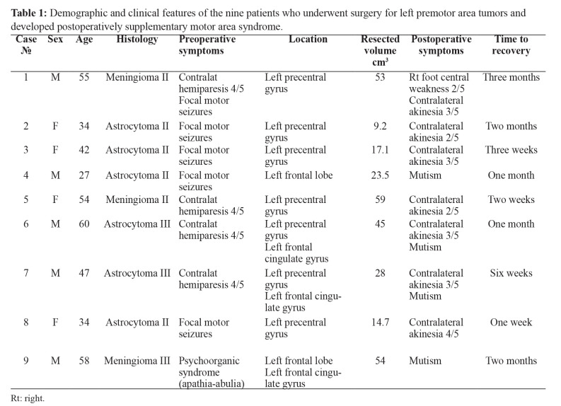
According to local regulations, the institutional review board (IRB) voided the need for ethics approval or informed consent for this retrospective case-series study, as the patients are fully anonymized and have received the standard care according to the institution guidelines. Nine patients with tumors engaging the premotor area of the left frontal hemisphere were operated on by the leading author over the period from 2010 to 2018, in the setting of two tertiary care centers. All patients were clinically defined as right-handed. Five of them were male and four female, with a median age of 47 years, range 27-60 years. Six patients harbored intra-axial and three extra-axial masses. The most common clinical presentation was that of focal motor seizures (five cases); three patients developed right-sided central hemiparesis, and one manifested with psycho-organic (frontal apathy-abulia) syndrome. The diagnosis was confirmed by contrast-enhanced magnetic resonance imaging (MRI). In all patients, we found tumor infiltration of the SMA cortex and gyrus cinguli involvement in three. Detailed neurology assessment preoperatively and immediately postoperatively (after waking the patient), then periodically as needed for at least one year, was performed with emphasis on the motor power, skills, and speech and language functions. A summary of the patients’ demographic and clinical data is available in Table 1.
Table 1. Demographic and clinical features of the nine patients who underwent surgery for left premotor area tumors and developed postoperatively supplementary motor area syndrome.
Rt: right.
Tumor excision was performed under general anesthesia, never using awake craniotomy. To define the SMA, we used the following morphologic criteria: the posterior border being the sulcus precentralis, the anterior border a transversal line at the level of the anterior part of the corpus callosum, the lower border in the medial plane, sulcus cinguli, the lateral border, in the field of gyrus frontalis superior without definite microsurgical border laterally. Volumetric microsurgical resection was performed using neuro-navigational systems: VectorVision-Brain LabTM (Munich, Germany) in three patients and ultrasound-guided system SonoWand InviteTM (Trondheim, Norway) in six. As “volumetric resection” is defined the excision of the tumor under neuronavigation control, using the preoperatively calculated tumor volume based on neuronavigation protocol DICOM (Digital Imaging and Communications in Medicine) images. We pursued gross total tumor excision. In all cases, the bridging veins and the integrity of the superior sagittal sinus were spared. All patients received anticonvulsant prophylactics (Valproate 20 mg/kg/24 hr or about 1,000-1,500 mg in two divided doses). With the onset of neurologic deficit, the patients received oral Dexamethasone at 12 mg daily for three days, tapered over ten days. Vasoactive medications were not used in the perioperative period. The minimal clinical follow-up period was one year; control computed tomography (CT) imaging was performed 24-48 hours postoperatively, and control MRI at three months and one year postoperatively. We compared the extent of resection, the involvement of gyrus cinguli, and the proximity to the sulcus (more than one cm or less than one cm) to the severity of SMA syndrome, duration of recovery, and the pattern of involvement. By severity, the SMA syndrome cases were stratified according to the degree and distribution of akinesia. We used descriptive and small-sample alternative and correlation statistics.
Gross total or nearly gross-total tumor excision was achieved in all the patients and confirmed by control imaging. Postoperatively, seven patients developed contralateral akinesia, that was severe [2/5 by the Medical Research Council (MRC) scale] in two, moderate to severe (3/5) in four, and mild (4/5) in one (2/5: a full range of movement with gravity eliminated, 3/5: active movement against gravity, 4/5: active movement against gravity and resistance). The motor deficit was accompanied by normal muscle tone and reflexes, without pyramidal tract signs, except for one patient described below. Four patients developed speech disorder/mutism (isolated in two and combined with akinesia in two). The exact clinical, surgical, and histological characteristics of the patients compared to the postoperative development are shown in Table 1. The imaging revealed normal postoperative appearance, with no evidence of hematoma or other collections, increased edema, or ischemia.
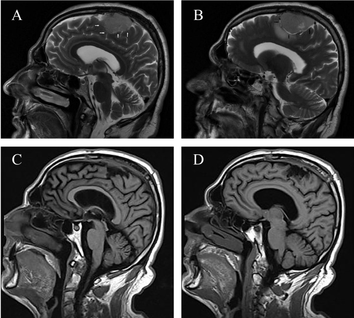
The small patient number precludes a rigorous statistical analysis; nevertheless, we will report test results and some tendencies that could not be statistically tested. SMA syndrome tended to be more severe and last longer with tumors closer to the precentral area, in the hemisphere’s convexital aspect, although this was not supported statistically. Illustrative are the cases of patients 1 and 2. Patient 1 was operated for an atypical meningioma (WHO grade II) with areas of undefined borders and cortical infiltration of the premotor cortex adjacent to the precentral gyrus (Figure 1). Postoperatively he developed contralateral moderate to severe right-sided akinesia with additional real pyramidal weakness 2/5 by MRC for the right foot only, with preserved muscle tone but hyperreflexia in the leg. The deficits resolved after three months. Patient 2 had an astrocytoma (WHO grade II) situated in the convexital premotor area close to the precentral area. During the microsurgical approach to the tumor, a small cortical incision was done across the medial frontal gyrus with an area of 1-1.5 cm2 between two cortical veins, and total tumor excision, assisted with 3-D ultrasound, was achieved (Figure 2). The patient developed a prominent contralateral motor deficit of 2/5 that resolved only after two months.
Figure 1. A, B) Preoperative magnetic resonance imaging (MRI) T2 images of reported patient 1, diagnosed with atypical meningioma WHO grade II showing border areas without a clear arachnoid plane with a tendency to invade the SMA cortex (image A, white arrows). There are also areas with a clear arachnoid plane with a distinct border, which had been easily dissected without affection of the cerebral cortex (image B, black arrows). C, D) Postoperative T1 MRI of patient 1, three months after the operation.
Figure 2. A, B) Preoperative images of reported patient 2, diagnosed with Astrocytoma WHO grade II located closely to precentral gyrus (image A, black arrow). A small cortical window was done between two cortical veins (image B, white arrows). C, D) Postoperative images three months after the operation.
In three patients with lesions located more anteriorly, farther from the precentral gyrus, the motor deficit was mild and resolved faster, within 1-3 weeks, with the distal muscle groups of the hands recovering first (cases 3, 5, and 8).
In the three patients with lesions of the medial surface of the left hemisphere and gyrus cinguli, the SMA syndrome presented mostly with a speech disorder (cases 4, 6, and 9). Speech disorder may also develop without the direct involvement of the cingular gyrus (case 7), but it is significantly more frequent with lesions of that structure (Fisher’s exact probability test value =0.047, p =0.041). It started as mutism that within days rapidly reverted to partial frontal dynamic aphasia, with preserved repetition skills but reduced speech initiative and paraphasias/perseverations. The speech disorder in our patients resolved entirely within three weeks to two months. The involvement of the gyrus cinguli was not related to greater severity or duration of SMA syndrome (Fisher’s exact probability test value =0.52, p =0.40).
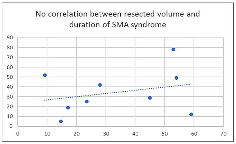
The resected tissue volume itself was not related to SMA syndrome severity and duration. The Pearson’s correlation coefficient between volume and duration was r =0.268, p =0.485 (Figure 3).
Figure 3. Resected volume and duration in postoperative SMA syndrome. In the x axis is illustrated the excised volume in cm2, and in the y axis the days to complete resolution, r =0.27, p =0.48.

Discussion
Our patient series confirms the main features of the postoperative SMA syndrome known from the earlier observations, namely its development after surgery on the premotor area of the dominant hemisphere, its transitory nature with full recovery in weeks to months, the relation of clinical manifestations to the topography of the resections, the possible role of gyrus cinguli and its connections in the development of speech disorder.
The SMA was defined as an area with specific architectonics and connections by the classics of neuroanatomy (Brodmann, Vogt, Kleist, and Foerster) while the cortical stimulation experiments of Penfield and Welch revealed some of its functions in initiation and control of movement and speech (review Nachev, Bozkurt)13,14. The initial description of the SMA syndrome by Laplane et al in 19771 corresponded to the expected deficits in movement and speech initiation but noted the unexpected features of fast reversibility with minimal or no residual sequelae confirmed by later series2-4. It is now established that the SMA has a somatotopic organization and consists of two separate areas; a rostral one (preSMA, F6), which gets projections from the prefrontal neocortex and the gyrus cinguli, and a caudal part (SMAproper, F3), which gives projections directly to the motor cortex and the spinal cord13-15.
The motor deficit after surgical SMA is due to disordered initiation and performance of the contralateral motor actions and may start as global akinesia that rapidly recedes to involve the contralateral limbs only. Our patients showed involvement of the contralateral limbs only. This feature may be due to the improved neuronavigation techniques, maximally sparing the cortex.
Our postoperative results would correspond to the premotor area’s somatotopic organization, with interventions in the posterior part closer than one cm to the precentral gyrus producing more prominent and longer-lasting deficits; similar observations are found in other recently reported series15-18.
The ideal surgical approach to minimize the risk of SMA syndrome would be awake craniotomy with cortical stimulation4,8,15,18,19. Cortical or subcortical stimulation carries some risk of intraoperative seizures20, so intraoperative somatosensory evoked potentials mapping for localization of the central sulcus was utilized in resections under general anesthesia4,21. Important developments that allow for precise tailoring of the resection are preoperative functional MRI (fMRI), tractography, and neuronavigation with ultrasound guidance22,23. All these are not, however, available in every institution, and we consider an actual strength of our report the “real-life setting” that is reported (volumetric resection based on the preoperative DICOM images but without the possibility of preoperative fMRI and tractography).
Transient postoperative symptoms may be due to the effects of retraction, edema, and arterial or venous damage. In our series, the early postoperative CT did not support such possibility, in line with others that interpret the postoperative SMA syndrome as a deficit compensated due to brain plasticity and reorganization7,10-12. In our patients, operating in the vicinity of gyrus cinguli could compromise the “frontal aslant tract” that runs close in this area and thus cause speech disorder, as seen in two cases.
SMA syndrome may also develop after non-dominant hemisphere surgery even after precise preoperative mapping22. In such cases, it usually does not include speech disorder3,17. We did not observe such cases, and handedness was defined clinically only.
Some authors reported a positive correlation between the resected volume and the duration of SMA syndrome17. A recent study by Nakajima et al15 noted increased severity and longer recovery with larger volume resections in the region of gyrus cinguli. In our patients, like in the series of Russel et al4, we could not find such a dependence.
Most descriptions have focused on SMA after removal of intra-axial tumors; one case series (five patients) focused on SMA after meningioma resection and commented on the rarity of such cases in literature16. Our series includes three meningioma grade II cases, with ingrowth to the cortex, in whom SMA syndrome developed and was not different from the features of SMA syndrome after intra-axial tumor removal.
The duration of SMA syndrome seems longer in our patients than in some other series6,9,16. Such difference may be due to this study’s retrospective character: very mild cases of SMA syndrome may have gone undetected in the early postoperative period, while in prospective studies with a high level of clinical suspicion, they would have been diagnosed.
The retrospective protocol, the lack of functional imaging, and the relatively small number of patients, are the main weaknesses of our study. Still, we believe it contributes to knowledge in the field. Our findings are in concert with the previous series, with some discrepancies easily explained by the small cohort size in most studies.
The possibility for the development of SMA syndrome and its transient nature should be discussed preoperatively with the patients at risk and their caregivers. The postoperative encounter with a mute and hemiplegic person who had only minor deficits before entering the theatre may be rather distressful; medical staff of all levels and specialties involved in postoperative care (anesthesiology, physiotherapy) should be well informed regarding the condition and reassure the patient and his relatives24.
Conclusion
The postoperative SMA syndrome may complicate the pursuit of total excision of the tumor even with precise neuronavigation microsurgical approaches. This entity with a favorable prognosis should be well known to all specialists involved in such patients’ postoperative care. The possibility of development of the SMA syndrome should be preoperatively discussed with patients undergoing SMA area surgery.
Conflicts of Interest
The authors declare no conflict of interest.
References
- 1.Laplane D, Talairach J, Meininger V, Bancaud J, Orgogozo J. Clinical consequences of corticectomies involving the supplementary motor area in man. J Neurol Sci. 1977;34:301–314. doi: 10.1016/0022-510x(77)90148-4. [DOI] [PubMed] [Google Scholar]
- 2.Rostomily RC, Berger MS, Ojemann GA, Lettich E. Postoperative deficits and functional recovery following removal of tumors involving the dominant hemisphere supplementary motor area. J Neurosurg. 1991;75:62–68. doi: 10.3171/jns.1991.75.1.0062. [DOI] [PubMed] [Google Scholar]
- 3.Russell SM, Kelly PJ. Incidence and clinical evolution of postoperative deficits after volumetric stereotactic resection of glial neoplasms involving the supplementary motor area. Neurosurgery. 2003;52:506–516. doi: 10.1227/01.neu.0000047670.56996.53. [DOI] [PubMed] [Google Scholar]
- 4.Zentner J, Hufnagel A, Pechstein U, Wolf HK, Schramm J. Functional results after resective procedures involving the supplementary motor area. J Neurosurg. 1996;85:542–549. doi: 10.3171/jns.1996.85.4.0542. [DOI] [PubMed] [Google Scholar]
- 5.Nakajima R, Kinoshita M, Yahata T, Nakada M. Recovery time from supplementary motor area syndrome: relationship to postoperative day 7 paralysis and damage of the cingulum. J Neurosurg. 2019:1–10. doi: 10.3171/2018.10.JNS182391. [DOI] [PubMed] [Google Scholar]
- 6.Duffau H, Lopes M, Denvil D, Capelle L. Delayed onset of the supplementary motor area syndrome after surgical resection of the mesial frontal lobe: a time course study using intraoperative mapping in an awake patient. Stereotact Funct Neurosurg. 2001;76:74–82. doi: 10.1159/000056496. [DOI] [PubMed] [Google Scholar]
- 7.Grønbæk J, Molinari E, Avula S, Wibroe M, Oettingen G, Juhler M. The supplementary motor area syndrome and the cerebellar mutism syndrome: a pathoanatomical relationship? Childs Nerv Syst. 2020;36:1197–1204. doi: 10.1007/s00381-019-04202-3. [DOI] [PubMed] [Google Scholar]
- 8.Oda K, Yamaguchi F, Enomoto H, Higuchi T, Morita A. Prediction of recovery from supplementary motor area syndrome after brain tumor surgery: preoperative diffusion tensor tractography analysis and postoperative neurological clinical course. Neurosurg Focus. 2018;44:E3. doi: 10.3171/2017.12.FOCUS17564. [DOI] [PubMed] [Google Scholar]
- 9.Baker CM, Burks JD, Briggs RG, Smitherman AD, Glenn CA, Conner AK, et al. The crossed frontal aslant tract: A possible pathway involved in the recovery of supplementary motor area syndrome. Brain Behav. 2018;8:e00926. doi: 10.1002/brb3.926. [DOI] [PMC free article] [PubMed] [Google Scholar]
- 10.Abel TJ, Buckley RT, Morton RP, Gabikian P, Silbergeld DL. Recurrent Supplementary Motor Area Syndrome Following Repeat Brain Tumor Resection Involving Supplementary Motor Cortex. Neurosurgery. 2015;11 Suppl 3:447–455. doi: 10.1227/NEU.0000000000000847. discussion 456. [DOI] [PMC free article] [PubMed] [Google Scholar]
- 11.Vassal M, Charroud C, Deverdun J, Le Bars E, Molino F, Bonnetblanc F, et al. Recovery of functional connectivity of the sensorimotor network after surgery for diffuse low-grade gliomas involving the supplementary motor area. J Neurosurg. 2017;126:1181–1190. doi: 10.3171/2016.4.JNS152484. [DOI] [PubMed] [Google Scholar]
- 12.Acioly MA, Cunha AM, Parise M, Rodrigues E, Tovar-Moll F. Recruitment of Contralateral Supplementary Motor Area in Functional Recovery Following Medial Frontal Lobe Surgery: An fMRI Case Study. J Neurol Surg A Cent Eur Neurosurg. 2015;76:508–512. doi: 10.1055/s-0035-1558408. [DOI] [PubMed] [Google Scholar]
- 13.Nachev P, Kennard C, Husain M. Functional role of the supplementary and pre-supplementary motor areas. Nat Rev Neurosci. 2008;9:856–869. doi: 10.1038/nrn2478. [DOI] [PubMed] [Google Scholar]
- 14.Bozkurt B, Yagmurlu K, Middlebrooks EH, Karadag A, Ovalioglu TC, Jagadeesan B, et al. Microsurgical and Tractographic Anatomy of the Supplementary Motor Area Complex in Humans. World Neurosurg. 2016;95:99–107. doi: 10.1016/j.wneu.2016.07.072. [DOI] [PubMed] [Google Scholar]
- 15.Fontaine D, Capelle L, Duffau H. Somatotopy of the supplementary motor area: evidence from correlation of the extent of surgical resection with the clinical patterns of deficit. Neurosurgery. 2002;50:297–303. doi: 10.1097/00006123-200202000-00011. [DOI] [PubMed] [Google Scholar]
- 16.Berg-Johnsen J, Høgestøl EA. Supplementary motor area syndrome after surgery for parasagittal meningiomas. Acta Neurochir (Wien) 2018;160:583–587. doi: 10.1007/s00701-018-3474-3. [DOI] [PubMed] [Google Scholar]
- 17.Rosenberg K, Nossek E, Liebling R, Fried I, Shapira-Lichter I, Hendler T, et al. Prediction of neurological deficits and recovery after surgery in the supplementary motor area: a prospective study in 26 patients. J Neurosurg. 2010;113:1152–1163. doi: 10.3171/2010.6.JNS1090. [DOI] [PubMed] [Google Scholar]
- 18.Ulu MO, Tanriöver N, Ozlen F, Sanus GZ, Tanriverdi T, Ozkara C, et al. Surgical treatment of lesions involving the supplementary motor area: clinical results of 12 patients. Turk Neurosurg. 2008;18:286–293. [PubMed] [Google Scholar]
- 19.Berger MS, Kincaid J, Ojemann GA, Lettich E. Brain mapping techniques to maximize resection, safety, and seizure control in children with brain tumors. Neurosurgery. 1989;25:786–792. doi: 10.1097/00006123-198911000-00015. [DOI] [PubMed] [Google Scholar]
- 20.Spena G, Roca E, Guerrini F, Panciani PP, Stanzani L, Salmaggi A, et al. Risk factors for intraoperative stimulation-related seizures during awake surgery: an analysis of 109 consecutive patients. J Neurooncol. 2019;145:295–300. doi: 10.1007/s11060-019-03295-9. [DOI] [PubMed] [Google Scholar]
- 21.Rowed DW, Houlden DA, Basavakumar DG. Somatosensory evoked potential identification of sensorimotor cortex in removal of intracranial neoplasms. Can J Neurol Sci. 1997;24:116–120. doi: 10.1017/s0317167100021430. [DOI] [PubMed] [Google Scholar]
- 22.Nelson L, Lapsiwala S, Haughton VM, Noyes J, Sadrzadeh AH, Moritz CH, et al. Preoperative mapping of the supplementary motor area in patients harboring tumors in the medial frontal lobe. J Neurosurg. 2002;97:1108–1114. doi: 10.3171/jns.2002.97.5.1108. [DOI] [PubMed] [Google Scholar]
- 23.Wongsripuemtet J, Tyan AE, Carass A, Agarwal S, Gujar SK, Pillai JJ, et al. Preoperative Mapping of the Supplementary Motor Area in Patients with Brain Tumor Using Resting-State fMRI with Seed-Based Analysis. AJNR Am J Neuroradiol. 2018;39:1493–1498. doi: 10.3174/ajnr.A5709. [DOI] [PMC free article] [PubMed] [Google Scholar]
- 24.Fontaine D, Capelle L, Duffau H. Somatotopy of the supplementary motor area: evidence from correlation of the extent of surgical resection with the clinical patterns of deficit. Neurosurgery. 2002;50:297–303. doi: 10.1097/00006123-200202000-00011. discussion 303-305. [DOI] [PubMed] [Google Scholar]


