Abstract
A novel anaerobic heterotrophic strain, designated strain sy52T, was isolated from a hydrothermal chimney at Suiyo Seamount in the Pacific Ocean. A 16S rRNA gene analysis revealed that the strain belonged to the family Petrotogaceae in the phylum Thermotogae. The strain was mesophilic with optimum growth at 48°C and the phylum primarily comprised hyperthermophiles and thermophiles. Strain sy52T possessed unique genomic characteristics, such as an extremely low G+C content and 6 copies of rRNA operons. Genomic analyses of strain sy52T revealed that amino acid usage in the predicted proteins resulted from adjustments to mesophilic environments. Genomic features also indicated independent adaptions to the mesophilic environment of strain sy52T and Mesotoga species, which belong to the mesophilic lineage in the phylum Thermotogae. Based on phenotypic and phylogenetic evidence, strain sy52T is considered to represent a novel genus and species in the family Petrotogaceae with the proposed name Tepiditoga spiralis gen. nov., sp. nov.
Keywords: Thermotogae, Tepiditoga spiralis, Mesophilic environment
Growth temperature is one of the important physiological features for characterizing bacteria and archaea, and psychrophiles, mesophiles, thermophiles and hyperthermophiles are categorized based on them. An optimum growth temperature of 45°C is typically the boundary that separates mesophiles and thermophiles, but may vary (Madigan et al., 2002; Wagner and Wiegel, 2008; Taylor and Vaisman, 2010). At the molecular level, growth temperature has been shown to correlate with genome and protein properties (Zheng and Wu, 2010).
Bacteria of the Thermotogae lineage primarily comprise hyperthermophiles and thermophiles isolated from high-temperature environments, and the phylum is phylogenetically placed in a deep-branched position in the domain Bacteria. Thirteen genera have been described since Thermotoga maritima was initially discovered in geothermally heated marine sediments (Huber et al., 1986), and Bhandari and Gupta recently categorized the phylum into 4 orders and 5 families using genome data (Bhandari and Gupta, 2014). They primarily grow by fermentation under strictly anaerobic and thermophilic conditions and have a characteristic outer sheath-like structure called a ‘toga’. These genomic features were found to be unique, and detailed analyses revealed lateral gene transfer from diverse lineages of both Bacteria and Archaea as well as the genomic machinery of adaptation to high-temperature environments (Nelson et al., 1999; Zhaxybayeva et al., 2009; Bhandari and Gupta, 2014).
In addition to hyperthermophilic and thermophilic species, previous studies predicted the presence of mesophiles known as “mesotoga” in the phylum based on examinations of 16S rRNA genes from mesophilic environments and enrichments (Chouari et al., 2005; Nesbo et al., 2006; Berlendis et al., 2010; Nesbo et al., 2010). “Mesotoga sulfurireducens” PhosAc3 was initially isolated as a mesophilic bacterium belonging to the phylum; however, a complete description of the strain is not yet available (Ben Hania et al., 2011; 2015). M. prima MesG1.Ag.4.2T isolated from a marine sediment was the first strain to have its characteristics formally described and grows optimally at 37°C (Nesbo et al., 2012). Mesotoga infera VNs100T was also retrieved from anoxic water in a deep aquifer, and the temperature range for growth was 30–50°C with an optimum at 45°C (Ben Hania et al., 2013). Species belonging to the genus Geotoga grow under relatively mesophilic conditions, and the optimum growth temperatures of Geotoga petraea T5T and Geotoga subterranea CC-1T were 50 and 45°C, respectively (Davey et al., 1993). Except for the genera Mesotoga and Geotoga, bacteria of the Thermotogae lineage generally comprise hyperthermophiles and thermophiles, with an optimum growth temperature of higher than 55°C.
Previous analyses of the G+C content of 16S rRNA genes and the amino acid composition of protein-coding genes suggested that thermophilic features are systematically original characteristics of bacteria of the Thermotogae lineage (Zhaxybayeva et al., 2009), and ‘mesotoga’ may have adapted to mesophilic environments. The genome of M. prima MesG1.Ag.4.2T was found to be markedly larger than that of other bacteria of the Thermotogae lineage, and 32% of predicted protein-coding genes were shown to be acquired by lateral gene transfer (Zhaxybayeva et al., 2012). Based on these findings, Zhaxybayeva et al. suggested that the genomic features of M. prima were indicative of its adaption to a new lifestyle, such as a mesophilic environment.
A novel mesophilic anaerobic heterotroph, designated strain sy52T and belonging to the phylum Thermotogae, was recently isolated from a deep-sea hydrothermal field. The present study focused on physiological and genomic analyses and discusses mesophilic adaptations by this strain. In addition, based on phenotypic characteristics as well as phylogenetic analyses, a novel taxon is proposed for the isolate with the name Tepiditoga spiralis gen. nov., sp. nov.
Materials and Methods
Sample collection, enrichment, and isolation
The sample for enrichment and isolation was collected from a deep-sea hydrothermal vent chimney in Suiyo Seamount, the Izu-Bonin Arc, the western Pacific Ocean by DSV Shinkai6500 during the YK11-06 scientific cruise aboard the R/V Yokosuka (JAMSTEC, Yokosuka, Kanagawa, Japan) in August 2012. The region has a submarine caldera with numerous hydrothermal vents at a depth of 1,390 m (Glasby et al., 2000). Chips of an active chimney were selected for enrichment and were immediately inoculated on board.
The medium under a N2/CO2 (80:20 [v/v]) atmosphere was added to a vial sealed with a butyl rubber stopper and aluminum cap for enrichment, and isolation comprised (L–1) 0.6 g KH2PO4, 0.1 g K2HPO4, 0.75 g MgCl2·6H2O, 0.15 g CaCl2·2H2O, 0.3 g NH4Cl, 30 g NaCl, 0.3 g Na2SO4, 1.6 g Na2S2O3, 3 g Bacto Yeast Extract (Difco), 2 mL trace element solution (Mori and Suzuki, 2008), 2 mL vitamin solution (Mori and Suzuki, 2008), 1 mg resazurin, 1 g Na2CO3, and 0.5 g Na2S·9H2O. After mixing ingredients, except for the vitamin solution, Na2CO3, and Na2S·9H2O, the medium was autoclaved under a N2/CO2 atmosphere. The vitamin and Na2CO3 solutions were sterilized with filtration. Na2S·9H2O solution autoclaved separately was then added to the medium. Anaerobic bacteria were cultivated at various temperatures for enrichment; after 1 week of cultivation, bacterial growth was confirmed at 30°C. Regarding single strain isolation, colonies were allowed to form on medium solidified with 1.5% (w/v) agar (Difco Agar Noble) in vials for approximately 2 months. After a second purification step with the same solid medium, a pure culture of strain sy52T was obtained.
Physiological characterization
Cell morphology was routinely observed using phase-contrast microscopy (model AX-70; Olympus). Optical density (A660) was measured with a spectrophotometer (model U-2800; Hitachi). A direct cell count was performed under a fluorescent microscope by 4',6-diamidino-2-phenylindole (DAPI) staining on a polycarbonate membrane filter (K020N025A; Advantec). The concentrations of sulfate, thiosulfate, and nitrate were assessed by HPLC (model 2695 with conductivity detector model 432 and an IC-Pac Anion column; Waters) (Mori et al., 2008).
The following substrates were examined as the sole energy and carbon sources: 10 mM D-glucose, 10 mM D-fructose, 10 mM D-mannose, 10 mM D-galactose, 10 mM maltose, 10 mM lactose, 10 mM D-trehalose, 10 mM sucrose, 10 mM D-cellobiose, 10 mM D-raffinose, 10 mM D-arabinose, 10 mM L-rhamnose, 10 mM D-xylose, 10 mM D-ribose, 10 mM ribitol, 10 mM D-mannitol, 10 mM D-sorbitol, 20 mM glycerol, 20 mM citrate, 20 mM pyruvate, 20 mM succinate, 20 mM malate, 20 mM L-glutamate, 20 mM butyrate, 20 mM lactate, 20 mM propionate, 5 g L–1 starch, 1 g L–1 yeast extract, 1 g L–1 polypeptone, and 1 g L–1 casamino acids. The substrate utilization test was also performed in the presence of 0.2 g L–1 yeast extract. The utilization of the following electron acceptors was evaluated in the presence of 3 g L–1 yeast extract as the substrate: 10 mM thiosulfate, 10 mM sulfate, 2 and 5 mM sulfite, 5 g L–1 elemental sulfur, 10 mM fumarate, 10 mM nitrate, 2 and 5 mM nitrite, and 2 and 5% (v/v) oxygen. H2S production from sulfur compounds as electron acceptors was confirmed by FeS precipitation after the addition of one drop of 0.1 M FeSO4 solution to cultures in media without sulfide as the reducing agent. The effects of temperature, initial pH, and NaCl concentrations on growth in the presence of 3 g L–1 yeast extract and 10 mM thiosulfate were assessed by examining the time course of optical density changes with a temperature gradient incubator (model TN-2612; Advantec). The initial pH of the medium was adjusted by adding Na2CO3 or HCl solution.
Cellular fatty acids were methylated using a 5% HCl/methanol solution (Sasser, 1990) and analyzed by the MIDI microbial identification system and GC-MS (gas chromatograph model GC-2010; gas chromatograph mass spectrometer model GCMS-QP2010Plus; Shimadzu).
Genome sequencing and analyses
Genomic DNA was extracted using the EZ1 Tissue kit according to the manufacturer’s instructions (Qiagen). Whole-genome shotgun sequencing was performed using the 454 GS FLX-Titanium system (Roche) and MiSeq (Illumina). Reads were assembled using the Newbler assembler version 2.8 (Roche). Primer walking on gap-spanning PCR products from genomic DNA closed the gaps between the assembled sequences. The genome was submitted to RAST (http://rast.nmpdr.org/) for automatic annotation.
The phylogenetic position was elucidated using the 16S rRNA gene sequence. Sequences were aligned using the ARB program (Ludwig et al., 2004), and a phylogenetic tree was reconstructed by the neighbor-joining method using the CLUSTAL_X program (Saitou and Nei, 1987; Thompson et al., 1997).
Absolute differences between charged and polar amino acid residues (CvP bias) and the Ile, Val, Tyr, Trp, Arg, Glu, and Leu amino acid bias (IVYWREL bias) of predicted proteins were calculated according to the methods described by Suhre and Claverie (2003) and Zeldovich et al. (2007), respectively. Proteins with less than 2 predicted trans-membrane helices (assessed using TMHMM Server v. 2.0 [Sonnhammer et al., 1998; Krogh et al., 2001]) were used for calculations. We analyzed the G+C content for every 10,000 bases on each genome and the codon usage of amino acids for predicted proteins using the G-language system (Arakawa et al., 2008; 2010).
Sequence accession numbers
The genome sequence of strain sy52T was deposited in DDBJ/EMBL/GenBank with the accession number AP018712 under the BioProject accession number PRJDB6802 and BioSample accession number SAMD00113976 using DFAST, the DDBJ Fast Annotation and Submission Tool (Tanizawa et al., 2016; 2018). The 16S rRNA gene sequence was also deposited with the accession number LC485113. All DDBJ/EMBL/GenBank accession numbers for analyses are shown in Fig. 3.
Fig. 3.
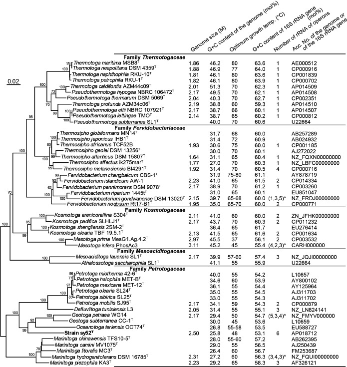
Neighbor-joining phylogenetic tree based on sequences of the 16S rRNA gene and genome size, G+C contents of the genome and 16S rRNA gene, optimum temperature for growth, and the number of ribosomal RNA operons of bacteria of the Thermotogae lineage. Bootstrap values are indicated at branch nodes. Genomic G+C contents calculated based on genome sequences are preferentially indicated. DDBJ/EMBL/GenBank accession numbers for analyses are shown in the rightmost column. Bar, 0.02 substitutions per nucleotide position. *They have some partial rRNA operons (numbered 5S, 16S, and 23S): F. gondwanense (1, 3, 5); M. infera, (4, 2, 3); G. petraea, (5, 3, 4); M. hydrogenitolerans, (3, 4, 3).
Results
Growth properties and chemotaxonomic characteristics
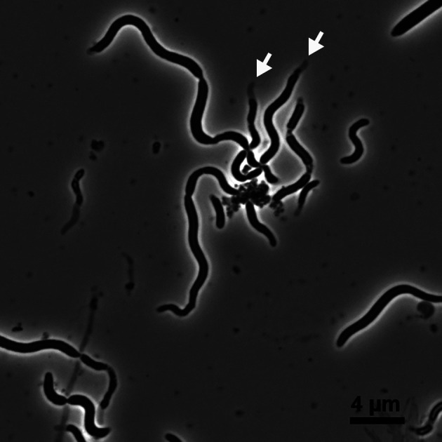
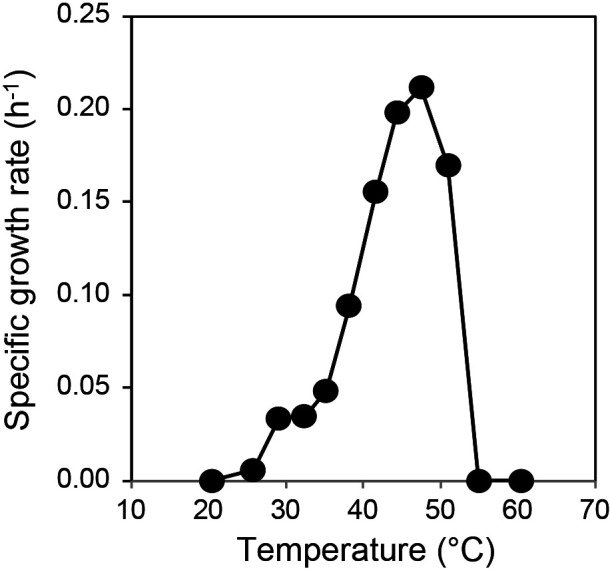
The cells of strain sy52T had a rod-shaped morphology with the presence of a toga structure, and motility was observed under a microscope. Spiral-shaped cells were detected under optimum growth conditions (Fig. 1). Strain sy52T is a strictly anaerobic bacterium and, thus, was unable to grow under aerobic conditions. It required yeast extract as a growth factor, which was not replaceable by a vitamin mixture. The vitamin solution was not required for its growth. In the presence of 0.2 g L–1 yeast extract, strain sy52T grew with yeast extract, polypeptone, and starch as energy and carbon sources. However, even in the presence of 0.2 g L–1 yeast extract, other organic substrates did not stimulate growth. In the presence of yeast extract as energy and carbon sources, strain sy52T used thiosulfate and elemental sulfur as electron acceptors and reduced them to hydrogen sulfide. Growth yield was two-fold higher following their addition than with fermentation. Strain sy52T grew at temperatures ranging between 26 and 51°C, with the optimum temperature being 48°C (Fig. 2). The initial pH range for growth was 5.0–7.0, with an optimum at pH 6.0. The strain grew in 1–5% (w/v) NaCl, with an optimum concentration of 2–4% NaCl. The doubling time under optimum growth conditions was 3 h, and growth yield reached approximately 5×10⁷ cells mL–1 in the presence of yeast extract and thiosulfate.
Fig. 1.

Phase-contrast micrograph of strain sy52T. The ‘toga’ structure is indicated by open arrows.
Fig. 2.

Effects of temperature on the growth of strain sy52T.
The cells of strain sy52T contained C16:0 (52% of all fatty acids) as the major fatty acid, and C16:1ω7c (13%), C16:1ω9c (12%), C18:1ω9c (6%), C18:0 (6%), C14:0 (5%), C12:0 (2%), C18:1ω7c/ω6c (2%), C17:1iso/anteiso (1%), and C10:0 (1%) were identified as minor fatty acids.
Genome sequencing
The complete genome sequence of strain sy52T was elucidated, resulting in a genome that consists of a 2,502,404-bp circular chromosome with a G+C content of 25.8 mol%. Six copies of an rRNA operon and 2,302 predicted protein-coding genes were identified. Six copies of complete rRNA operons obtained from the genome sequence of strain sy52T and their 16S rRNA gene sequences showed slight differences and a similarity of 99.8–100%.
Phylogenetic position
The phylogenetic position of strain sy52T was identified using the 16S rRNA gene sequence. The neighbor-joining tree (Fig. 3) revealed that the strain belonged to the family Petrotogaceae in the phylum Thermotogae. The 16S rRNA gene sequence of strain sy52T had a similarity of less than 90% with that of species in the phylum Thermotogae, and the closest relatives were Oceanotoga teriensis (sequence similarity of 87.8%) and G. subterranea (87.2%).
Genome characteristics
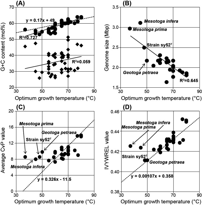
The G+C contents of the 16S rRNA gene sequences of bacteria of the Thermotogae lineage and strain sy52T plotted against their optimum growth temperatures revealed a correlation (Fig. 4A). On the other hand, the G+C content of the whole genome sequence did not correlate with optimum growth temperatures (Fig. 4A). The genome size of strain sy52T was larger than those of the thermophilic and hyperthermophilic species in the Thermotogae lineage, but not as large as those of Mesotoga species (Fig. 4B). Genome sizes were related to optimum growth temperatures, and species with lower optimum growth temperatures generally had a larger genome. Regarding average CvP values (Fig. 4C), the plots of strain sy52T, M. prima, M. infera, and G. petraea were distant from those of species of the Thermotogae lineage. The IVYWREL value of strain sy52T against the optimum temperature may harmonize with the linear regression reported by Zeldovich (Zeldovich et al., 2007), whereas those of Mesotoga species showed marked deviations (Fig. 4D).
Fig. 4.
Relationship between the optimum temperature and various parameters of bacteria of the Thermotogae lineage: (A) correlations with the G+C content of the 16S rRNA gene (circle) and genome (diamond), the dot-linear mathematical formula based on 406 prokaryotes was from Kimura et al. (2006); (B) correlation with genome sizes; (C) correlation with average CvP values, the dot-linear mathematical formula based on 4 bacteria of the Thermotogae lineage was from Zhaxybayeva et al. (2009); (D) correlation with IVYWREL values, the dot-linear mathematical formula based on 86 prokaryotes denoted by Zeldovich et al. (2007).
Discussion
Enrichment and isolation procedures resulted in the successful isolation of strain sy52T from the hydrothermal vent chimney at Suiyo Seamount. According to the phylogenetic analysis based on 16S rRNA gene sequences, the isolated strain belonged to the family Petrotogaceae in the phylum Thermotogae (Fig. 3). However, sequence similarities with known species were less than 90%, indicating that the strain was phylogenetically independent at the genus level. Characteristics such as the presence of a toga structure (Fig. 1) and energy acquisition by fermentation were similar to bacteria of the Thermotogae lineage. On the other hand, strain sy52T grew at temperatures lower than 51°C with an optimum temperature of 48°C (Fig. 2), which is lower than the growth temperature of most bacteria of the Thermotogae lineage, with a few exceptions. Based on the optimum growth temperature, it may not be reasonable to state that strain sy52T is a typical mesophile or “mesotoga”; however, it is not a thermophile and displayed better adaptation to a mesophilic environment than other bacteria of the Thermotogae lineage. The optimum growth temperature of G. subterranea was previously reported to be 45°C (Davey et al., 1993), and, thus, strain sy52T and G. subterranea of the family Petrotogaceae appear to belong to a lineage that is adapted to mesophilic environments, in contrast to the lineage of the genus Mesotoga in the family Kosmotogaceae. Therefore, we investigated the relationship between the genome and adaptation to mesophilic environments using genomic information from two lineages.
The genome of strain sy52T is 2.50 Mb in length and has a G+C content of 25.8 mol%. The size of its genome was markedly larger than that of other bacteria of the Thermotogae lineage (Fig. 3), similar to that observed for genomes of the genus Mesotoga, and genome sizes negatively correlated with optimum growth temperatures among bacteria of the Thermotogae lineage (Fig. 4B). A relationship may exist between habitat changes and genome sizes; however, adaptations to mesophilic environments only do not indicate habitat changes in the bacterial lineage. The G+C content of strain sy52T was lower than those of any other bacteria of the Thermotogae lineage (Fig. 3). This low G+C content may be attributed to the high use of adenine and thymine as the 3rd base of the amino acid code (data not shown). Although a low G+C content occurred in some species, such as those in the family Petrotogaceae (Fig. 3), it was not associated with optimum growth temperatures (Fig. 4A). Therefore, the low G+C content of the genome may be due to other factors as well as adaptations to mesophilic environments.
Previous studies proposed an inverse correlation between the G+C contents of 16S rRNA sequences and optimal growth temperatures in bacteria and archaea (Khachane et al., 2005; Kimura et al., 2006; 2007). A similar relationship was observed for bacteria of the Thermotogae lineage (Fig. 4A), and the thermodynamic stability of 16S rRNA secondary structures also reflects their habitats. On the other hand, 6 copies of rRNA operons were identified in the genome of strain sy52T, and although the possession of multiple rRNA operons may be of significance in the family Petrotogaceae (Fig. 3), the number of rRNA operons in strain sy52T is marked compared to others in the phylum. Several divergent/identical 16S rRNA genes were previously shown to be harbored in the genomes of bacteria and archaea, and one base or more dissimilar 16S rRNA genes were detected in almost 50% of genomes (Sun et al., 2013). Although we considered multiple aspects of the possession of a high number of rRNA operons, its significance remains unclear, and its relationship with adaptations to mesophilic environments has not yet been elucidated.
Overrepresentations of charged amino acid residues over polar ones (CvP bias) and IVYWREL amino acids in predicted proteins have been suggested as indicators of the optimal growth temperatures of bacteria and archaea (Suhre and Claverie, 2003; Zeldovich et al., 2007; Taylor and Vaisman, 2010). Previous analyses of some bacteria of the Thermotogae lineage revealed that average CvP and IVYWREL values were linearly related to optimal growth temperatures and also that the proteins of M. prima were not suitable for a thermophilic environment (Zhaxybayeva et al., 2009; 2012). Although many of the average CvP values are on the line calculated based on four bacteria of the Thermotogae lineage reported by Zhaxybayeva et al. (2009), the values of strain sy52T, M. prima, M. infera, and G. petraea are not on the line (Fig. 4C), indicating that their proteins remain thermophilic. On the other hand, except for two Mesotoga species, IVYWREL values were on the line calculated based on 86 bacteria and archaea (Zeldovich et al., 2007). The two indicators of Mesotoga species suggest that they still possess thermo-adapted proteins, while proteins of strain sy52T and G. petraea adapt to mesophilic environments.
Thermophilic features were previously suggested to be the original characteristics of bacteria of the Thermotogae lineage (Zhaxybayeva et al., 2009), and based on the amino acids in predicted proteins, strain sy52T and G. petraea showed better adaptation to a mesophilic environment than any other known species in the phylum Thermotogae. In mesophilic species in the phylum Thermotogae, the essential factor for growth temperature is the G+C content of the 16S rRNA sequence rather than its amino acid composition. In addition, we focused on the extremely low genomic G+C content and high number of rRNA operons in strain sy52T; however, it is pure speculation that they are traits of adaptation to a different environment. More isolates of “mesotoga” and further details of their genomic characteristics are needed to completely clarify adaptations to mesophilic environments by the phylum Thermotogae.
A phylogenetic analysis based on 16S rRNA gene sequences (Fig. 3) revealed that strain sy52T belonged to the family Petrotogaceae, and the characteristics of strain sy52T, such as it being a moderate thermophile, requiring NaCl for growth, possessing C16:0 as a major cellular fatty acid, and having a low genomic G+C content, were similar to those of genera in the family (Table 1). However, sequence similarities between the strain and species in the family were less than 90%, and the difference was sufficient to denote a new genus for the strain (Stackebrandt and Goebel, 1994). Based on physiological and phylogenetic evidence, a novel taxon, Tepiditoga spiralis gen. nov., sp. nov., belonging to the family Petrotogaceae, is proposed.
Table 1.
Characteristics of strain sy52T and genera in the family Petrotogaceae.
| Characteristics | strain sy52T | Petrotoga | Defluviitoga | Geotoga | Oceanotoga | Marinitoga |
|---|---|---|---|---|---|---|
| Optima for growth | ||||||
| temperature (°C) | 48 | 55–60 | 55 | 45–50 | 55–58 | 55–65 |
| pH | 6.0 | 6.5–8.0 | 6.9 | 6.5 | 7.3–7.8 | 5.5–7.0 |
| NaCl (w/v [%]) | 2–4 | 1–6 | 0.5 | 3 | 4–4.5 | 2–4 |
| Growth temperature range (°C) | 26–51 | 30–65 | 37–65 | 30–60 | 25–70 | 25–70 |
| Reduction of sulfur compounds | + | +/– | + | + | + | + |
| Major fatty acids | C16:0 | C16:0, C18:1 | C16:0, C18:1ω9c | C16:0, C16:1 | C16:0 | C16:0, C18:0 |
| Genomic G+C content (mol%) | 25.8 | 33.0–40.0 | 31.4 | 29.4–30.0 | 26.8 | 26.4–29.2 |
| Reference | this study | (Davey et al., 1993; Miranda-Tello et al., 2007) | (Ben Hania et al., 2012) | (Davey et al., 1993) | (Jayasinghearachchi and Lal, 2011) | (Nunoura et al., 2007; Postec et al., 2010) |
+, positive; –, negative.
Description of Tepiditoga gen. nov.
Tepiditoga (Te.pi.di.to’ga. L. adj. tepidus moderately warm; L. fem. n. toga outer garment; N.L. fem. n. Tepiditoga a moderately warm garment).
Cells are rods with a sheath-like outer structure. Spiral rod-shaped cells are observed under optimum growth conditions. It is obligately anaerobic and chemoorganotrophic. It is moderately thermophilic and moderately halophilic. It grows by fermentation and reduces sulfur compounds. The major cellular fatty acid is C16:0. Its phylogenetic position based on the 16S rRNA gene sequence is in the family Petrotogaceae. The type species is Tepiditoga spiralis.
Description of Tepiditoga spiralis sp. nov.
Tepiditoga spiralis (spi.ra’lis. L. adj. spiralis spiral).
It has the following characteristics in addition to those given in the genus description. Under optimum growth conditions, cells are spiral rod-shaped and motile. It reduces thiosulfate and elemental sulfur to sulfide. Yeast extract, polypeptone, and starch are used as growth substrates. It does not grow on a sole substrate and yeast extract is necessary for growth. It grows at temperatures ranging between 26 and 51°C, with optimal growth at 48°C. The initial pH for growth is pH 5.0–7.0, with an optimum at pH 6.0. The NaCl concentration for growth ranges between 1 and 5% (w/v), with an optimum at 2–4%. The predominant cellular fatty acid is C16:0. C16:1ω7c, C16:1ω9c, C18:1ω9c, C18:0, C14:0, C12:0, C18:1ω7c/ω6c, C17:1iso/anteiso, and C10:0 are minor fatty acids.
The type strain, sy52T (=NBRC 112788T=DSM 105848T), was isolated from a hydrothermal chimney in Suiyo Seamount, the Izu-Bornin Arc, the western Pacific Ocean. The genomic G+C content of the type strain is 25.8 mol%.
Citation
Mori, K., Sakurai, K., Hosoyama, A., Kakegawa, T., and Hanada, S. (2020) Vestiges of Adaptation to the Mesophilic Environment in the Genome of Tepiditoga spiralis gen. nov., sp. nov.. Microbes Environ 35: ME20046.
https://doi.org/10.1264/jsme2.ME20046
Acknowledgements
We are grateful to the crew of the R/V Yokosuka, the operational team of the DSV Shinkai6500, and the scientists who joined the YK11-06 scientific cruise for their cooperation with sample collection. We also thank Shinobu Iwasaki for his technical support, and Keiko Tsuchikane and Mami Igarashi for genome elucidation.
References
- Arakawa K., Suzuki H., and Tomita M. (2008) Computational genome analysis using the G-language system. Genes, Genomes and Genomics 2: 1–13. [Google Scholar]
- Arakawa K., Kido N., Oshita K., and Tomita M. (2010) G-language genome analysis environment with REST and SOAP web service interfaces. Nucleic Acids Res 38: W700–705. [DOI] [PMC free article] [PubMed] [Google Scholar]
- Ben Hania W., Ghodbane R., Postec A., Brochier-Armanet C., Hamdi M., Fardeau M.L., et al. (2011) Cultivation of the first mesophilic representative (“mesotoga”) within the order Thermotogales. Syst Appl Microbiol 34: 581–585. [DOI] [PubMed] [Google Scholar]
- Ben Hania W., Godbane R., Postec A., Hamdi M., Ollivier B., and Fardeau M.-L. (2012) Defluviitoga tunisiensis gen. nov., sp. nov., a thermophilic bacterium isolated from a mesothermic and anaerobic whey digester. Int J Syst Evol Microbiol 62: 1377–1382. [DOI] [PubMed] [Google Scholar]
- Ben Hania W., Postec A., Aüllo T., Ranchou-Peyruse A., Erauso G., Brochier-Armanet C., et al. (2013) Mesotoga infera sp. nov., a mesophilic member of the order Thermotogales, isolated from an underground gas storage aquifer. Int J Syst Evol Microbiol 63: 3003–3008. [DOI] [PubMed] [Google Scholar]
- Ben Hania W., Fadhlaoui K., Brochier-Armanet C., Persillon C., Postec A., Hamdi M., et al. (2015) Draft genome sequence of Mesotoga strain PhosAC3, a mesophilic member of the bacterial order Thermotogales, isolated from a digestor treating phosphogypsum in Tunisia. Stand Genomic Sci 10: 12. [DOI] [PMC free article] [PubMed] [Google Scholar]
- Berlendis S., Lascourreges J.F., Schraauwers B., Sivadon P., and Magot M. (2010) Anaerobic biodegradation of BTEX by original bacterial communities from an underground gas storage aquifer. Environ Sci Technol 44: 3621–3628. [DOI] [PubMed] [Google Scholar]
- Bhandari V., and Gupta R.S. (2014) Molecular signatures for the phylum (class) Thermotogae and a proposal for its division into three orders (Thermotogales, Kosmotogales ord. nov. and Petrotogales ord. nov.) containing four families (Thermotogaceae, Fervidobacteriaceae fam. nov., Kosmotogaceae fam. nov. and Petrotogaceae fam. nov.) and a new genus Pseudothermotoga gen. nov. with five new combinations. Antonie van Leeuwenhoek 105: 143–168. [DOI] [PubMed] [Google Scholar]
- Chouari R., Le Paslier D., Daegelen P., Ginestet P., Weissenbach J., and Sghir A. (2005) Novel predominant archaeal and bacterial groups revealed by molecular analysis of an anaerobic sludge digester. Environ Microbiol 7: 1104–1115. [DOI] [PubMed] [Google Scholar]
- Davey M.E., Wood W.A., Key R., Nakamura K., and Stahl D.A. (1993) Isolation of three species of Geotoga and Petrotoga: two new genera, representing a new lineage in the bacterial line of descent distantly related to the “Thermotogales”. Syst Appl Microbiol 16: 191–200. [Google Scholar]
- Glasby G.P., Iizasa K., Yuasa M., and Usui A. (2000) Submarine hydrothermal mineralization on the Izu-Bonin Arc, south of Japan: an overview. Mar Georesour Geotechnol 18: 141–176. [Google Scholar]
- Huber R., Langworthy T., König H., Thomm M., Woese C., Sleytr U., et al. (1986) Thermotoga maritima sp. nov. represents a new genus of unique extremely thermophilic eubacteria growing up to 90°C. Arch Microbiol 144: 324–333. [Google Scholar]
- Jayasinghearachchi H.S., and Lal B. (2011) Oceanotoga teriensis gen. nov., sp. nov., a thermophilic bacterium isolated from offshore oil-producing wells. Int J Syst Evol Microbiol 61: 554–560. [DOI] [PubMed] [Google Scholar]
- Khachane A.N., Timmis K.N., and dos Santos V.A. (2005) Uracil content of 16S rRNA of thermophilic and psychrophilic prokaryotes correlates inversely with their optimal growth temperatures. Nucleic Acids Res 33: 4016–4022. [DOI] [PMC free article] [PubMed] [Google Scholar]
- Kimura H., Sugihara M., Kato K., and Hanada S. (2006) Selective phylogenetic analysis targeted at 16S rRNA genes of thermophiles and hyperthermophiles in deep-subsurface geothermal environments. Appl Environ Microbiol 72: 21–27. [DOI] [PMC free article] [PubMed] [Google Scholar]
- Kimura H., Ishibashi J., Masuda H., Kato K., and Hanada S. (2007) Selective phylogenetic analysis targeting 16S rRNA genes of hyperthermophilic archaea in the deep-subsurface hot biosphere. Appl Environ Microbiol 73: 2110–2117. [DOI] [PMC free article] [PubMed] [Google Scholar]
- Krogh A., Larsson B., von Heijne G., and Sonnhammer E.L. (2001) Predicting transmembrane protein topology with a hidden Markov model: application to complete genomes. J Mol Biol 305: 567–580. [DOI] [PubMed] [Google Scholar]
- Ludwig W., Strunk O., Westram R., Richter L., Meier H., Yadhukumar, et al. (2004) ARB: a software environment for sequence data. Nucleic Acids Res 32: 1363–1371. [DOI] [PMC free article] [PubMed] [Google Scholar]
- Madigan, M.T., Martinko, J.M., and Parker, J. (2002) Microbial growth. In Brock Biology of Microorganisms. New York, NY: Pearson Education, Inc., pp. 137–166. [Google Scholar]
- Miranda-Tello E., Fardeau M.-L., Joulian C., Magot M., Thomas P., Tholozan J.-L., et al. (2007) Petrotoga halophila sp. nov., a thermophilic, moderately halophilic, fermentative bacterium isolated from an offshore oil well in Congo. Int J Syst Evol Microbiol 57: 40–44. [DOI] [PubMed] [Google Scholar]
- Mori K., Maruyama A., Urabe T., Suzuki K., and Hanada S. (2008) Archaeoglobus infectus sp. nov., a novel thermophilic, chemolithoheterotrophic archaeon isolated from a deep-sea rock collected at Suiyo Seamount, Izu-Bonin Arc, western Pacific Ocean. Int J Syst Evol Microbiol 58: 810–816. [DOI] [PubMed] [Google Scholar]
- Mori K., and Suzuki K. (2008) Thiofaba tepidiphila gen. nov., sp. nov., a novel obligately chemolithoautotrophic, sulfur-oxidizing bacterium of the Gammaproteobacteria isolated from a hot spring. Int J Syst Evol Microbiol 58: 1885–1891. [DOI] [PubMed] [Google Scholar]
- Nelson K.E., Clayton R.A., Gill S.R., Gwinn M.L., Dodson R.J., Haft D.H., et al. (1999) Evidence for lateral gene transfer between Archaea and Bacteria from genome sequence of Thermotoga maritima. Nature 399: 323–329. [DOI] [PubMed] [Google Scholar]
- Nesbo C.L., Dlutek M., Zhaxybayeva O., and Doolittle W.F. (2006) Evidence for existence of “mesotogas,” members of the order Thermotogales adapted to low-temperature environments. Appl Environ Microbiol 72: 5061–5068. [DOI] [PMC free article] [PubMed] [Google Scholar]
- Nesbo C.L., Kumaraswamy R., Dlutek M., Doolittle W.F., and Foght J. (2010) Searching for mesophilic Thermotogales bacteria: “mesotogas” in the wild. Appl Environ Microbiol 76: 4896–4900. [DOI] [PMC free article] [PubMed] [Google Scholar]
- Nesbø C.L., Bradnan D.M., Adebusuyi A., Dlutek M., Petrus A.K., Foght J., et al. (2012) Mesotoga prima gen. nov., sp. nov., the first described mesophilic species of the Thermotogales. Extremophiles 16: 387–393. [DOI] [PubMed] [Google Scholar]
- Nunoura T., Oida H., Miyazaki M., Suzuki Y., Takai K., and Horikoshi K. (2007) Marinitoga okinawensis sp. nov., a novel thermophilic and anaerobic heterotroph isolated from a deep-sea hydrothermal field, Southern Okinawa Trough. Int J Syst Evol Microbiol 57: 467–471. [DOI] [PubMed] [Google Scholar]
- Postec A., Ciobanu M., Birrien J.-L., Bienvenu N., Prieur D., and Le Romancer M. (2010) Marinitoga litoralis sp. nov., a thermophilic, heterotrophic bacterium isolated from a coastal thermal spring on Île Saint-Paul, Southern Indian Ocean. Int J Syst Evol Microbiol 60: 1778–1782. [DOI] [PubMed] [Google Scholar]
- Saitou N., and Nei M. (1987) The neighbor-joining method—a new method for reconstructing phylogenetic trees. Mol Biol Evol 4: 406–425. [DOI] [PubMed] [Google Scholar]
- Sasser, M. (1990) Identification of Bacteria by Gas Chromatography of Cellular Fatty Acids, MIDI Technical Note 101. Newark, DE: MIDI Inc. [Google Scholar]
- Sonnhammer E.L., von Heijne G., and Krogh A. (1998) A hidden Markov model for predicting transmembrane helices in protein sequences. Proc Int Conf Intell Syst Mol Biol 6: 175–182. [PubMed] [Google Scholar]
- Stackebrandt E., and Goebel B.M. (1994) Taxonomic note: a place for DNA-DNA reassociation and 16S rRNA sequence analysis in the present species definition in bacteriology. Int J Syst Bacteriol 44: 846–849. [Google Scholar]
- Suhre K., and Claverie J.M. (2003) Genomic correlates of hyperthermostability, an update. J Biol Chem 278: 17198–17202. [DOI] [PubMed] [Google Scholar]
- Sun D.L., Jiang X., Wu Q.L., and Zhou N.Y. (2013) Intragenomic heterogeneity of 16S rRNA genes causes overestimation of prokaryotic diversity. Appl Environ Microbiol 79: 5962–5969. [DOI] [PMC free article] [PubMed] [Google Scholar]
- Tanizawa Y., Fujisawa T., Kaminuma E., Nakamura Y., and Arita M. (2016) DFAST and DAGA: web-based integrated genome annotation tools and resources. Biosci Microbiota, Food Health 35: 173–184. [DOI] [PMC free article] [PubMed] [Google Scholar]
- Tanizawa Y., Fujisawa T., and Nakamura Y. (2018) DFAST: a flexible prokaryotic genome annotation pipeline for faster genome publication. Bioinformatics 34: 1037–1039. [DOI] [PMC free article] [PubMed] [Google Scholar]
- Taylor T.J., and Vaisman I.I. (2010) Discrimination of thermophilic and mesophilic proteins. BMC Struct Biol 10 Suppl 1: S5. [DOI] [PMC free article] [PubMed] [Google Scholar]
- Thompson J.D., Gibson T.J., Plewniak F., Jeanmougin F., and Higgins D.G. (1997) The CLUSTAL_X windows interface: flexible strategies for multiple sequence alignment aided by quality analysis tools. Nucleic Acids Res 25: 4876–4882. [DOI] [PMC free article] [PubMed] [Google Scholar]
- Wagner I.D., and Wiegel J. (2008) Diversity of thermophilic anaerobes. Ann N Y Acad Sci 1125: 1–43. [DOI] [PubMed] [Google Scholar]
- Zeldovich K.B., Berezovsky I.N., and Shakhnovich E.I. (2007) Protein and DNA sequence determinants of thermophilic adaptation. PLoS Comput Biol 3: e5. [DOI] [PMC free article] [PubMed] [Google Scholar]
- Zhaxybayeva O., Swithers K.S., Lapierre P., Fournier G.P., Bickhart D.M., DeBoy R.T., et al. (2009) On the chimeric nature, thermophilic origin, and phylogenetic placement of the Thermotogales. Proc Natl Acad Sci U S A 106: 5865–5870. [DOI] [PMC free article] [PubMed] [Google Scholar]
- Zhaxybayeva O., Swithers K.S., Foght J., Green A.G., Bruce D., Detter C., et al. (2012) Genome sequence of the mesophilic Thermotogales bacterium Mesotoga prima MesG1.Ag.4.2 reveals the largest Thermotogales genome to date. Genome Biol Evol 4: 700–708. [DOI] [PMC free article] [PubMed] [Google Scholar]
- Zheng H., and Wu H. (2010) Gene-centric association analysis for the correlation between the guanine-cytosine content levels and temperature range conditions of prokaryotic species. BMC Bioinformatics 11: S7. [DOI] [PMC free article] [PubMed] [Google Scholar]


