Abstract
Background
The efficacy and safety of high flow nasal therapy (HFNT) in patients with acute hypercapnic exacerbation of chronic obstructive pulmonary disease (AECOPD) are unclear. Our aim was to evaluate the short-term effect of HFNT versus NIV in patients with mild-to-moderate AECOPD, with the hypothesis that HFNT is non-inferior to NIV on CO2 clearance after 2 h of treatment.
Methods
We performed a multicenter, non-inferiority randomized trial comparing HFNT and noninvasive ventilation (NIV) in nine centers in Italy. Patients were eligible if presented with mild-to-moderate AECOPD (arterial pH 7.25–7.35, PaCO2 ≥ 55 mmHg before ventilator support). Primary endpoint was the mean difference of PaCO2 from baseline to 2 h (non-inferiority margin 10 mmHg) in the per-protocol analysis. Main secondary endpoints were non-inferiority of HFNT to NIV in reducing PaCO2 at 6 h in the per-protocol and intention-to-treat analysis and rate of treatment changes.
Results
Seventy-nine patients were analyzed (80 patients randomized). Mean differences for PaCO2 reduction from baseline to 2 h were − 6.8 mmHg (± 8.7) in the HFNT and − 9.5 mmHg (± 8.5) in the NIV group (p = 0.404). By 6 h, 32% of patients (13 out of 40) in the HFNT group switched to NIV and one to invasive ventilation. HFNT was statistically non-inferior to NIV since the 95% confidence interval (CI) upper boundary of absolute difference in mean PaCO2 reduction did not reach the non-inferiority margin of 10 mmHg (absolute difference 2.7 mmHg; 1-sided 95% CI 6.1; p = 0.0003). Both treatments had a significant effect on PaCO2 reductions over time, and trends were similar between groups. Similar results were found in both per-protocol at 6 h and intention-to-treat analysis.
Conclusions
HFNT was statistically non-inferior to NIV as initial ventilatory support in decreasing PaCO2 after 2 h of treatment in patients with mild-to-moderate AECOPD, considering a non-inferiority margin of 10 mmHg. However, 32% of patients receiving HFNT required NIV by 6 h. Further trials with superiority design should evaluate efficacy toward stronger patient-related outcomes and safety of HFNT in AECOPD.
Trial registration: The study was prospectively registered on December 12, 2017, in ClinicalTrials.gov (NCT03370666).
Keywords: High flow nasal therapy, High flow nasal cannula, Noninvasive ventilation, Chronic obstructive pulmonary disease, Acute respiratory failure
Introduction
Chronic obstructive pulmonary disease (COPD) patients may require respiratory support and hospitalization due to an acute exacerbation of their disease (AECOPD) [1, 2]. To date, noninvasive ventilation (NIV) represents the cornerstone treatment for the management of patients with AECOPD with associated respiratory acidosis [3]. However, several factors may determine NIV failure like discomfort related to the interface, patient–ventilator interaction, airway secretions, the severity of the disease, and the skill of the team of caregivers [4–7].
High flow nasal therapy (HFNT) [8] has been shown to provide potential beneficial effects for patients with stable COPD: It creates a distending pressure generating a positive end-expiratory pressure (PEEP) effect that may counterbalance intrinsic PEEP [9], a washout of nasopharyngeal dead space optimizing ventilatory efficiency and facilitating carbon dioxide removal [10], a reduced inspiratory resistance providing adequate flow and warm gases preventing bronchoconstriction response to dry air [11, 12] enhancing lung mucociliary clearance [13, 14], and finally decreasing diaphragmatic effort in a similar way to NIV [15]. Recent data also showed that HFNT might have a role in managing patients with AECOPD [16]. It has been shown to reduce arterial partial pressure of carbon dioxide (PaCO2) [17, 18] and to improve inspiratory effort when used in the NIV interval periods as compared to conventional oxygen therapy [19, 20]. In non-randomized trials, HFNT showed to be equivalent to NIV in avoiding intubation in mild-to-moderate AECOPD patients with respiratory acidosis with similar failure rates, but with better comfort and fewer complications for the patients using HFNT [21, 22]. Thus, there is a reasonable physiological and clinical rationale for using HFNT in AECOPD, but its efficacy and safety are unclear [23].
Our aim was to evaluate the short-term effect of HFNT versus NIV in patients with mild-to-moderate AECOPD, with the hypothesis that HFNT is non-inferior to NIV on CO2 clearance after 2 h of treatment.
Methods
Study design and patients
This was an investigator-initiated randomized, unblinded, multicenter, non-inferiority, controlled trial conducted from February 15, 2018, to March 25, 2020. The study was conducted in the Emergency Department, Intensive Care Units (ICU), or Respiratory Unit of 9 centers in Italy. The study protocol was approved by “Comitato Etico Sezione Area Centro” Ethics Committee (approval no. 245—October 24, 2017, Catanzaro, Italy) and by the local ethics committee of all the study centers. This study was conducted in accordance with the amended Declaration of Helsinki. Written informed consent was obtained from all patients or legal representatives. The study has been prospectively registered in ClinicalTrials.gov in December 2017 (Identifier: NCT03370666), and the study protocol has been published [24].
We considered eligible adults (i.e., > 18 years/old) patients with a diagnosis of COPD exacerbation according to GOLD criteria [25] admitted for a mild-to-moderate acute hypercapnic respiratory failure, with an arterial pH between 7.25 and 7.35 and a PaCO2 ≥ 55 mmHg. Exclusion criteria were: (1) received HFNT or NIV before the study enrolment; (2) long-term domiciliary NIV; (3) clinical cardiovascular instability, as defined by the need for vasopressors, acute coronary syndrome or life-threatening arrhythmias [26]; (4) treatment refusal; 5) agitation, characterized by a Richmond Agitation Sedation Scale (RASS) ≥ 2 or lack of collaboration, defined by a Kelly Matthay score ≥ 5 [26]; (6) acute failure of more than two organs [26]; (7) cardiac arrest; (8) respiratory arrest deeming immediate intubation; (9) recent facial or neck trauma, burns or skin breakdown; (10) pregnancy; (11) consent withdrawal; and (12) enrolment in other research protocols.
Interventions
In the intervention group, patients received HFNT (Optiflow and MR850 or AIRVO™ 2, Fisher & Paykel Healthcare, Auckland, New Zealand), initially set at 60 L/min, at a temperature of 37 °C. In case of discomfort, flow and/or temperature were down-regulated to the most tolerated setting.
In the control group, patients received NIV through a total full-face or oro-nasal mask. The ventilator was set in Pressure Support Ventilation (PSV) mode, with a PEEP titrated between 3 and 5 cm H2O. The inspiratory pressure was titrated to achieve a measured or estimated expiratory tidal volume equal to 6–8 mL kg−1 of ideal body weight [26]. The attending physician, based on local availability, selected the ventilators used to deliver NIV.
In both groups, therapeutic management other than ventilatory support was according to current guidelines [25]. Sedation was allowed to improve patients’ comfort and tolerance of the interfaces (target RASS between 0 and -2). The inspired oxygen fraction (FiO2) was set to maintain a peripheral oxygen saturation (SpO2) target between 88–92% [25]. During study interventions, patients were monitored with continuous SpO2, electrocardiogram, and noninvasive blood pressure.
Study endpoints
The primary endpoint was the mean difference of PaCO2 to evaluate the non-inferiority of HFNT to NIV from baseline to 2 h after the randomization. The secondary endpoints were: (1) non-inferiority of HFNT to NIV in reducing PaCO2 at 6 h after randomization; (2) treatment change rates (switch to the other study intervention, to IMV, to no support or no change); (3) dyspnea score and proportion of patients who did not improve the dyspnea score; (4) discomfort score and proportion of patients showing poor tolerance to treatment; (5) the proportion of patients who had PaCO2 worsening or reduction < 10 mmHg from baseline assessment, or worsening or no improvement of the dyspnea; (6) respiratory rate; (7) change in arterial blood gases; 8) time spent under mechanical ventilation (both IMV and NIV); (9) hospital length of stay; and (10) hospital mortality.
Data collection and outcome assessment
We collected anthropometric and clinical baseline characteristics, i.e., the Simplified Acute Physiology Score (SAPS II), the Kelly-Matthay Score [27], the Charlson index [28], and the Richmond Agitation Sedation Scale (RASS) [29]. Furthermore, soon before the randomization, we collected the vital parameters, the presence of dyspnea (Borg scale) [30], and the arterial blood gases at patients’ inclusion. All these relevant variables and endpoints were evaluated at 2 and 6 h after randomization. In particular, we recorded: the NIV and HFNT settings; the discomfort related to the interface, as assessed through a ten-point numeric rating scale [31], the proportion of patients reporting poor tolerance to the treatment (defined as a patient-reported complaint to the assigned treatment that did not cause treatment interruption) due to flow, temperature, noise, claustrophobia, sweating, tightness, airway dryness, vomiting gastric distension, ocular irritation, or skin breakdown [31].
The decision to change the assigned treatment was left up to the clinical judgment of the attending physician and was motivated (in unfavorable cases) by any of the following: failure to improve or worsening of clinical signs of respiratory failure or gas exchange (i.e., PaCO2 > 20% baseline and/or pH < 7.25) in the case of change from HFNT to NIV; intolerance to the assigned intervention defined as a patient-reported complaint that compromised the pursuit of the treatment (i.e., subject refusal). The decision to intubate was based on the physician’s clinical judgment and on the presence of at least one of the following criteria: respiratory arrest, respiratory apnea or pauses with loss of consciousness, severe agitation, bradycardia (< 50 beats/min) with loss of consciousness, hemodynamic instability with systolic arterial pressure < 70 mmHg, need for invasive mechanical ventilation due to worsening in arterial blood gases and pH decline or pH < 7.25, management of abundant respiratory secretions, intolerance to all the interfaces (including the eventual change of the treatment). Data were collected on a dedicated case report form.
Randomization and statistical analysis
We computed a sample size of 56 patients, given an alpha error of 5% (one-sided) and a power of 80%, with a standard deviation for the primary outcome equal to 15 mmHg and a non-inferiority limit of 10 mmHg. Thus, the non-inferiority would be demonstrated if the upper boundary of the 95% CI for the mean difference was lower than 10 mmHg. A non-inferiority margin of 10 mmHg was set as a clinically relevant difference in change of PaCO2 between the groups by consensus among the investigators, based on clinical judgment and available data at the time of trial design [17, 26, 32, 33]. After considering potential dropouts (30%) and an increase in sample size for nonparametric analysis (15%), the final computed sample size was 80 patients (40 per group). No imputation was planned for missing data.
Randomization was achieved using a computer-generated randomization sequence, generated by an independent investigator, not otherwise involved in the trial, with an allocation ratio of 1:1 and with permuted block method. A single randomization list for all participants was created. Allocation concealment was maintained using sequentially numbered sealed opaque envelopes. Each envelope contained the patient’s allocation to either control (NIV) or intervention (HFNT), with a unique patient identifier code. The randomization was based on a centralized phone call system. Due to the research design, neither the individual collecting data nor the patient can be blinded to treatment allocation. The baseline was defined as the time of randomization.
An independent statistician (FM), blinded to treatment allocation, performed all statistical analyses on per-protocol and intention-to-treat bases as recommended [34]. The per-protocol analysis included only patients who received the randomly assigned intervention till the 2-h (per-protocol analysis at 2 h) and the 6-h assessment (per-protocol analysis at 6 h), excluding those who changed the treatment. The intention-to-treat analysis included all patients according to the randomization, whether they changed the intervention or not at the study timepoints [34].
After checking the skewness of distribution, continuous data will be presented as mean (standard deviation) or median [25th–75th percentile]. Categorical data were expressed as counts and percentages. Differences between treatments in continuous variables were evaluated by the Mann–Whitney U-test or the Student t test according to Normal distribution. Categorical data were compared with the Chi-square test or Fisher exact test. Paired sample t test or paired samples Wilcoxon test were used to assess the difference between timepoints in continuous variables for each group. Change over time was modeled using mixed-effects linear regression with a random intercept and slope (time) in order to account for non-independence among measures. PaCO2 values were regressed on time (baseline, 2 h, 6 h), group treatment (HFNT, NIV), and the time per group interaction.
For the primary outcome, a one-sided two-sample Student t test was performed while taking into account the non-inferiority margin (10 mmHg) to test whether PaCO2 reduction with HFNT was non-inferior to NIV. The same approach was used to evaluate the non-inferiority of HFNT compare to NIV in the PaCO2 decrease after 6 h.
A p value < 0.05 will be considered significant. Statistical analyses were performed with SAS software, version 9.4 (SAS Institute Inc., Cary, NC, USA) and R, version 3.5.2 (The R Foundation for Statistical Computing) was used for data visualization. Data monitoring was done by two investigators (LB, PC) querying the final database after collecting CRFs from enrolling centers.
Results
CONSORT checklist for this non-inferiority trial is reported in the Additional file 1.
Patients and interventions
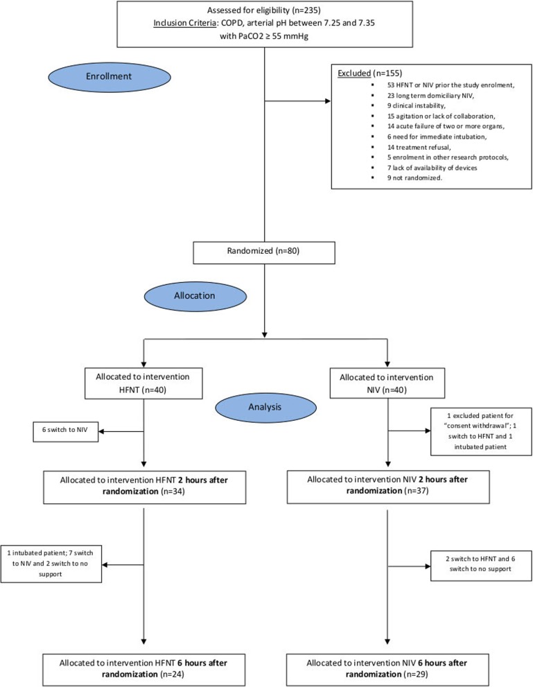
From 235 eligible patients, we randomized 80 patients, 40 in both groups (Fig. 1). The most frequent reason for exclusion that we registered was the prior use of NIV or HFNT before enrollment (53 out of 155 patients excluded). One patient withdrew consent after randomization; therefore, data from 79 patients was analyzed.
Fig. 1.
Flow diagram of the trial according to CONSORT
Baseline characteristics and blood gases of patients were evenly distributed between the two groups (Table 1). Mean age was 74 (± 13) and 77 (± 12), SAPS II was 30 (± 9.0), and 33 (± 10), the mean arterial pH was 7.30 (± 0.03) and 7.29 (± 0.03), and the mean PaCO2 was 73.7 mmHg (± 12.8) and 72.0 mmHg (± 13.0) in the HFNT and NIV group, respectively (Table 1).
Table 1.
Patients’ characteristics in the high flow nasal therapy (HFNT) and noninvasive ventilation (NIV) groups at baseline
| HFNT group | NIV group | p value | |
|---|---|---|---|
| N | 40 | 39 | |
| Females, n (%) | 19 (47.5) | 20 (51.3) | 0.7368 |
| Age (years), mean ± SD | 74 ± 13 | 77. ± 12 | 0.2273 |
| Weight (kg)a, mean ± SD | 85 ± 23 | 76 ± 13 | 0.0964 |
| Height (m)a, mean ± SD | 1.7 ± 0.1 | 1.7 ± 0.1 | 0.5494 |
| BMI (kg/m2)a, mean ± SD | 30.5 ± 8.7 | 26.7 ± 5.5 | 0.0622 |
| Ward of admission, n (%) | 0.5807 | ||
| Emergency room | 24 (60) | 21 (53.8) | |
| ICU or respiratory unit | 16 (40) | 18 (46.1) | |
| SAPS II, mean ± SD | 30 ± 9 | 33 ± 10 | 0.1497 |
| Charlson index, mean ± SD | 4 ± 2 | 5 ± 3 | 0.4709 |
| Systolic blood pressure (mmHg), mean ± SD | 131 ± 27 | 137 ± 24 | 0.3629 |
| Diastolic blood pressure (mmHg), mean ± SD | 71 ± 17 | 70 ± 13 | 0.7089 |
| Heart rate (per min), mean ± SD | 91 ± 20 | 92 ± 19 | 0.9609 |
| Respiratory rate (per min), mean ± SD | 27 ± 7 | 28 ± 7 | 0.5544 |
| Body temperatureb (C°), mean ± SD | 36.6 ± 0.7 | 36.8 ± 0.5 | 0.0489 |
| Kelly Matthay score, n (%) | 0.0694 | ||
| Alert, follows complex command (1) | 19 (47.5) | 25 (64.1) | |
| Alert, follows simple commands (2) | 9 (22.5) | 8 (20.5) | |
| Lethargie (3) | 12 (30) | 4 (10.3) | |
| Stuporous (4) | 0 (0) | 2 (5.1) | |
| Borg dyspnea scoreb, mean ± SD | 5 ± 2 | 5 ± 2 | 0.4463 |
| RASS, n (%) | 0.4813 | ||
| Light sedation (− 2) | 2 (5) | 2 (5.1) | |
| Drowsy (− 1) | 13 (32.5) | 7 (17.9) | |
| Alert and calm (0) | 21 (52.5) | 26 (66.7) | |
| Restless (+ 1) | 4 (10) | 4 (10.3) | |
| Secretion, n (%) | 0.2163 | ||
| Normal | 23 (57.5) | 17 (43.6) | |
| Abundant | 17 (42.5) | 22 (56.4) | |
| PaCO2 (mmHg), mean ± SD | 73.7 ± 12.8 | 72.0 ± 13.0 | 0.5270 |
| Arterial pH, mean ± SD | 7.30 ± 0.03 | 7.29 ± 0.03 | 0.7450 |
| PaO2 (mmHg), mean ± SD | 64.3 ± 17.6 | 73.3 ± 27.9 | 0.1480 |
| SpO2 (%), median [IQR] | 90.1 [87.0–94.1] | 92.0 [88.0–96.0] | 0.1835 |
| HCO3− (mmol L−1), mean ± SD | 34.3 ± 5.9 | 33.1 ± 6.30 | 0.3680 |
| PaO2/FiO2, mean ± SD | 203.2 ± 45.5 | 222.4 ± 71.0 | 0.4801 |
| Lactatec (mmol L−1), median [IQR] | 1.1 [0.7–1.6] | 1.1 [0.9–1.5] | 0.7376 |
BMI body mass index, FiO2 fraction of inspired oxygen, HCO3− bicarbonate, HFNT high-flow nasal therapy, ICU intensive care unit, IQR interquartile range (first and third quartile), NIV noninvasive ventilation, PaO2arterial partial pressure, PaCO2 partial pressure of carbon dioxide, RASS Richmond agitation-sedation scale, SAPS simplified acute physiology score, SD standard deviation
aData was not available for five patients (three in HFNT and two NIV group)
bData was not available for one patient in NIV group
cData was not available for one patient in HFNT group
All patients received the assigned treatment after randomization. By 2 h, six patients in the HFNT group had switched to NIV (n = 5 worsening/no improvement of respiratory failure; n = 1 intolerance to the intervention) while one patient switched to HFNT (intolerance to the intervention) and one to IMV in the NIV group (worsening of respiratory failure). By 6 h, seven patients had switched to NIV (worsening/no improvement of respiratory failure), one to IMV (worsening of respiratory failure) in the HFNT group. Due to the improvement of respiratory failure, one patient switched to no support in the HFNT group while two patients switched to HFNT and six to no support in the NIV group.
Seventy-one patients (HFNT n = 34 [85%] vs. NIV n = 37 [95%]; p = 0.2633) and 53 patients (HFNT n = 24 [60%] vs. NIV n = 29 [74.4%]; p = 0.1745) continued the allocated interventions after 2 and 6 h, respectively (Fig. 1, Table 2). Characteristics of interventions are reported in Additional file 2: Table S1. During the 6 h of treatment, three patients in the HFNT (7.5%) and in the NIV (7.7%) received sedatives according to the study protocol.
Table 2.
Clinical outcomes in the high flow nasal therapy (HFNT) and noninvasive ventilation (NIV) groups
| HFNT group | NIV group | p value | |
|---|---|---|---|
| N | 40 | 39 | |
| Treatment changes from baseline to 2 h, n (%) | |||
| Switching to NIV or HFNT | 6 (15) | 1 (2.6) | 0.1084 |
| IMV | 0 (0 | 1 (2.6) | 0.4937 |
| No treatment change | 34 (85) | 37 (94.9) | 0.2633 |
| Treatment changes from baseline to 6 h, n (%) | |||
| Switch to NIV or HFNT | 13 (32.5) | 3 (7.7) | 0.0061 |
| Switch to IMV | 1 (2.5) | 1 (2.6) | 1.0000 |
| Switch to no support | 2 (5) | 6 (15.4) | 0.1543 |
| No treatment change | 24 (60) | 29 (74.4) | 0.1745 |
| Poor treatment tolerance/intolerance from baseline to 6 hoursa, n (%) | |||
| In patients switching to NIV or HFNT or IMV | 5 (35.7) | 3 (75) | 0.2745 |
| In patients switching to no support or with no treatment change | 9 (34.6) | 26 (74.3) | 0.0019 |
| Discomfort, median [IQR] | |||
| At 2 ha | 1 [0–2] | 3 [1–5] | 0.0010 |
| At 6 hb | 0 [0–2] | 2 [1–4] | 0.0003 |
| Borg dyspnea score, mean ± SD | |||
| At 2 ha | 3 ± 2 | 3 ± 2 | 0.2509 |
| At 6 hb | 5 ± 2 | 5 ± 2 | 0.4865 |
| No improvement of Borg dyspnea score at 6 h, n (%) | 6 (15) | 6 (15.4) | 0.9620 |
| Respiratory rate (per min), mean ± SD | |||
| At 2 hb | 22 ± 5 | 22 ± 4 | 0.5789 |
| At 6 hc | 20 ± 4 | 21 ± 4 | 0.5573 |
| PaCO2 worsening or reduction < 10 mmHg after 6 h, n (%) | 23 (57.5) | 14 (35.9) | 0.0544 |
| IMV during hospitalizationd | |||
| Subjects, n (%) | 2 (5) | 1 (2.6) | 1.0000 |
| Length of IMV (hours), median [IQR] |
123 [45.5–200] |
166 [166–166] |
1.0000 |
| NIV during hospitalization | |||
| Subjects, n (%) | 23 (57.5) | 39 (100.0) | < .0001 |
| Length of NIV (hours), median [IQR] |
70 [14–142] |
48 [18–75] |
0.3007 |
| Length of hospital stay (days), median [IQR] | |||
| All patients | 10 [9–19] | 13 [9–16] | 0.6579 |
| Survivors at hospital discharge | 10 [9–19] | 13 [9–16] | 0.5510 |
| Dead at hospital discharge | 16 [9–22] | 15 [3–19] | 0.6150 |
| In-hospital mortality, n (%) | 2 (5) | 6 (15.4) | 0.1543 |
HFNT high flow nasal therapy, IQR interquartile range [first and third quartile], IMV invasive mechanical ventilation, NIV noninvasive ventilation
aPoor tolerance was defined as patient-reported complaint to the assigned treatment that did not cause treatment interruption. Intolerance was defined as patient-reported complaint that compromised the pursuit of the treatment (i.e., subject refusal)
bOutcome evaluated on patients still receiving the assigned treatment at 2 h (34 HFNT, 37 NIV)
cOutcome evaluated on patients still receiving the assigned treatment at 6 h (24 HFNT, 29 NIV)
dIMV during hospitalization was calculated from baseline from hospital discharge or death
Per-protocol analysis
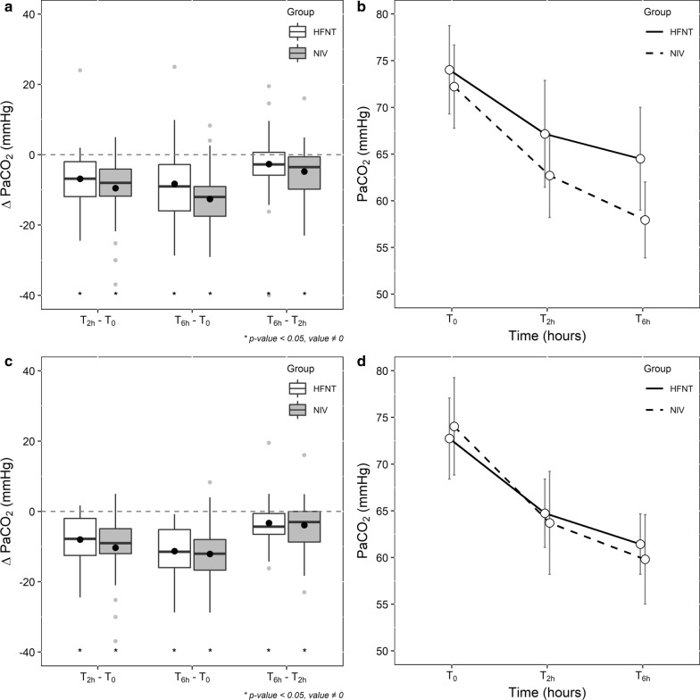
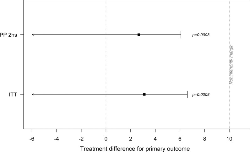
At baseline, clinical characteristics and blood gases were similar between the two groups in patients who completed the treatment initially allocated after 2 h (see Additional file 2: Table S2). Mean differences for PaCO2 from baseline to 2 h were − 6.8 mmHg (± 8.7) in the HFNT and − 9.5 mmHg (± 8.5) in the NIV group (p = 0.404) (Table 3). Figure 2a, b shows the differences and trends in PaCO2. Both treatments were able to significantly lower the PaCO2 over the study timepoints (Fig. 2a). As regards to the primary outcome, absolute PaCO2 difference was 2.7 mmHg (1-sided 95% CI − ∞; 6.1) and HFNT was non-inferior to NIV since the upper boundary of 95% CI did not reach the non-inferiority margin of 10 mmHg (p = 0.0003, Fig. 3). Both treatments had a significant effect on PaCO2 reduction over time (time effect, p < 0.0001), and trends were similar between groups (interaction term, p = 0.5864), (Fig. 2b).
Table 3.
Modifications in PaCO2 values during follow-up period in study population stratified by intervention (high flow nasal therapy and noninvasive ventilation)
| HFNT group | NIV group | p value | |
|---|---|---|---|
| Per-protocol 2 h | |||
| PaCO2 (mmHg) on patients who completed the treatment originally allocated at 2 h, mean ± SD | |||
| Subjects, n | 34 | 37 | – |
| At baseline ( T0) | 74.0 ± 13.5 | 72.2 ± 13.3 | 0.5845 |
| After 2 h ( T2h ) | 67.2 ± 16.4 | 62.7 ± 13.5 | 0.1933 |
| After 6 h ( T6h ) | 64.5 ± 15.8 | 57.9 ± 12.2 | 0.0630 |
| ΔT2h − T0 | − 6.8 ± 8.7 | − 9.5 ± 8.5 | 0.4040 |
| ΔT6h − T0 | − 9.5 ± 13.0 | − 14.3 ± 11.1 | 0.0962 |
| ΔT6h − T2h | − 2.7 ± 9.7 | − 4.8 ± 7.1 | 0.1637 |
| Per-protocol 6 h | |||
| PaCO2 (mmHg) on patients who completed the treatment originally allocated at 6 h, mean ± SD | |||
| Subjects, n | 24 | 29 | − |
| At baseline ( T0) | 72.7 ± 10.3 | 74.0 ± 13.7 | 0.7955 |
| After 2 h ( T2h ) | 64.7 ± 8.7 | 63.7 ± 14.5 | 0.7493 |
| After 6 h ( T6h ) | 61.4 ± 7.7 | 59.8 ± 12.6 | 0.5632 |
| ΔT2h − T0 | − 8.0 ± 6.5 | − 10.3 ± 8.9 | 0.5200 |
| ΔT6h − T0 | − 11.3 ± 7.3 | − 14.2 ± 12.0 | 0.4475 |
| ΔT6h − T2h | − 3.3 ± 6.9 | − 3.9 ± 7.6 | 0.8163 |
| Intention-to-treat analysis | |||
| PaCO2 (mmHg) of enrolled patients, mean ± SD | |||
| Subjects, n | 40 | 39 | |
| At baseline ( T0) | 73.7 ± 12.8 | 72.0 ± 13.0 | 0.5270 |
| After 2 h ( T2h ) | 68.2 ± 15.6 | 63.4 ± 13.6 | 0.1387 |
| After 6 h ( T6h ) | 64.0 ± 14.9 | 58.1 ± 12.4 | 0.0610 |
| ΔT2h − T0 | − 5.5 ± 9.3 | − 8.6 ± 9.3 | 0.2940 |
| ΔT6h − T0 | − 9.7 ± 13.2 | − 13.9 ± 11.3 | 0.1329 |
| ΔT6h − T2h | − 4.2 ± 11.1 | − 5.3 ± 7.5 | 0.3670 |
HFNT high flow nasal therapy, NIV noninvasive ventilation, PaCO2 partial pressure of carbon dioxide, SD standard deviation, Δ difference in PaCO2 values between timepoints
Fig. 2.
a, c Report boxplots showing median, interquartile range and mean (full dot) for PaCO2 differences (ΔPaCO2) between baseline (T0), 2 h (T2h) and 6 h (T6h) in HFNT and NIV groups (*p value < 0.05, difference ≠ 0) in per-protocol analysis at 2 h and at 6 h, respectively; b, d report mean PaCO2 (and 95% confidence interval) observed at T0, T2h, T6h in HFNT and NIV groups in per-protocol analysis at 2 h and at 6 h, respectively
Fig. 3.
Absolute difference between HFNT and NIV treatment in mean PaCO2 reduction after 2 h (and 1-sided 95% confidence interval), according to conducted analyses: per-protocol on patients who completed the treatment originally allocated after 2 h (PP 2 h) and intention-to-treat (ITT). The black box indicates the mean PaCO2 reduction after 2 h, full lines indicate 95% confidence interval and dashed line the pre-planned non-inferiority margin of 10 mmHg
Similar results were found in the per-protocol analysis at 6 h (see Fig. 2c, d; Table 3, Additional file 2: Table S3 and Figure S1). However, in this analysis, there was a lower baseline SAPS II score in the HFNT group.
Intention-to-treat analysis and secondary outcomes
In Additional file 2: Figure S2 (panel A and B) shows the differences and trends in PaCO2 for this analysis. Both interventions significantly lower PaCO2 over the study timepoints in both groups. Criteria for the non-inferiority of HFNT versus NIV were also met in this analysis at 2 and 6 h (Fig. 3, Additional file 2: Figure S1)
The other secondary outcomes, including dyspnea score, discomfort, length of mechanical ventilation, respiratory rate, the proportion of patients with worsening or no significant reduction in PaCO2 values after 6 h, length of hospital stay, and hospital mortality are reported in Table 2.
A higher proportion of patients in the NIV group showed poor tolerance to the intervention by 6 h (74%) compare to HFNT (35%) (p = 0.0019). The discomfort score was slightly higher in the NIV group at 2 and 6 h of treatment. For the other secondary outcomes, no differences were found between groups.
Changes in arterial blood gases and vital parameters over the study timepoints for the per-protocol analysis and intention-to-treat analysis were reported in Additional file 2: Tables S4–S6. We found no difference in mean change in arterial pH from baseline to 2 h (HFNT group: 0.04 ± 0.04, NIV group: 0.05 ± 0.03, p = 0.39) in the per-protocol analysis at 2 h. In the same analysis, we found a significant difference in mean change of PaO2/FiO2 ratio (HFNT group: −16.5 ± 52.2 mmHg, NIV group: 7.2 ± 56.3 mmHg, p = 0.0357) (see Additional file 2: Table S4).
Discussion
The main findings of this trial are that HFNT was non-inferior to NIV as initial ventilatory support in mean PaCO2 reduction in patients with mild-to-moderate AECOPD considering a non-inferiority margin of 10 mmHg; HFNT was able to reduce PaCO2 over both study timepoints significantly, but 32% (14/40) of patients were switched to NIV within 6 h.
To the best of our knowledge, this is the first multicenter randomized controlled non-inferiority trial comparing NIV and HFNT in mild-to-moderate AECOPD. In our study, we included patients with mild-to-moderate AECOPD for two reasons: (1) the efficacy and safety of HFNT in AECOPD have not been demonstrated so far [16]; (2) NIV is considered the gold standard respiratory support for managing patients with AECOPD with high-quality evidence supporting its use [3]. However, HFNT has shown several valuable effects in COPD patients [16] and there are some drawbacks of using NIV, such as reduced comfort and poor patient–ventilator interaction, which is often challenging to recognize and manage [4, 5, 35]. We intended to determine whether HFNT was not inferior to NIV in achieving relevant changes in physiologic outcomes in AECOPD patients (i.e., PaCO2) and explore its safety as the first step for future superiority trials.
Previously published studies had shown the promising results on the efficacy and safety of HFNT use for treating AECOPD. A retrospective study performed in a single ICU compared HFNT and NIV in 82 patients with mild-to-moderate AECOPD. No difference was found in the proportion of patients who switched treatment or received IMV (28% HFNT vs. 39% NIV). Of note, patients were assigned to study treatments if they received HFNT or NIV for at least 4 h within the first 24 h from admission [22]. In an observational study performed in one respiratory unit, no difference was found between HFNT and NIV with regard to the 30-day intubation rate (25% vs. 27%) and mortality rate (16% vs. 18%) in 92 patients with moderate AECOPD. The investigators also compared blood gases (including PaCO2) at 6- and 24-h, finding no difference between groups [21]. The results included between-group analysis at each timepoint, and no comparison of trends or differences were reported. A randomized study performed in one respiratory unit concluded that HFNT and NIV were both effective in improving blood gases in 168 patients with AECOPD and the HFNT group had a lower rate of complications and higher comfort. No sample size calculation and protocol registration have been reported and the outcomes assessment was done after 12 h and 5 days [36].
We showed that HFNT might be a feasible initial ventilator strategy in the management of mild-to-moderate AECOPD, finding a mean difference in PaCO2 reduction of 2.66 mmHg at 2 h, and the 95% CI upper boundary of absolute difference in mean PaCO2 reduction not reaching the set non-inferiority margin of 10 mmHg. Nevertheless, we acknowledge that a different non-inferiority margin and a larger sample size would have changed the study conclusions. From a clinical point of view, a clinician may prefer a technique allowing a greater and faster decarboxylation. In fact, 32% of patients treated with HFNT were switched to NIV, even if patients had a slightly lower severity of illness (SAPS II) than those in the NIV group (Table 1 and Additional file 2). Moreover, in our study cohort, patients randomized to HFNT worsened the oxygenation during the 6 h (Additional file 2: Table S4), and those who subsequently underwent NIV during hospitalization had a longer length of NIV than those originally allocated to the NIV group (Table 2). Altogether, these findings should incite caution in declaring that HFNT is non-inferior to NIV from a clinical perspective, all the most because NIV is a true standard of care in this context.
The external validity of our findings is supported by the multicenter design, but the results may have changed according to different expertise in the use of devices, settings and management protocols of AECOPD. Of note, all patients were continuously monitored with standard tools and serial blood gases check, so our findings cannot be extrapolated to settings with different levels of care. Larger clinical trials comparing HFNT and NIV in this patient population are currently ongoing, focusing on other outcomes such as endotracheal intubation (NCT03014869) and treatment failure (NCT03466385) or with crossover design (NCT03033251).
Strengths of the study were the multicenter randomized design, the prospective registration, and protocol publication and the analyses, both per-protocol and intention-to-treat, according to CONSORT recommendations for non-inferiority trials. However, our study has limitations. First, due to the nature of the interventions, blinding was not possible. This study was a non-inferiority trial with a primary physiologic outcome, leaving uncertainty on stronger patient-related outcomes. Moreover, due to the study design, the variables associated with the need for escalation of treatments cannot be adequately evaluated. Although we found lower baseline oxygenation in HFNT patients who escalated the treatment to NIV or IMV after 6 h compared to patients who did not switch (see Additional file 2: Table S7), further studies should assess predictors for HFTN success in this patient population. Our patient cohort was old and the mean BMI in the HFNT group was 30 kg/m2 (± 8.7). These factors may limit the external validity of our findings. Furthermore, we cannot exclude the co-presence of obesity-related hypoventilation. It is difficult to screen patients for this condition during the first management AECOPD, but we excluded patients on domiciliary NIV. We did not register the tidal volume generated or estimated by the ventilators in the NIV group. Thus, we cannot exclude that the applied level of pressure support might have affected patients’ comfort and NIV tolerance. Moreover, the PEEP values applied were slightly higher than the range stated in the protocol.
Our study had a parallel-group design with short-term timepoints of assessment, so we cannot evaluate the efficacy and safety of sequential use of the study treatments. The findings that 32% of patients after 6 h and more than 50% of patients during hospitalization needed NIV suggest that the two treatments may have a complementary role in AECOPD that should be further studied [19]. We collected data about the primary outcome, blood gases and secondary outcomes at 6 h as the longest follow-up without further assessments. This can limit the evaluation of the long-term efficacy of HFNT. However, we registered patients’ safety-related outcomes until hospital discharge or death.
At 6 h, the number of patients actually treated as randomly allocated was slightly lower (n = 53) than the calculated sample size (n = 56) for the primary outcome at 2 h. The power of our per-protocol analysis after 6 h was 76.5%, and therefore, the estimate of the compared effect at this timepoint should be considered with caution. Finally, it has to take into account also the documented clinical heterogeneity of COPD exacerbations [37]; therefore, although we used strict criteria to define mild-to-moderate AECOPD, we may have enrolled patients with different trajectories of the diseases and responses to ventilator support and medical treatments.
Conclusions
In this trial, HFNT was statistically non-inferior to NIV as initial ventilatory support in decreasing PaCO2 after 2 h of treatment in patients with mild-to-moderate AECOPD, considering a non-inferiority margin of 10 mmHg. However, 32% of patients receiving HFNT required NIV by 6 h. Further trials with superiority design should evaluate efficacy toward stronger patient-related outcomes and safety of HFNT in AECOPD.
Supplementary information
Additional file 1: Description of data: CONSORT (consolidated standards of reporting trials) Checklist for Non-inferiority and Equivalence Trials Checklist for Non-inferiority and Equivalence Trials.
Additional file 2: Table S1: Characteristics of interventions in the high flow nasal therapy (HFNT) and noninvasive ventilation group (NIV); Table S2: Per-protocol 2 h. Patients’ characteristics in the noninvasive ventilation (NIV) and high flow nasal therapy (HFNT) groups at baseline; Table S3: Per-protocol 6 h. Patients’ characteristics in the noninvasive ventilation (NIV) and high flow nasal therapy (HFNT) groups at baseline; Figure S1: Absolute difference between HFNT and NIV treatment in mean PaCO2 reduction after 6 h (and 1-Sided 95% confidence interval), according to conducted analyses: intention-to-treat (ITT) and per-protocol on patients who completed the treatment originally allocated after 6 h (PP 6hs). Figure S2: Changes in PaCO2 values during time (intention to treat analysis). Table S4: Per-protocol 2 h. Differences during follow-up in clinical characteristics in the noninvasive ventilation (NIV) and high flow nasal therapy (HFNT) groups; Table S5: Per-protocol 6 h. Differences during follow-up in clinical characteristics in the noninvasive ventilation (NIV) and high flow nasal therapy (HFNT) groups. Table S6: Intention-to-treat analysis. Differences during follow-up in clinical characteristics in the noninvasive ventilation (NIV) and high flow nasal therapy (HFNT) groups. Table S7: reports patients’ characteristics at baseline in the high flow nasal therapy (HFNT) group stratified by success by 6 h.
Acknowledgements
The list of HF-AECOPD study investigators is reported in the acknowledgment section. List of The HF-AECOPD study investigators: Lorenzo Ball (Anesthesia and Intensive Care, Ospedale Policlinico San Martino; Department of Surgical Sciences and Integrated Diagnostic, University of Genoa, Genoa, Italy); Tiziana Bove (Anesthesia and Intensive Care Clinic, Department of Anesthesia and Intensive Care, Azienda Sanitaria Integrata di Udine; Department of Medicine, University of Udine, Udine, Italy); Raffaele Campisi (Respiratory Medicine Unit, A.O.U. “Policlinico-Vittorio Emanuele”, Catania, Italy); Paola Chirco (Department of Surgical, Oncological and Oral Science University of Palermo, Italy); Maria Stella Dionisi (Emergency Department, “S. Maria della Misericordia” Hospital, Perugia, Italy); Mariachiara Ippolito (Department of Surgical, Oncological and Oral Science, University of Palermo, Italy); Riccardo Fantini (Respiratory Diseases Unit, University Hospital of Modena, Italy); Luca Guidelli (Pulmonology and Respiratory Intensive Care Unit, S. Donato Hospital, Arezzo, Italy); Uberto Maccari (Pulmonology and Respiratory Intensive Care Unit, S. Donato Hospital, Arezzo, Italy); Luca Tabbì (Respiratory Diseases Unit, University Hospital of Modena, Italy); Maria Rita Taliani (Emergency Department, “S. Maria della Misericordia” Hospital, Perugia, Italy).
Abbreviations
- ABGs
Arterial blood gases
- AECOPD
Acute exacerbation of chronic obstructive pulmonary disease
- CI
Confidence interval
- CONSORT
Consolidated standards of reporting trials
- COPD
Chronic obstructive pulmonary disease
- FiO2
Inspired oxygen fraction
- HFNT
High flow nasal therapy
- ICU
Intensive care unit
- NIV
Noninvasive ventilation
- PaCO2
Arterial partial pressure of carbon dioxide
- PEEP
Positive end-expiratory pressure
- RASS
Richmond Agitation Sedation Scale
- SAPS II
Simplified Acute Physiology Score
- SpO2
Peripheral oxygen saturation
Authors’ contributions
AC, FL, FM, PN, and CG had full access to all the data in the study and take responsibility for the integrity of the data and the accuracy of the data analysis, including and especially any adverse effects. AC, FM, CC, and CG wrote the manuscript; AC, FL, FM, PG, RS, CC, ACa, AB, EG, SMR, RT, VC, EL, LV, EC, AG, SN, PN, and CG contributed substantially to the study design, data analysis and interpretation, and the writing of the manuscript. All authors approved the final version of the manuscript and agreed to be accountable for all aspects of the work in ensuring that questions related to the accuracy or integrity of any part of the work are appropriately investigated and resolved. All authors read and approved the final manuscript.
Funding
No funding has been received for this trial. Some centers received material support (devices and cannula) by Fisher and Paykel that has no role in protocol design, data management, interpretation and analysis.
Availability of data and materials
The dataset consisting of de-identified participants' data is available from the corresponding author upon reasonable requests.
Ethics approval and consent to participate
The study protocol was approved by “Comitato Etico Sezione Area Centro” Ethics Committee (approval no. 245—October 24, 2017, Catanzaro, Italy) and by the local ethics committee of all the study centers.
Consent for publication
Written informed consent for publication was obtained from each participant enrolled or legal representatives.
Competing interests
Dr. Cortegiani, Prof. Giarratano and Prof. Gregoretti declare a patent pending, in association with the University of Palermo—Italy (No. 102019000020532—Italian Ministry of Economic Development), not discussed in the present study. Prof. Gregoretti received honoraria for lectures or consultancies from Philips, Resmed, Vivisol, OrionPharma, Origin (not relevant to this protocol). Prof. Enrico Clini received fees for lectures from Chiesi, Astra-Zeneca, Menarini, Guidotti_Malesci, and AlfaSigma (not relevant to this protocol). Prof. Stefano Nava’s institution received unrestricted research grant from Fisher and Paykel. Prof. Paolo Navalesi’s institution received funding from Maquet Critical Care, Draeger, and Intersurgical S.p.A.; he received honoraria/speaking fees from Maquet Critical Care, Orionpharma, Philips, Resmed, Merck Sharp & Dome, and Novartis; he disclosed that he contributed to the development of the helmet Next, whose license for patent belongs to Intersurgical S.P.A., and receives royalties for that invention. Prof. Federico Longhini and Prof. Paolo Navalesi contributed to the development of a device not discussed in the present study whose patent is in progress (European Patent application number EP20170199831). Dr. Paolo Groff received honoraria for lectures from Aspen Pharmaceuticals and Menarini Pharmaceuticals (not relevant to this study). Dr. Luigi Vetrugno received travel support for congress lecture by Cook Medical. The remaining authors have disclosed that they do not have any conflicts of interest.
Footnotes
Publisher's Note
Springer Nature remains neutral with regard to jurisdictional claims in published maps and institutional affiliations.
Contributor Information
Andrea Cortegiani, Email: andrea.cortegiani@unipa.it.
Federico Longhini, Email: longhini.federico@gmail.com.
Fabiana Madotto, Email: fabiana.madotto@unimib.it.
Paolo Groff, Email: groff.paolo64@gmail.com.
Raffaele Scala, Email: raffaele_scala@hotmail.com.
Claudia Crimi, Email: dott.claudiacrimi@gmail.com.
Annalisa Carlucci, Email: annalisa.carlucci@icsmaugeri.it.
Andrea Bruni, Email: andreabruni87@gmail.com.
Eugenio Garofalo, Email: eugenio.garofalo@gmail.com.
Santi Maurizio Raineri, Email: maurizio.raineri@unipa.it.
Roberto Tonelli, Email: roberto.tonelli@me.com.
Vittoria Comellini, Email: vittoria.comellini@gmail.com.
Enrico Lupia, Email: enrico.lupia@unito.it.
Luigi Vetrugno, Email: luigi.vetrugno@asuiud.sanita.fvg.it.
Enrico Clini, Email: enrico.clini@unimore.it.
Antonino Giarratano, Email: antonino.giarratano@unipa.it.
Stefano Nava, Email: stefanava@gmail.com.
Paolo Navalesi, Email: pnavalesi@gmail.com.
Cesare Gregoretti, Email: cesare.gregoretti@unipa.it.
the H. F.-AECOPD study investigators:
Lorenzo Ball, Tiziana Bove, Raffaele Campisi, Paola Chirco, Maria Stella Dionisi, Mariachiara Ippolito, Riccardo Fantini, Luca Guidelli, Uberto Maccari, Luca Tabbì, and Maria Rita Taliani
Supplementary information
Supplementary information accompanies this paper at 10.1186/s13054-020-03409-0.
References
- 1.Singh D, Agusti A, Anzueto A, Barnes PJ, Bourbeau J, Celli BR, et al. Global strategy for the diagnosis, management, and prevention of chronic obstructive lung disease: the GOLD science committee report 2019. Eur Respir J Engl. 2019;2019:53. doi: 10.1183/13993003.00164-2019. [DOI] [PubMed] [Google Scholar]
- 2.Celli BR, Barnes PJ. Exacerbations of chronic obstructive pulmonary disease. Eur Respir J Engl. 2007;29:1224–1238. doi: 10.1183/09031936.00109906. [DOI] [PubMed] [Google Scholar]
- 3.Rochwerg B, Brochard L, Elliott MW, Hess D, Hill NS, Nava S, et al. Official ERS/ATS clinical practice guidelines: noninvasive ventilation for acute respiratory failure. Eur Respir J. 2017;50:1602426. doi: 10.1183/13993003.02426-2016. [DOI] [PubMed] [Google Scholar]
- 4.Gregoretti C, Pisani L, Cortegiani A, Ranieri VM. Noninvasive ventilation in critically ill patients. Crit Care Clin US. 2015;31:435–457. doi: 10.1016/j.ccc.2015.03.002. [DOI] [PubMed] [Google Scholar]
- 5.Bruni A, Garofalo E, Pelaia C, Messina A, Cammarota G, Murabito P, et al. Patient-ventilator asynchrony in adult critically ill patients. Minerva Anestesiol Italy. 2019;85:676–688. doi: 10.23736/S0375-9393.19.13436-0. [DOI] [PubMed] [Google Scholar]
- 6.Cortegiani A, Russotto V, Antonelli M, Azoulay E, Carlucci A, Conti G, et al. Ten important articles on noninvasive ventilation in critically ill patients and insights for the future: a report of expert opinions. BMC Anesthesiol. 2017;17:122. doi: 10.1186/s12871-017-0409-0. [DOI] [PMC free article] [PubMed] [Google Scholar]
- 7.Garofalo E, Bruni A, Pelaia C, Liparota L, Lombardo N, Longhini F, et al. Recognizing, quantifying and managing patient-ventilator asynchrony in invasive and noninvasive ventilation. Expert Rev Respir Med Engl. 2018;12:557–567. doi: 10.1080/17476348.2018.1480941. [DOI] [PubMed] [Google Scholar]
- 8.Spoletini G, Cortegiani A, Gregoretti C. Physiopathological rationale of using high-flow nasal therapy in the acute and chronic setting: a narrative review. Trends Anaesth Crit Care. 2019;26–27:22–29. doi: 10.1016/j.tacc.2019.02.001. [DOI] [Google Scholar]
- 9.Groves N, Tobin A. High flow nasal oxygen generates positive airway pressure in adult volunteers. Aust Crit Care. 2007;20:126–131. doi: 10.1016/j.aucc.2007.08.001. [DOI] [PubMed] [Google Scholar]
- 10.Ricard JD, Roca O, Lemiale V, Corley A, Braunlich J, Jones P, et al. Use of nasal high flow oxygen during acute respiratory failure. Intensive Care Med. 2020 doi: 10.1007/s00134-020-06228-7. [DOI] [PMC free article] [PubMed] [Google Scholar]
- 11.Fontanari P, Zattara-Hartmann MC, Burnet H, Jammes Y. Nasal eupnoeic inhalation of cold, dry air increases airway resistance in asthmatic patients. Eur Respir J. 1997;10:2250–2254. doi: 10.1183/09031936.97.10102250. [DOI] [PubMed] [Google Scholar]
- 12.On LS, Boonyongsunchai P, Webb S, Davies L, Calverley PMA, Costello RW. Function of pulmonary neuronal M2 muscarinic receptors in stable chronic obstructive pulmonary disease. Am J Respir Crit Care Med. 2001;163:1320–1325. doi: 10.1164/ajrccm.163.6.2002129. [DOI] [PubMed] [Google Scholar]
- 13.Kilgour E, Rankin N, Ryan S, Pack R. Mucociliary function deteriorates in the clinical range of inspired air temperature and humidity. Intensive Care Med. 2004;30:1491–1494. doi: 10.1007/s00134-004-2235-3. [DOI] [PubMed] [Google Scholar]
- 14.Crimi C, Noto A, Cortegiani A, Campisi R, Heffler E, Gregoretti C, et al. High flow nasal therapy use in patients with acute exacerbation of COPD and bronchiectasis: a feasibility study. COPD J Chronic Obstr Pulm Dis. 2020;17:184–190. doi: 10.1080/15412555.2020.1728736. [DOI] [PubMed] [Google Scholar]
- 15.Pisani L, Fasano L, Corcione N, Comellini V, Musti MA, Brandao M, et al. Change in pulmonary mechanics and the effect on breathing pattern of high flow oxygen therapy in stable hypercapnic COPD. Thorax. 2017;72:373–375. doi: 10.1136/thoraxjnl-2016-209673. [DOI] [PubMed] [Google Scholar]
- 16.Pisani L, Astuto M, Prediletto I, Longhini F. High flow through nasal cannula in exacerbated COPD patients: a systematic review. Pulmonology. 2019;25:348–354. doi: 10.1016/j.pulmoe.2019.08.001. [DOI] [PubMed] [Google Scholar]
- 17.Pilcher J, Eastlake L, Richards M, Power S, Cripps T, Bibby S, et al. Physiological effects of titrated oxygen via nasal high-flow cannulae in COPD exacerbations: a randomized controlled cross-over trial. Respirology. 2017;22:1149–1155. doi: 10.1111/resp.13050. [DOI] [PubMed] [Google Scholar]
- 18.Bräunlich J, Wirtz H. Nasal high-flow in acute hypercapnic exacerbation of COPD. Int J COPD. 2018;13:3995–3897. doi: 10.2147/COPD.S185001. [DOI] [PMC free article] [PubMed] [Google Scholar]
- 19.Longhini F, Pisani L, Lungu R, Comellini V, Bruni A, Garofalo E, et al. High-flow oxygen therapy after noninvasive ventilation interruption in patients recovering from hypercapnic acute respiratory failure: a physiological crossover trial. Crit Care Med. 2019;47:e506–e511. doi: 10.1097/CCM.0000000000003740. [DOI] [PubMed] [Google Scholar]
- 20.Rittayamai N, Phuangchoei P, Tscheikuna J, Praphruetkit N, Brochard L. Effects of high-flow nasal cannula and non-invasive ventilation on inspiratory effort in hypercapnic patients with chronic obstructive pulmonary disease: a preliminary study. Ann Intensive Care. 2019;9:122. doi: 10.1186/s13613-019-0597-5. [DOI] [PMC free article] [PubMed] [Google Scholar]
- 21.Lee MK, Choi J, Park B, Kim B, Lee SJ, Kim SH, et al. High flow nasal cannulae oxygen therapy in acute-moderate hypercapnic respiratory failure. Clin Respir J. 2018;12:2046–2056. doi: 10.1111/crj.12772. [DOI] [PubMed] [Google Scholar]
- 22.Sun J, Li Y, Ling B, Zhu Q, Hu Y, Tan D, et al. High flow nasal cannula oxygen therapy versus non-invasive ventilation for chronic obstructive pulmonary disease with acute-moderate hypercapnic respiratory failure: an observational cohort study. Int J COPD. 2019;14:1229–1239. doi: 10.2147/COPD.S206567. [DOI] [PMC free article] [PubMed] [Google Scholar]
- 23.Pantazopoulos I, Daniil Z, Moylan M, Gourgoulianis K, Chalkias A, Zakynthinos S, et al. Nasal high flow use in COPD patients with hypercapnic respiratory failure: treatment algorithm & review of the literature. COPD J Chronic Obstr Pulm Dis. 2020;7:101–111. doi: 10.1080/15412555.2020.1715361. [DOI] [PubMed] [Google Scholar]
- 24.Cortegiani A, Longhini F, Carlucci A, Scala R, Groff P, Bruni A, et al. High-flow nasal therapy versus noninvasive ventilation in COPD patients with mild-to-moderate hypercapnic acute respiratory failure: study protocol for a noninferiority randomized clinical trial. Trials. 2019;20:450. doi: 10.1186/s13063-019-3514-1. [DOI] [PMC free article] [PubMed] [Google Scholar]
- 25.Vogelmeier CF, Criner GJ, Martinez FJ, Anzueto A, Barnes PJ, Bourbeau J, et al. Global strategy for the diagnosis, management, and prevention of chronic obstructive lung disease 2017 report. Am J Respir Crit Care Med. 2017;195:557–582. doi: 10.1164/rccm.201701-0218PP. [DOI] [PubMed] [Google Scholar]
- 26.Pisani L, Mega C, Vaschetto R, Bellone A, Scala R, Cosentini R, et al. Oronasal mask versus helmet in acute hypercapnic respiratory failure. Eur Respir J. 2015;45:691–699. doi: 10.1183/09031936.00053814. [DOI] [PubMed] [Google Scholar]
- 27.Kelly BJ, Matthay MA. Prevalence and severity of neurologic dysfunction in critically ill patients: Influence on need for continued mechanical ventilation. Chest. 1993;104:1818–1824. doi: 10.1378/chest.104.6.1818. [DOI] [PubMed] [Google Scholar]
- 28.Quan H, Li B, Couris CM, Fushimi K, Graham P, Hider P, et al. Updating and validating the charlson comorbidity index and score for risk adjustment in hospital discharge abstracts using data from 6 countries. Am J Epidemiol. 2011;173:676–682. doi: 10.1093/aje/kwq433. [DOI] [PubMed] [Google Scholar]
- 29.Sessler CN, Gosnell MS, Grap MJ, Brophy GM, O’Neal PV, Keane KA, et al. The Richmond Agitation-Sedation Scale: validity and reliability in adult intensive care unit patients. Am J Respir Crit Care Med. 2002;166:1338–1344. doi: 10.1164/rccm.2107138. [DOI] [PubMed] [Google Scholar]
- 30.Kendrick KR, Baxi SC, Smith RM. Usefulness of the modified 0–10 Borg scale in assessing the degree of dyspnea in patients with COPD and asthma. J Emerg Nurs. 2000;26:216–222. doi: 10.1016/S0099-1767(00)90093-X. [DOI] [PubMed] [Google Scholar]
- 31.Maggiore SM, Idone FA, Vaschetto R, Festa R, Cataldo A, Antonicelli F, et al. Nasal high-flow versus venturi mask oxygen therapy after extubation: effects on oxygenation, comfort, and clinical outcome. Am J Respir Crit Care Med. 2014;190:282–288. doi: 10.1164/rccm.201402-0364OC. [DOI] [PubMed] [Google Scholar]
- 32.Bräunlich J, Wirtz H. NHF and hypercapnia: how brief can you look? Respirology. 2017;22:1049–50. doi: 10.1111/resp.13092. [DOI] [PubMed] [Google Scholar]
- 33.Brochard L, Mancebo J, Wysocki M, Lofaso F, Conti G, Rauss A, et al. Noninvasive ventilation for acute exacerbations of chronic obstructive pulmonary disease. N Engl J Med. 1995;333:817–22. doi: 10.1056/NEJM199509283331301. [DOI] [PubMed] [Google Scholar]
- 34.Piaggio G, Elbourne DR, Pocock SJ, Evans SJW, Altman DG. Reporting of noninferiority and equivalence randomized trials: extension of the CONSORT 2010 statement. JAMA - J Am Med Assoc. 2012;308:2594–2604. doi: 10.1001/jama.2012.87802. [DOI] [PubMed] [Google Scholar]
- 35.Longhini F, Liu L, Pan C, Xie J, Cammarota G, Bruni A, et al. Neurally-adjusted ventilatory assist for noninvasive ventilation via a helmet in subjects with COPD exacerbation: a physiologic study. Respir Care US. 2019;64:582–589. doi: 10.4187/respcare.06502. [DOI] [PubMed] [Google Scholar]
- 36.Cong L, Zhou L, Liu H, Wang J. Outcomes of high-flow nasal cannula versus non-invasive positive pressure ventilation for patients with acute exacerbations of chronic obstructive pulmonary disease. Int J Clin Exp Med. 2019;12:10863–10867. [Google Scholar]
- 37.Lopez-Campos JL, Agustí A. Heterogeneity of chronic obstructive pulmonary disease exacerbations: a two-axes classification proposal. Lancet Respir Med Engl. 2015;3:729–734. doi: 10.1016/S2213-2600(15)00242-8. [DOI] [PubMed] [Google Scholar]
Associated Data
This section collects any data citations, data availability statements, or supplementary materials included in this article.
Supplementary Materials
Additional file 1: Description of data: CONSORT (consolidated standards of reporting trials) Checklist for Non-inferiority and Equivalence Trials Checklist for Non-inferiority and Equivalence Trials.
Additional file 2: Table S1: Characteristics of interventions in the high flow nasal therapy (HFNT) and noninvasive ventilation group (NIV); Table S2: Per-protocol 2 h. Patients’ characteristics in the noninvasive ventilation (NIV) and high flow nasal therapy (HFNT) groups at baseline; Table S3: Per-protocol 6 h. Patients’ characteristics in the noninvasive ventilation (NIV) and high flow nasal therapy (HFNT) groups at baseline; Figure S1: Absolute difference between HFNT and NIV treatment in mean PaCO2 reduction after 6 h (and 1-Sided 95% confidence interval), according to conducted analyses: intention-to-treat (ITT) and per-protocol on patients who completed the treatment originally allocated after 6 h (PP 6hs). Figure S2: Changes in PaCO2 values during time (intention to treat analysis). Table S4: Per-protocol 2 h. Differences during follow-up in clinical characteristics in the noninvasive ventilation (NIV) and high flow nasal therapy (HFNT) groups; Table S5: Per-protocol 6 h. Differences during follow-up in clinical characteristics in the noninvasive ventilation (NIV) and high flow nasal therapy (HFNT) groups. Table S6: Intention-to-treat analysis. Differences during follow-up in clinical characteristics in the noninvasive ventilation (NIV) and high flow nasal therapy (HFNT) groups. Table S7: reports patients’ characteristics at baseline in the high flow nasal therapy (HFNT) group stratified by success by 6 h.
Data Availability Statement
The dataset consisting of de-identified participants' data is available from the corresponding author upon reasonable requests.


