. 2020 Dec 8:203496. doi: 10.1148/radiol.2020203496

Figure 4b:

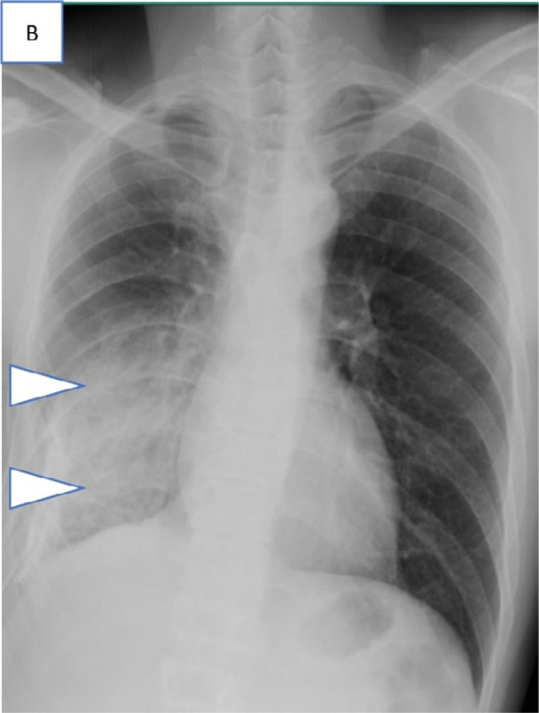
Series of chest radiograph in patients with COVID-19 pneumonia who were clinically asymptomatic and transferred out of the community care facility, Singapore Expo, to acute hospitals in view of findings of pulmonary infection on chest radiographs. Admission chest radiographs, A, in 36-year-old man on day 6 of illness showing bilateral lower zone patchy GGOs suspicious for infective foci (arrowheads), B, in 28-year-old man on day 2 of illness showing extensive consolidation (arrowheads) in the right mid and lower zone, and, C, in 55-year-old man on day 24 of illness with consolidation in the right upper zone (arrowhead).

Series of chest radiograph in patients with COVID-19 pneumonia who were clinically asymptomatic and transferred out of the community care facility, Singapore Expo, to acute hospitals in view of findings of pulmonary infection on chest radiographs. Admission chest radiographs, A, in 36-year-old man on day 6 of illness showing bilateral lower zone patchy GGOs suspicious for infective foci (arrowheads), B, in 28-year-old man on day 2 of illness showing extensive consolidation (arrowheads) in the right mid and lower zone, and, C, in 55-year-old man on day 24 of illness with consolidation in the right upper zone (arrowhead).