PLOS ONE logoLink to PLOS ONE
. 2020 Dec 14;15(12):e0243033. doi: 10.1371/journal.pone.0243033

Effect of intentional restriction of venous return on tissue oxygenation in a porcine model of acute limb ischemia

Wonho Kim 1,2,*,#, Donghoon Choi 3, Yangsoo Jang 3, Chung Mo Nam 4, Seung-Ho Hur 5, Myeong-Ki Hong 3,*,#
Editor: Yoshihiro Fukumoto6
PMCID: PMC7735909  PMID: 33318709

Abstract

Introduction

A sufficient oxygen supply to ischemic limb tissue is the most important requirement for wound healing and limb salvage. We investigated whether partial venous occlusion in the common iliac vein (CIV) causes a further increase of venous oxygenation in a porcine model of acute hindlimb ischemia.

Materials and methods

In 7 pigs, the model of acute hindlimb ischemia was created with intra-vascular embolization of the common iliac artery (CIA). The arterial and venous oxygen saturation was evaluated at different moments. Oxygen saturation was evaluated at baseline (T0), just after the arterial embolization (T1), at 10 minutes (T2), at 20 minutes (T3), and at 40 minutes (T4). Next, an intentional partial venous occlusion was achieved by inflating the vascular balloon at the level of the right CIV. Then, blood sampling was repeated at 5 minutes (T5), at 15 minutes (T6), and at 25 minutes (T7).

Results

The arterial oxygen saturation in the right SFA was similar during all phases. In contrast, after arterial embolization, an immediate reduction of venous oxygen saturation was observed (from 85.57 ± 1.72 at T0 to 71.86 ± 7.58 at T4). After the partial venous occlusion, interestingly, the venous oxygen saturations (T5-T7) were significantly increased, again. The venous oxygen saturations evaluated in the hindlimb ischemia with partial venous occlusion and in the control limb (without partial venous occlusion) were significantly over time. Venous oxygen saturations in the experimental limbs were higher than those in the control limbs (79.28 ± 4.82 vs 59.00 ± 2.82, p-value <0.001, 79.71 ± 4.78 vs 60.00 ± 4.24 at T7, p-value <0.001).

Conclusions

Partial venous occlusion results in an increase of venous oxygen saturation in the ischemic limb, while significant changes in venous oxygen saturation are not observed in the control limb. An explanation for this may be that the oxygen consumption in the limb tissue is increased because it gets congested with the partial venous occlusion in the right CIV.

Introduction

Peripheral artery disease (PAD) is a cause of significant morbidity, mortality, and disability due to limb loss [1]. In particular, acute limb ischemia (ALI) occurs when there is a sudden decrease in limb perfusion that threatens the viability of the limb and requires urgent treatment to prevent loss of limb [2]. A lack of oxygen due to an interruption of the blood supply to an acutely occluded limb causes the accumulation of various metabolites, production of reactive oxygen species (ROS) and an inflammatory reaction in conjugation with tissue swelling, threatening the possibility of limb loss and even death [3]. Subsequently, a sufficient oxygen supply to the ischemic limb tissue is the most important requirement for the promotion of wound healing and limb salvage. For limb salvage and restoring ambulatory function, therapies targeting ALI are directed primarily towards mechanical revascularization with an endovascular or surgical approach because essentially no effective medical therapies exist. Unfortunately, revascularization is often of limited benefit, and the clinical outcomes of the revascularization are not always satisfactory [2,4].

Interestingly, previous investigators showed that an intentional narrowing of the coronary sinus lumen elevates the coronary venous pressure, leading to an increase in capillary and arteriolar dilatation, a lower resistance to blood flow, and the restoration of the normal endocardial/epicardial blood flow ratio in patients with refractory angina [5,6]. As a consequence of the enhanced blood flow to the ischemic myocardium, this process increases the oxygen supply and its metabolism in ischemic myocardium.

In this study, a porcine model of acute hindlimb ischemia was created by intra-vascular embolization of the common iliac artery (CIA). By using models, we investigated if partial venous occlusion in the common iliac vein (CIV) might cause a further increase of venous oxygenation.

Materials and methods

This study was approved by the Yonsei University Institutional Animal Care and Use Committee. Sexually mature female swine (N = 8, 6 to 8 months of age) were obtained from a local vendor. The animals used in this study were miniature pigs originated from Korean Jeju island native (Cronex Co., Ltd., Osong, Republic of Korea). All the animals received humane care in compliance with the Animal Welfare Act and “The Guide for the Care and Use of Laboratory Animals” formulated by the Institute of Laboratory Animal Research [7]. The animals were housed individually following standard laboratory conditions (temperature: 19–25 ˚C; humidity: 30–70%; ventilation: 10–15 per hour; light: 150–300 Lux; light cycle: twice per day (8 AM- 8PM); nose: 45 dB) and fed a standard laboratory pellet diet and water ad libitum. A veterinarian dedicated to our laboratory supervised and helped with the study. At the end of this study, animals were euthanized, according to the American Veterinary Medical Association guidelines for the euthanasia of animals. Briefly, under deep anesthesia, the animals received bolus injections of potassium chloride.

Procedures

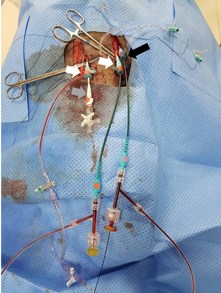
Anesthesia was induced by using intra-muscular injections of ketamine (20 mg/kg) and xylazine (2 mg/kg). After adequate systemic anesthesia was attained, the animals were placed in the supine position under mechanical ventilation, and isoflurane (1–2%) was delivered with a precision vaporizer and a circle absorption breathing system with periodic arterial blood gas monitoring. Cardiovascular monitoring was used during the whole procedure to control the heart rate and blood pressure. After surgical exposure of both carotid arteries and one carotid vein under sterile conditions, two 8 Fr arterial sheathes and one 9 Fr vein sheath were inserted (Fig 1). Intra-venous heparin (150 units/kg) was injected to maintain an activated clotting time of ≥ 250 seconds. First, a 0.014-inch hydrophilic guidewire was carefully inserted into the left carotid artery and navigated down to the right CIA. An 8 Fr multi-purpose guiding catheter was then advanced over the guidewire into the right CIA. A micro-catheter through the right carotid artery was placed in the right superficial femoral artery (SFA). The role of micro-catheter is to obtain arterial blood sampling in the right SFA when the right CIA is embolized to induce hindlimb ischemia. Second, a 0.014-inch hydrophilic guidewire through the left carotid vein was advanced in the inferior vena cava (IVC) and selectively, down into the right CIV. A 9 Fr multi-purpose guiding catheter was moved down over the guidewire and positioned in the CIV.

Fig 1. Preparation of an animal model of ALI.

Fig 1

After surgical exposure of both carotid arteries (white arrows) and one carotid vein (black arrow) under sterile conditions, two 8 Fr arterial sheaths (white arrows) and one 9 Fr vein sheath (black arrow) were inserted. Note that a micro-catheter (grey arrow) is inserted in the right carotid artery, an 8 Fr multi-purpose guiding catheter in the left carotid artery, and a 9 Fr multi-purpose guiding catheter in the left carotid vein. Abbreviations: ALI, acute limb ischemia.

Lower limb ischemia model & blood sampling

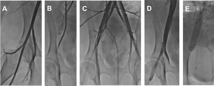
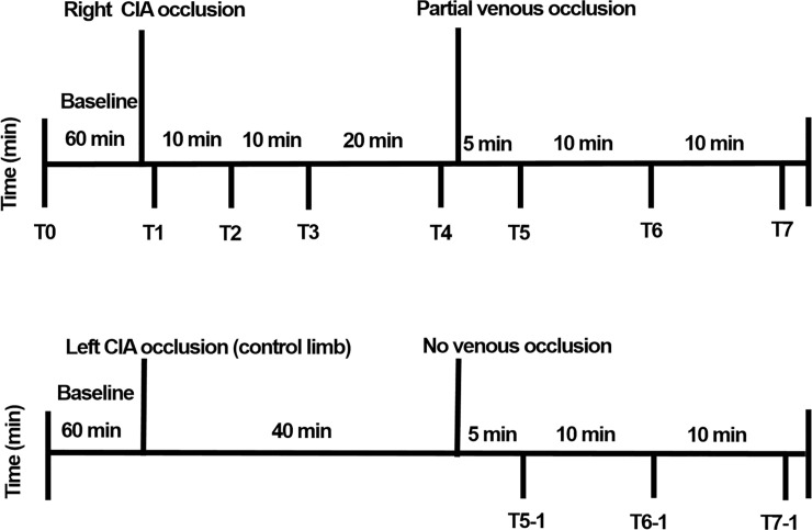
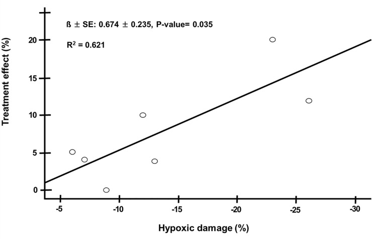
Baseline angiography was performed in the right leg (Fig 2). Embolization to induce hindlimb ischemia in the right CIA was performed using an Amplatzer Vascular Plug (AVP; St/ Jude Medical, St. Paul, MN), which acts as an embolic agent by promoting clot formation. To increase the thrombogenicity of the AVP, it was oversized by 30–50% of the estimated diameter of the right CIA. The occlusion time is highly variable depending on the caliber of the vessel, the coagulation status, the flow dynamics, and the size of the AVP. Therefore, repeated angiography was frequently performed to see if complete stasis of the injected contrast was noted. Blood sampling (Fig 3) for evaluating oxygen saturation was obtained from the right SFA and CIV at baseline (T0), just after the arterial occlusion with embolization (T1), at 10 minutes (T2), at 20 minutes (T3), and at 40 minutes (T4). Next, an intentional partial (90%) venous occlusion was achieved by inflating the vascular balloon at the level of the right CIV. Subsequently, blood sampling was repeated 5 minutes (T5) after the partial venous occlusion, at 15 minutes (T6), and at 25 minutes (T7). The left leg in two pigs served as a control limb; the left CIA was embolized while the left CIV was not occluded. In those legs, blood sampling was obtained 40 minutes after the left CIA occlusion in the left CIV with the same method as above (T5-1, T6-1, and T7-1). Blood gas analysis was performed to measure oxygen saturation, carbon dioxide, bicarbonate, and acid-base balance (pH). A proper blood sample for blood gas analysis consists of a 2 to 3 ml blood specimen collected anaerobically in a 3 ml plastic syringe fitted with a small-bore needle. Heparin was added to the syringe as an anticoagulant. Any air bubbles inadvertently introduced during sampling were promptly evacuated. Hypoxic damage by arterial embolization was defined as T4 minus T0 in vein saturation, and treatment effect with partial venous occlusion was defined as T7 minus T4 in vein saturation, respectively. Mean venous pressure was measured during the study period. All the mean venous pressure values were calculated over a stable period.

Fig 2. Representative images of the porcine model of ALI.

Fig 2

The pigs underwent intravascular induction of ALI. Baseline angiography (A), Embolization of the CIA. ALI was generated by the deployment of an Amplatzer Vascular Plug II in the CIA, followed by an angiography to verify occlusion (B), representative angiography after CIA occlusion (C), Baseline venography (D), Venous blood flow restriction with partial balloon inflation (E). Abbreviations: ALI, acute limb ischemia; CIA, common iliac artery.

Fig 3. Experimental study protocol and blood sampling for evaluating oxygen saturation (right SFA and the right CIV).

Fig 3

Blood sampling for evaluating oxygen saturation was obtained from the right SFA and the right CIV at baseline (T0), just after the arterial occlusion with embolization (T1), at 10 minutes (T2), at 20 minutes (T3), and at 40 minutes (T4). Next, an intentional partial (90%) venous occlusion was achieved by inflating the vascular balloon at the level of the right CIV. Subsequently, blood sampling was repeated at 5 minutes (T5) after the partial venous occlusion, at 15 minutes (T6), and at 25 minutes (T7). The left leg in two pigs served as a control limb; the left CIA was embolized while the left CIV was not occluded. In those legs, blood sampling was obtained 40 minutes after the left CIA occlusion in the left CIV with the same method as above (T5-1, T6-1, and T7-1). Abbreviations: CIA, common iliac artery; SFA, superficial femoral artery; CIV, common iliac vein.

Statistical analysis

Dichotomous data were reported as a number and percentage. Continuous data were reported as the mean and the standard deviation. Student t test was used to analyze continuous data and a chi-square test for dichotomous data. Arterial and venous oxygen saturations at different momentums were submitted to a repeated measures analysis of variance (ANOVA). All tests were two-sided, and statistical significance was defined as a p-value of less than 0.05. The analyses were performed using the SPSS version 20 software (SPSS Inc., Chicago, Illinois).

Results

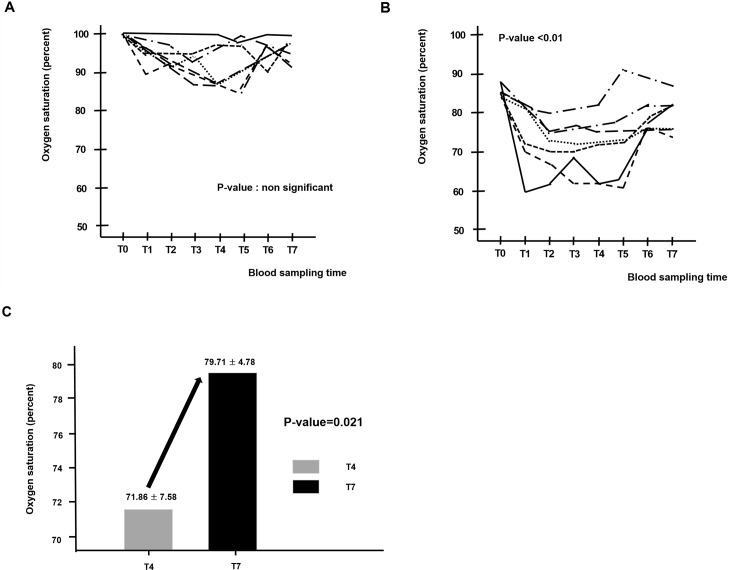
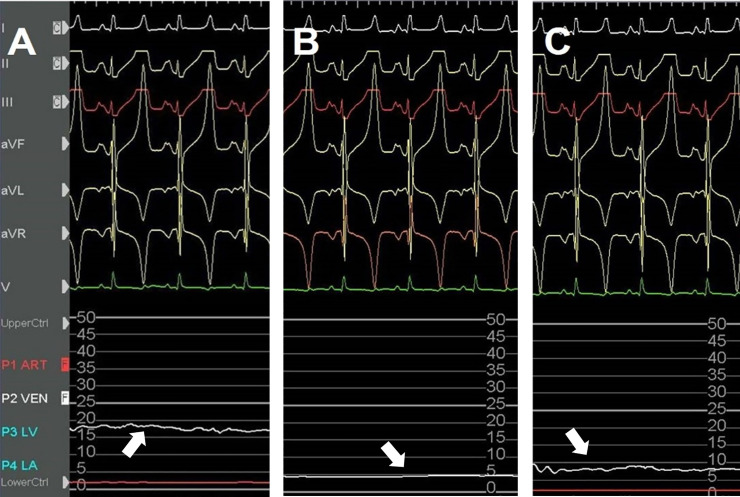
The model of acute hindlimb ischemia was successfully created in all 8 pigs. One pig died of sudden cardiac death due to iatrogenic catheter induced ventricular fibrillation. The seven surviving pigs developed acute hindlimb ischemia. The arterial oxygen saturation in the right SFA was similar during all phases. There were no significant differences between the values observed at T1-T4 and T5-T7 (Fig 4A and Table 1). In contrast, after arterial embolization, an immediate reduction of venous oxygen saturation was observed (from 85.57 ± 1.72 at T0 to 71.86 ± 7.58 at T4). After partial venous occlusion, interestingly, the venous oxygen saturations (T5-T7) were significantly increased, again. Repeated measures ANOVA show a significant difference after the partial venous occlusion concerning venous oxygen saturation (p-value < 0.001) (Fig 4B and Table 1). The venous oxygen saturations evaluated in the hindlimb ischemia with partial venous occlusion and in the control limb (without partial venous occlusion) were significant over time. Venous oxygen saturations in the experimental limb were higher than those in the control limb (79.28 ± 4.82 vs 59.00 ± 2.82, p-value <0.001, 79.71 ± 4.78 vs 60.00 ± 4.24 at T7, p-value <0.001) (Table 2). The largest changes in venous oxygen saturation during partial venous occlusion were observed at T6 (from 74.14 ± 9.56 at T5 to 79.28 ± 4.82 at T6). Overall, venous oxygenation was significantly increased after partial venous occlusion (from 74.14 ± 9.56 at T5 to 79.71 ± 4.78 at T7, p-value <0.001) (Fig 4C), while its largest change was observed at T6 (from 74.14 ± 9.56 at T5 to 79.28 ± 4.82 at T6). Fig 5 shows a representative image of the mean venous blood pressure response during the study period. A significant decline after arterial embolization (from 18.9 ± 1.3 at T0 to 3.0 ± 1.2 mmHg at T1, p-value <0.001) and elevation were observed (from 3.0 ± 1.2 at T0 to 7.1 ± 1.3 mmHg at T1, p-value <0.001) in the mean venous pressure. Fig 6 shows that hypoxic damage by arterial embolization is positively associated with treatment effect with venous partial occlusion (r = 0.788, P-value = 0.035). This strong positive correlation might be interpreted that the more severe hypoxic damage, the greater would be the treatment effect with partial venous occlusion. On the other hand, we did not detect a significant change in the level of carbon dioxide, bicarbonate or pH during the study period.

Fig 4.

Fig 4

Blood sampling of the right SFA (A) and right CIV (B) at different moments (From T0 to T7). Effect of partial venous occlusion on venous oxygen saturation over time (from T4 to T7) (C). Abbreviations: SFA, superficial femoral artery; CIV, common iliac vein.

Table 1. Blood sampling of the SFA and CIV at different moments (From T0 to T7).

SFA (percent) CIV (percent)
T0 100 T0 85.57 ± 1.72
T1 98.43 ± 1.27 T1 75.43 ± 8.44
T2 98.29 ± 1.11 T2 71.71 ± 5.93
T3 97.57 ± 1.51 T3 72.57 ± 6.35
T4 97.71 ± 1.97 T4 71.86 ± 7.58
T5 98.29 ± 2.13 T5 74.14 ± 9.56
T6 98.29 ± 1.60 T6 79.29 ± 4.82
T7 98.57 ± 0.97 T7 79.71 ± 4.78

Values are n (%) or mean ± SD, as appropriate. Abbreviations: SFA, supercifical femoral artery; CIV, common iliac vein.

Table 2. Comparision of the venous oxygen saturations between the experimental limbs with arterial embolization and the control limbs.

Experimental limb Control limb p-value
T5 74.14 ± 9.56 57.50 ± 7.77 0.061
T6 79.28 ± 4.82 59.00 ± 2.82 < 0.001
T7 79.71 ± 4.78 60.00 ± 4.24 < 0.001

Values are n (%) or mean ± SD, as appropriate.

Fig 5. Representative image of the venous blood pressure response in the ischemic limb.

Fig 5

White arrows show a recording of the mean venous pressure, at baseline (T0), just after the arterial occlusion (T1), and 25 minutes after partial vein occlusion (T7). A decline after arterial occlusion (B) and an elevation after partial vein occlusion were observed in the mean venous pressure (C).

Fig 6. Spearman correlation was performed for bivariate correlations between hypoxic damage by arterial embolization (defined as T4 minus T0 in vein saturation) and treatment effect with partial venous occlusion (defined as T7 minus T4 in vein saturation) to establish a relationship.

Fig 6

Discussion

In this porcine model of acute hindlimb ischemia, arterial embolization decreases venous oxygen saturation over time. However, intentional partial venous occlusion results in an increase of venous oxygen saturation in the ischemic limb, while significant changes in venous oxygen saturation are not observed in the control limb. An explanation for this may be that the oxygen consumption in the limb tissue is increased because it gets congested with the partial venous occlusion in the right CIV. Long term effects of the partial venous occlusion are beyond the spectrum of this study. However, our result may lead to investigating an optional therapy targeted at increasing tissue oxygenation, which could be used alone or in combination with mechanical revascularization to improve clinical care.

Mechanical revascularization is a fundamental strategy for limb preservation. However, this strategy is occasionally unsuccessful at improving limb salvage. Investigational approaches derived from animal models or preclinical studies, like stem cell-based or gene therapies, have also been largely ineffective [8,9]. Because there is still much debate on the best clinical and therapeutic strategies, it remains unclear just how good is good enough when it comes to increasing the blood supply to an ischemic limb. In clinical scenarios, there are patients for whom the revascularization method is not feasible for them. The intentional partial venous restriction in the CIV for oxygenation in the ischemic limb may assist in wound healing and decrease the risk of major amputation.

Our experimental idea got inspiration from Beck’s study, which demonstrated in a dog model of acute coronary artery ligation that the partial narrowing of the coronary sinus is associated with a reduction in the infarct size and mortality by a doubled degree of retrograde backflow from the distal stump of the occluded coronary artery [10]. Coronary sinus narrowing leads to the development of an upstream pressure gradient that results in the redistribution of blood from the less ischemic epicardium to the ischemic endocardium [11].

An increase in oxygenated venous blood might be seen in the ischemic limb with both partial and complete venous occlusion. Enriched oxygen in tissue is considered beneficial to the metabolism in the muscle. The obstruction of arterial blood flow causes impaired oxygen delivery, tissue hypoxia and leads to the dysfunction of the electron transport chain in mitochondria. That induces anaerobic metabolism in cells, and the retention of lactic acid may lead to metabolic acidosis. Because tissue oxygen extraction/utilization is impaired in the ischemic limb, an increase in oxygen consumption in the ischemic tissue might not decrease the venous oxygen saturation [12]. Partial venous occlusion in the CIV induces local stasis of highly oxygenated blood in the ischemic area. It causes a slight venous pressure elevation in the vein (Fig 5), and the sustaining high oxygen level would be helpful for oxygen uptake by ischemic limb tissue, restoring cell metabolism and limb perfusion.

A total venous occlusion may develop a fatal deep vein thrombosis (DVT); there is a risk that a venous thrombosis starts as a partial venous occlusion and progresses over time. In animal experiments, the methods of creating a DVT model are mainly based on flow impairment induced by surgical ligation, endovascular balloon occlusion, or the assistance of specifically designed endovascular devices [13]. However, venous stasis alone, unless prolonged, does not consistently induce venous thrombosis in an intact vein [14]. Nevertheless, we often check if venous blood flow is patent during balloon occlusion to avoid a total venous thrombosis. On the other hand, there is concern that a venous ulcer or venous stasis related life-threatening condition may occur due to venous congestion by an intentional partial venous occlusion, causing pressure in the veins to increase. However, those catastrophic events are unlikely to develop because the intentional partial venous occlusion would be applied, temporarily.

The majority of studies evaluating susceptibility to tissue necrosis in hindlimb ischemia have focused on vascular contributions, such as angiogenesis and collateral artery growth [15,16]. In those studies, limb salvage is logically assumed to be dependent on perfusion and subsequent oxygen delivery. A critical component of limb tissue regeneration after ischemic injury is a sufficient oxygen concentration. On the other hand, oxygen has been used as a target of non-invasive measurements of tissue perfusion in limb ischemia. In clinical practice, the transcutaneous measurement of oxygen partial pressure is a good tool to assess the perfusion status of the foot in limb ischemia [17]. One preliminary study showed that hyperbaric oxygen therapy has anti-microbial and neovascularization effects in patients with successful endovascular revascularization for the affected limb [18].

The most common model of PAD is the hindlimb ischemia model, which is most frequently performed in small animals like rodents and rabbits [19,20]. Generally, the model entails surgical ligation of the femoral artery and its intervening side-branches followed by excision of the vessel, resulting in occlusion of the blood flow and induction of ALI. One might argue that the most important weakness in this study is to use a model of ALI. A criticism of this model is that ALI does not represent the natural history of the most common PAD with gradual development of progressive atherosclerotic lesions. A chronic limb ischemic model with ischemic changes similar to those seen in patients must be ideal, but this work needs several weeks after induction of hindlimb ischemia. They are logistically more complex to handle and not cost effective [21]. Compared to small animals, pigs have a size and physiology that more closely mimic humans.

Meanwhile, previous studies with the porcine hindlimb ischemic model have been limited by rapid collateralization [22,23]. Surgical CIA ligation would completely exclude the possibility of collateralization to the ischemic hindlimb and make a more durable and reproducible ischemic state in the porcine model [21]. However, the human peripheral limb vascular system has numerous collaterals and inter-individual disparities. Generally, irreversible tissue damage in ALI usually occurs within 6 hours after complete arterial occlusion. Although endovascular CIA embolization leads to a stronger reduction in the total amount of blood flow into the ipsilateral limb, it would seem barely feasible to occlude all collateral branches at the initial stage of ALI. Tissue blood flow and perfusion to the skin or limb muscles are significantly decreased, whereas intraluminal arterial saturation in the occluded artery may not be decreased and still has highly saturated blood. In addition, arterial anatomy of the porcine hind limb is different from that in human peripheral limb. In the pig, the distal abdominal aorta generally trifurcates into the right and left iliac arteries, and the common internal iliac trunk which then bifurcates into the right and left internal iliac arteries [24]. Development of collateral vasculature via those internal iliac arteries might be one of the important mechanisms in compensating for arterial occlusion in the porcine limb ischemic model. Our model was induced with arterial embolization in the CIA, not at the level of distal abdominal aorta. Thus, the patent common internal iliac trunk might facilitate the development of collateral blood flow into the ischemic limb. However, we did not actually show the limb perfusion through collaterals, which could be assessed with peripheral angiography, non-invasive imaging modalities, or histological staining of the ischemic tissues. Swine models fulfilling all ALI characteristics, with evaluating the adequacy of limb perfusion through collaterals intraprocedurally, seem to be lacking in the scientific literature. When it comes to further researches following this study, a major challenge may be having technologies available that can show numerous collaterals in the ischemic limb.

Abrupt total occlusion of the CIA, like surgical CIA ligation, is a rare medical scenario and is hardly seen in PAD patients clinically. Considering the mentioned points, our model is likely to show ischemic changes similar to those seen in patients with ALI. We focused on a straight-forward reliable and reproducible endovascular method. Moreover, our method is well equipped for performing our study which is designed to investigate the effect of partial venous occlusion on the oxygenation in porcine hindlimb ischemia.

It is important to recognize several other limitations of this study. First, our model is based on the non-atherosclerotic and normo-lipidemic hindlimb ischemia model. Because the model is done in a swine without any cardiovascular risk factors or comorbidities, the induction of hindlimb ischemia will never perfectly replicate the pathophysiology of clinical PAD. Although the diseased porcine models provide more similar biological responses, the normo-lipemic porcine artery is still recommended as the choice of the porcine model to evaluate pathophysiologic responses in consensus documents [21,25]. This study aimed to investigate the effect of partial venous occlusion on oxygenation in porcine hindlimb ischemia. The main target is not atherosclerotic arteries, but the vein. The presence of cardiovascular risk factors or the onset of ischemia may not influence our expected study outcomes. Second, there are concerns about the small number of experimental animals and a relatively short period of observation time. With the current amount of observations, however, there was an obvious trend towards improvement of the venous oxygen saturation over time, following intentional partial venous occlusion in the CIV. Possibly, the inclusion of an additional time point, e.g. 6 hours after the partial venous occlusion, may have revealed more information about the effect of the intentional restriction of venous return on tissue oxygenation in the ischemic limb. Third, partial venous occlusion was created with percutaneous balloon inflation, causing a 90% stenosis artificially in the CIV by visual estimation. The designated percutaneous device, like a coronary sinus reducer, may provide a more accurate way in which to achieve a stable and durable venous blood flow. However, those devices are not yet available, and it is not likely to be cost effective. Future investigators should clarify the question concerning the designated vein narrowing device. In our opinion, our porcine model with percutaneous balloon inflation could prove suitable for evaluating the effect of partial venous occlusion on venous oxygenation. Finally, our study was insufficient to demonstrate an effect of partial venous occlusion on the inflammatory marker, oxidative stress, and acid-base balance in the ischemic limb. However, this study was not designed or powered to look at the impact on those parameters, although those are likely of great interest. The study period may not have been long to detect significant changes.

Conclusions

Given the limited evidence-based research to date, the clinical efficacy of using a non-revascularization strategy in limb ischemia is still questionable. Our results suggest a potential positive therapeutic effect of partial venous occlusion to induce the enhancement of oxygen uptake in the ischemic limb tissue. The effect of a partial venous occlusion on tissue oxygenation in an ischemic limb may deserve to be investigated in patients for whom revascularization therapy is unsuccessful.

Supporting information

S1 Table. Blood sampling of the SFA and CIV at different moments (From T0 to T7).

(PDF)

S2 Table. Comparision of the venous oxygen saturations between the experimental limbs with arterial embolization and the control limbs.

(PDF)

S1 File. Analysis file.

(XLS)

Data Availability

All relevant data are within the manuscript and its Supporting Information files.

Funding Statement

The authors received no specific funding for this work.

References

  • 1.Fowkes FG, Rudan D, Rudan I, Aboyans V, Denenberg JO, McDermott MM, et al. Comparison of global estimates of prevalence and risk factors for peripheral artery disease in 2000 and 2010: a systematic review and analysis. Lancet 2013;382:1329–40. 10.1016/S0140-6736(13)61249-0 . [DOI] [PubMed] [Google Scholar]
  • 2.Aboyans V, Ricco JB, Bartelink MEL, Björck M, Brodmann M, Cohnert T, et al. 2017 ESC Guidelines on the Diagnosis and Treatment of Peripheral Arterial Diseases, in collaboration with the European Society for Vascular Surgery (ESVS): Document covering atherosclerotic disease of extracranial carotid and vertebral, mesenteric, renal, upper and lower extremity arteriesEndorsed by: the European Stroke Organization (ESO)The Task Force for the Diagnosis and Treatment of Peripheral Arterial Diseases of the European Society of Cardiology (ESC) and of the European Society for Vascular Surgery (ESVS). Eur Heart J 2018;39:763–816. http://doi.org.ssl.ymlproxy.yonsei.ac.kr:8000/10.1093/eurheartj/ehx095. . [DOI] [PubMed] [Google Scholar]
  • 3.Tang GL, Chang DS, Sarkar R, Wang R, Messina LM. The effect of gradual or acute arterial occlusion on skeletal muscle blood flow, arteriogenesis, and inflammation in rat hindlimb ischemia. J Vasc Surg 2005;41:312–20. 10.1016/j.jvs.2004.11.012 . [DOI] [PubMed] [Google Scholar]
  • 4.Romiti M, Albers M, Brochado-Neto FC, Durazzo AE, Pereira CA, De Luccia N. Meta-analysis of infrapopliteal angioplasty for chronic critical limb ischemia. J Vasc Surg 2008;47:975–81. 10.1016/j.jvs.2008.01.005 . [DOI] [PubMed] [Google Scholar]
  • 5.Verheye S, Jolicśur EM, Behan MW, Pettersson T, Sainsbury P, Hill J, et al. Efficacy of a device to narrow the coronary sinus in refractory angina. N Engl J Med 2015;372:519–27. PMCID: PMC6647842. 10.1056/NEJMoa1402556 [DOI] [PMC free article] [PubMed] [Google Scholar]
  • 6.Ielasi A, Todaro MC, Grigis G, Tespili M. Coronary Sinus Reducer system™: A new therapeutic option in refractory angina patients unsuitable for revascularization. Int J Cardiol 2016;209:122–30. 10.1016/j.ijcard.2016.02.018 . [DOI] [PubMed] [Google Scholar]
  • 7.Institute for Laboratory Animal Resources (US), Guide for the Care and Use of Laboratory Animals, Institute for Laboratory Animal Resources (US), Washington, DC, USA, 1996. [Google Scholar]
  • 8.Rajagopalan S, Mohler ER 3rd, Lederman RJ, Mendelsohn FO, Saucedo JF, Goldman CK, et al. Regional angiogenesis with vascular endothelial growth factor in peripheral arterial disease: a phase II randomized, double-blind, controlled study of adenoviral delivery of vascular endothelial growth factor 121 in patients with disabling intermittent claudication. Circulation 2003;108:1933–8. 10.1161/01.CIR.0000093398.16124.29 . [DOI] [PubMed] [Google Scholar]
  • 9.Fadini GP, Agostini C, Avogaro A. Autologous stem cell therapy for peripheral arterial disease meta-analysis and systematic review of the literature. Atherosclerosis 2010;209:10–7. 10.1016/j.atherosclerosis.2009.08.033 . [DOI] [PubMed] [Google Scholar]
  • 10.Beck CS, Leighninger DS. Scientific basis for the surgical treatment of coronary artery disease. JAMA 1955;159:1264–71. 10.1001/jama.1955.02960300008003 . [DOI] [PubMed] [Google Scholar]
  • 11.Verheye S, Jolicśur EM, Behan MW, Pettersson T, Sainsbury P, Hill J, et al. Efficacy of a device to narrow the coronary sinus in refractory angina. N Engl J Med 2015;372:519–27. 10.1056/NEJMoa1402556 . [DOI] [PMC free article] [PubMed] [Google Scholar]
  • 12.Kapanadze T, Bankstahl JP, Wittneben A, Koestner W, Ballmaier M, Gamrekelashvill J. Multimodal and Multiscale Analysis Reveals Distinct Vascular, Metabolic and Inflammatory Components of the Tissue Response to Limb Ischemia. Theranostics 2019;9:152–66. 10.7150/thno.27175 . [DOI] [PMC free article] [PubMed] [Google Scholar]
  • 13.Diaz JA, Obi AT, Myers DD Jr, Wrobleski SK, Henke PK, Mackman N, et al. Critical Review of Mouse Models of Venous Thrombosis. Arterioscler Thromb Vasc Biol 2012;32:556–62. 10.1161/ATVBAHA.111.244608 . [DOI] [PMC free article] [PubMed] [Google Scholar]
  • 14.Shi WY, Hu LY, Wu S, Liu CJ, Gu JP. Two swine models of iliac vein occlusion: Which form most contributes to venous thrombosis? Thromb Res 2015;135:1172–8. 10.1016/j.thromres.2015.03.006 . [DOI] [PubMed] [Google Scholar]
  • 15.Dokun AO, Keum S, Hazarika S, Li Y, Lamonte GM, Wheeler F, et al. A quantitative trait locus (LSq-1) on mouse chromosome 7 is linked to the absence of tissue loss after surgical hindlimb ischemia. Circulation 2008;117:1207–15. 10.1161/CIRCULATIONAHA.107.736447 . [DOI] [PMC free article] [PubMed] [Google Scholar]
  • 16.Sealock R, Zhang H, Lucitti JL, Moore SM, Faber JE. Congenic fine-mapping identifies a major causal locus for variation in the native collateral circulation and ischemic injury in brain and lower extremity. Circ Res 2014;114:660–71. 10.1161/CIRCRESAHA.114.302931 . [DOI] [PMC free article] [PubMed] [Google Scholar]
  • 17.Beckert S et al. The impact of the MicroLightguide O2C for the quantification of tissue ischemia in diabetic foot ulcers. Diabetes Care 2004;27:2863–86. 10.2337/diacare.27.12.2863 . [DOI] [PubMed] [Google Scholar]
  • 18.Nakamura H, Makiguchi T, Atomura D, Yamatsu Y, Shirabe K, Yokoo S. Changes in Skin Perfusion Pressure After Hyperbaric Oxygen Therapy Following Revascularization in Patients With Critical Limb Ischemia: A Preliminary Study. Int J Low Extrem Wounds 2020;19:57–62. 10.1177/1534734619868925 . [DOI] [PubMed] [Google Scholar]
  • 19.McClung JM, McCord TJ, Southerland K, Schmidt CA, Padgett ME, Ryan TE, Kontos CD. Subacute limb ischemia induces skeletal muscle injury in genetically susceptible mice independent of vascular density. J Vasc Surg 2016;64:1101–11. 10.1016/j.jvs.2015.06.139 . [DOI] [PMC free article] [PubMed] [Google Scholar]
  • 20.Chen M, Fan H, Ledford BT, Farah Z, Barron C, Liu Z, He JQ. Impacts of femoral artery and vein excision versus femoral artery excision on the hindlimb ischemic model in CD-1 mice. Microvasc Res 2017;110:48–55. 10.1016/j.mvr.2016.12.006 . [DOI] [PubMed] [Google Scholar]
  • 21.Long CA, Timmins LH, Koutakis P, Goodchild TT, Lefer DJ, Pipinos II, et al. An endovascular model of ischemic myopathy from peripheral arterial disease. J Vasc Surg 2017;66:891–901. 10.1016/j.jvs.2016.07.127 . [DOI] [PMC free article] [PubMed] [Google Scholar]
  • 22.Naehle CP, Steinberg VA, Schild H, Mommertz G. Assessment of peripheral skeletal muscle microperfusion in a porcine model of peripheral arterial stenosis by steady-state contrast-enhanced ultrasound and Doppler flow measurement. J Vasc Surg 2015;61:1312–20. 10.1016/j.jvs.2013.11.094 . [DOI] [PubMed] [Google Scholar]
  • 23.Ziegler MA, Distasi MR, Bills RG, Miller SJ, Alloosh M, Murphy MP, et al. Marvels, mysteries, and misconceptions of vascular compensation to peripheral artery occlusion. Microcirculation 2010;17:3–20. 10.1111/j.1549-8719.2010.00008.x . [DOI] [PMC free article] [PubMed] [Google Scholar]
  • 24.Gao Y, Aravind S, Patel NS, Fuglestad MA, Ungar JS, Mietus CJ, et al. Collateral Development and Arteriogenesis in Hindlimbs of Swine After Ligation of Arterial Inflow. J Surg Res 2020;249:168–79. 10.1016/j.jss.2019.12.005 . [DOI] [PMC free article] [PubMed] [Google Scholar]
  • 25.Rushing AM, Donnarumma E, Polhemus DJ, Au KR, Victoria SE, Schumacher JD, et al. Effects of a novel hydrogen sulfide prodrug in a porcine model of acute limb ischemia. J Vasc Surg 2019;69:1924–35. 10.1016/j.jvs.2018.08.172 . [DOI] [PMC free article] [PubMed] [Google Scholar]

Decision Letter 0

Yoshihiro Fukumoto

21 Aug 2020

PONE-D-20-19360

Effect of intentional restriction of venous return on tissue oxygenation in a porcine model of acute limb ischemia

PLOS ONE

Dear Dr. Kim,

Thank you for submitting your manuscript to PLOS ONE. After careful consideration, we feel that it has merit but does not fully meet PLOS ONE’s publication criteria as it currently stands. Therefore, we invite you to submit a revised version of the manuscript that addresses the points raised during the review process.

Please submit your revised manuscript by Oct 05 2020 11:59PM. If you will need more time than this to complete your revisions, please reply to this message or contact the journal office at plosone@plos.org. When you're ready to submit your revision, log on to https://www.editorialmanager.com/pone/ and select the 'Submissions Needing Revision' folder to locate your manuscript file.

Please include the following items when submitting your revised manuscript:

  • A rebuttal letter that responds to each point raised by the academic editor and reviewer(s). You should upload this letter as a separate file labeled 'Response to Reviewers'.

  • A marked-up copy of your manuscript that highlights changes made to the original version. You should upload this as a separate file labeled 'Revised Manuscript with Track Changes'.

  • An unmarked version of your revised paper without tracked changes. You should upload this as a separate file labeled 'Manuscript'.

If you would like to make changes to your financial disclosure, please include your updated statement in your cover letter. Guidelines for resubmitting your figure files are available below the reviewer comments at the end of this letter.

If applicable, we recommend that you deposit your laboratory protocols in protocols.io to enhance the reproducibility of your results. Protocols.io assigns your protocol its own identifier (DOI) so that it can be cited independently in the future. For instructions see: http://journals.plos.org/plosone/s/submission-guidelines#loc-laboratory-protocols

We look forward to receiving your revised manuscript.

Kind regards,

Yoshihiro Fukumoto

Academic Editor

PLOS ONE

Journal Requirements:

When submitting your revision, we need you to address these additional requirements.

1. Please ensure that your manuscript meets PLOS ONE's style requirements, including those for file naming. The PLOS ONE style templates can be found at

https://journals.plos.org/plosone/s/file?id=wjVg/PLOSOne_formatting_sample_main_body.pdf and

https://journals.plos.org/plosone/s/file?id=ba62/PLOSOne_formatting_sample_title_authors_affiliations.pdf

2.At this time, we request that you  please report additional details in your Methods section regarding animal care, as per our editorial guidelines:

(1) Please state the source of the pigs used in the study  

(2) Please provide details of animal welfare (e.g., shelter, food, water, environmental enrichment)

(3) Please include the method of euthanasia

(4) Please describe the post-operative care received by the animals, including the frequency of monitoring and the criteria used to assess animal health and well-being.  

Thank you for your attention to these requests.

3.Thank you for stating the following financial disclosure:

 [The funders had no role in study design, data collection and analysis, decision to publish, or preparation of the manuscript.].

At this time, please address the following queries:

  1. Please clarify the sources of funding (financial or material support) for your study. List the grants or organizations that supported your study, including funding received from your institution.

  2. State what role the funders took in the study. If the funders had no role in your study, please state: “The funders had no role in study design, data collection and analysis, decision to publish, or preparation of the manuscript.”

  3. If any authors received a salary from any of your funders, please state which authors and which funders.

  4. If you did not receive any funding for this study, please state: “The authors received no specific funding for this work.”

Please include your amended statements within your cover letter; we will change the online submission form on your behalf.

4. Please amend your manuscript to include your abstract after the title page.

[Note: HTML markup is below. Please do not edit.]

Reviewers' comments:

Reviewer's Responses to Questions

Comments to the Author

1. Is the manuscript technically sound, and do the data support the conclusions?

The manuscript must describe a technically sound piece of scientific research with data that supports the conclusions. Experiments must have been conducted rigorously, with appropriate controls, replication, and sample sizes. The conclusions must be drawn appropriately based on the data presented.

Reviewer #1: No

Reviewer #2: Yes

**********

2. Has the statistical analysis been performed appropriately and rigorously?

Reviewer #1: I Don't Know

Reviewer #2: I Don't Know

**********

3. Have the authors made all data underlying the findings in their manuscript fully available?

The PLOS Data policy requires authors to make all data underlying the findings described in their manuscript fully available without restriction, with rare exception (please refer to the Data Availability Statement in the manuscript PDF file). The data should be provided as part of the manuscript or its supporting information, or deposited to a public repository. For example, in addition to summary statistics, the data points behind means, medians and variance measures should be available. If there are restrictions on publicly sharing data—e.g. participant privacy or use of data from a third party—those must be specified.

Reviewer #1: No

Reviewer #2: Yes

**********

4. Is the manuscript presented in an intelligible fashion and written in standard English?

PLOS ONE does not copyedit accepted manuscripts, so the language in submitted articles must be clear, correct, and unambiguous. Any typographical or grammatical errors should be corrected at revision, so please note any specific errors here.

Reviewer #1: Yes

Reviewer #2: Yes

**********

5. Review Comments to the Author

Please use the space provided to explain your answers to the questions above. You may also include additional comments for the author, including concerns about dual publication, research ethics, or publication ethics. (Please upload your review as an attachment if it exceeds 20,000 characters)

Reviewer #1: Kim et al. have reported that an intentional partial venous occlusion in the common iliac vein (CIV) of porcine with acute hindlimb ischemia created by embolization of the ipsilateral common iliac artery (CIA) increased the venous oxygen saturation. The report is interesting; however, there are several issues to be resolved for the next review.

1. There is no abstract in this manuscript.

2. Although the CIA occlusion remained for 65 minutes in total, why did not the arterial oxygen saturation in the ipsilateral superficial femoral artery (SFA) decrease?

3. The authors hypothesized that the intentional partial occlusion of the CIV congested the whole vein, subsequently increased the oxygen consumption in the ischemic limb tissue, and thereby increased the venous oxygen saturation. However, I cannot understand the hypothesis because an increase in the oxygen consumption in the ischemic tissue should further decrease the venous oxygen saturation. Could you clear the doubts away?

4. The authors showed an increase in venous oxygen saturation after the intentional partial occlusion of the CIV. However, the authors should additionally test whether the CIV occlusion could increase arterial oxygen saturation and tissue oxygenation in the peripheral arteries of the occluded CIA.

5. Please show if the intentional partial occlusion of the CIV can reduce ischemic damage (e.g. necrosis, inflammatory, oxidative stress, and apoptosis) in the ischemic limb.

6. The authors should additionally test the effect of the intentional partial occlusion of the CIV in porcine with chronic hindlimb ischemia, which is created by the CIA occlusion for several days. If impossible, the authors should not describe a clinical perspective regarding the CIV occlusion method for treating patients with critical limb ischemia.

7. As for the statistical analysis of changes in the oxygen saturation following the intentional partial occlusion of the CIV, was the analysis adjusted by the baseline oxygen saturation before the occlusion?

Reviewer #2: The authors present a single institution study looking at the effect of intentional restriction of venous return on tissue oxygenation in a porcine model of acute limb ischemia and investigate whether partial venous occlusion in the common iliac vein (CIV) causes a further increase of venous oxygenation in a porcine model of acute hindlimb ischemia.

. Just unter 10 porcines were analyzed. While this is interesting work, I have some questions and suggestions for revisions:

I think the number of the study is to small to draw conculsion, the authors should soften their statements

In Discussion part line 15 “..An expectation for this may be that the oxygen consumption in the limb tissue is increased..” do you mean expectation or explanation . I think you mean explanation.

The pathophysiological reasons of venous occlusion and increased oxygen saturation is not becoming clear. Please discuss it better. How do you explain this inthe peripheral system?

We know in the peripheral system a venous congestion results in trophic skin disorder and venus ulcera and is contraindicated for wound healing.

How do you measure the saturation, this should be clarified.

Page 3 line 21-23 : The authors try to transferr the pathophysiological process in the heart to the perpheral vessels. I think this need to be discussed much deeper.

Arterial occlusion combined with venous occlusion results in the peripheral system to the situation phlegmasia coerlia dolens, which is a life threating condition. Who was lactat, do you measure any labor parameters.

**********

6. PLOS authors have the option to publish the peer review history of their article (what does this mean?). If published, this will include your full peer review and any attached files.

If you choose “no”, your identity will remain anonymous but your review may still be made public.

Do you want your identity to be public for this peer review? For information about this choice, including consent withdrawal, please see our Privacy Policy.

Reviewer #1: No

Reviewer #2: No

[NOTE: If reviewer comments were submitted as an attachment file, they will be attached to this email and accessible via the submission site. Please log into your account, locate the manuscript record, and check for the action link "View Attachments". If this link does not appear, there are no attachment files.]

While revising your submission, please upload your figure files to the Preflight Analysis and Conversion Engine (PACE) digital diagnostic tool, https://pacev2.apexcovantage.com/. PACE helps ensure that figures meet PLOS requirements. To use PACE, you must first register as a user. Registration is free. Then, login and navigate to the UPLOAD tab, where you will find detailed instructions on how to use the tool. If you encounter any issues or have any questions when using PACE, please email PLOS at figures@plos.org. Please note that Supporting Information files do not need this step.

Attachment

Submitted filename: Review.docx

PLoS One. 2020 Dec 14;15(12):e0243033. doi: 10.1371/journal.pone.0243033.r002

Author response to Decision Letter 0


18 Sep 2020

Journal Requirements:

When submitting your revision, we need you to address these additional requirements.

1. Please ensure that your manuscript meets PLOS ONE's style requirements, including those for file naming. The PLOS ONE style templates can be found at

https://journals.plos.org/plosone/s/file?id=wjVg/PLOSOne_formatting_sample_main_body.pdf and

https://journals.plos.org/plosone/s/file?id=ba62/PLOSOne_formatting_sample_title_authors_affiliations.pdf

2.At this time, we request that you please report additional details in your Methods section regarding animal care, as per our editorial guidelines:

(1) Please state the source of the pigs used in the study

A sentence was added in the method section as below :

The animals used in this study were miniature pigs originated from Korean Jeju island native (Cronex Co., Ltd., Osong, Republic of Korea).

(2) Please provide details of animal welfare (e.g., shelter, food, water, environmental enrichment) : Sentences were added in the method section as below :

The animals were housed individually following standard laboratory conditions (temperature: 19-25 ˚C; humidity: 30-70%; ventilation: 10-15 per hour; light: 150-300 Lux; light cycle: twice per day (8 AM- 8PM); nose: 45 dB) and fed a standard laboratory pellet diet and water ad libitum.

(3) Please include the method of euthanasia & (4) Please describe the post-operative care received by the animals, including the frequency of monitoring and the criteria used to assess animal health and well-being;

To address this reviewer’s comment, we added some sentences in the method section as below :

At the end of this study, the animals were euthanized, according to the American Veterinary Medical Association guidelines for the euthanasia of animals. Briefly, under the deep anesthesia, the animals received bolus injections of potassium chloride.

3.Thank you for stating the following financial disclosure:

[The funders had no role in study design, data collection and analysis, decision to publish, or preparation of the manuscript.].

At this time, please address the following queries:

Please clarify the sources of funding (financial or material support) for your study. List the grants or organizations that supported your study, including funding received from your institution. State what role the funders took in the study. If the funders had no role in your study, please state: “The funders had no role in study design, data collection and analysis, decision to publish, or preparation of the manuscript.” If any authors received a salary from any of your funders, please state which authors and which funders. If you did not receive any funding for this study, please state: “The authors received no specific funding for this work.”

A) Following the conclusion, we inserted the sentence “The authors received no specific funding for this work”

Please include your amended statements within your cover letter; we will change the online submission form on your behalf.

A) The amended statements included within the cover letter

4. Please amend your manuscript to include your abstract after the title page.

A) Abstract is included

5. Review Comments to the Author

Please use the space provided to explain your answers to the questions above. You may also include additional comments for the author, including concerns about dual publication, research ethics, or publication ethics. (Please upload your review as an attachment if it exceeds 20,000 characters)

Reviewer #1: Kim et al. have reported that an intentional partial venous occlusion in the common iliac vein (CIV) of porcine with acute hindlimb ischemia created by embolization of the ipsilateral common iliac artery (CIA) increased the venous oxygen saturation. The report is interesting; however, there are several issues to be resolved for the next review.

1. There is no abstract in this manuscript.

A) Abstract is included

2. Although the CIA occlusion remained for 65 minutes in total, why did not the arterial oxygen saturation in the ipsilateral superficial femoral artery (SFA) decrease?

A) To address this reviewer’s comment, we added some explanations and modified the previous paragraphs in the discussion section as below :

Meanwhile, previous studies with the porcine hindlimb ischemic model have been limited by rapid collateralization [21,22]. Surgical CIA ligation would completely exclude the possibility of collateralization to the ischemic hindlimb and make a more durable and reproducible ischemic state in porcine model [20]. However, human peripheral limb vascular system has numerous collaterals and inter-individual disparities. Generally, irreversible tissue damage in acute limb ischemia usually occurs within 6 hours after complete arterial occlusion. Although endovascular CIA embolization lead to a stronger reduction in the total amount of blood flow into the ipsilateral limb, it would seem barely feasible to occlude all collateral branches at the initial stage of acute limb ischemia. Tissue blood flow and perfusion to the skin or limb muscles are significantly decreased, whereas intraluminal arterial saturation in the occluded artery may not be decreased and still has highly saturated blood. Abrupt total occlusion of the CIA, like surgical CIA ligation, is a rare medical scenario and is hardly seen in PAD patients clinically. Considering the mentioned points, our model is likely to show ischemic changes similar to those seen in patients with ALI. We focused on a straight-forward reliable and reproducible endovascular method. Moreover, our method is well equipped for performing our study which is designed to investigate the effect of partial venous occlusion on the oxygenation in porcine hindlimb ischemia.

3. The authors hypothesized that the intentional partial occlusion of the CIV congested the whole vein, subsequently increased the oxygen consumption in the ischemic limb tissue, and thereby increased the venous oxygen saturation. However, I cannot understand the hypothesis because an increase in the oxygen consumption in the ischemic tissue should further decrease the venous oxygen saturation. Could you clear the doubts away?

A) To address the reviewer comments and support our hypothesis, we add a paragraph in the discussion as below :

Enriched oxygen in tissue is considered beneficial to the metabolism in the muscle. The obstruction of arterial blood flow causes impaired oxygen delivery, tissue hypoxia and leads to the dysfunction of the electron transport chain in mitochondria. That induces anaerobic metabolism in cells, and the retention of lactic acid may lead to metabolic acidosis. Because tissue oxygen extraction/utilization is impaired in the ischemic limb, an increase in oxygen consumption in the ischemic tissue might not decrease the venous oxygen saturation [12]. Partial venous occlusion in the CIV induces local stasis of highly oxygenated blood in the ischemic area. It causes a slight venous pressure elevation in the vein, and the sustaining high oxygen level would be helpful for oxygen uptake by ischemic limb tissue, restoring cell metabolism and limb perfusion.

A) Figure 5 regarding venous pressure was added in the method (1), result (2) as below :

(1) Mean venous pressure were measured during the study period. All the mean venous pressure values were calculated over the stable period.

(2). Fig 5 shows a representative image of the mean venous blood pressure response during the study period. A significant decline after arterial embolization (from 18.9 ± 1.3 at T0 to 3.0 ± 1.2 mmHg at T1, p-value <0.001) and an obvious elevation were observed (from 3.0 ± 1.2 at T0 to 7.1 ± 1.3 mmHg at T1, p-value <0.001) in the mean venous pressure.

4. The authors showed an increase in venous oxygen saturation after the intentional partial occlusion of the CIV. However, the authors should additionally test whether the CIV occlusion could increase arterial oxygen saturation and tissue oxygenation in the peripheral arteries of the occluded CIA.

A) Arterial oxygen saturation was not changed significantly during the study. We mentioned about the aformentioned question, and inserted the explanation in the discussion as below:

Although endovascular CIA embolization lead to a stronger reduction in the total amount of blood flow into the ipsilateral limb, it would seem barely feasible to occlude all collateral branches at the initial stage of acute limb ischemia. Tissue blood flow and perfusion to skin or limb muscles are significantly decreased, whereas intraluminal arterial saturation in the occluded artery may not be decreased and still has highly saturated blood.

5. Please show if the intentional partial occlusion of the CIV can reduce ischemic damage (e.g. necrosis, inflammatory, oxidative stress, and apoptosis) in the ischemic limb.

A) We added a sentence in the result :

We did not detect a significant change in the level of carbon dioxide, bicarbonate or pH during the study period.

A) We added a response in the limitation :

Finally, our study was insufficient to demonstrate an effect of partial venous occlusion on the inflammatory marker, oxidative stress, and acid-base balance in the ischemic limb. However, this study was not designed or powered to look at the impact on those parameters, although those are likely of great interest. The study period may not have been long to detect significant changes.

6. The authors should additionally test the effect of the intentional partial occlusion of the CIV in porcine with chronic hindlimb ischemia, which is created by the CIA occlusion for several days. If impossible, the authors should not describe a clinical perspective regarding the CIV occlusion method for treating patients with critical limb ischemia.

A) Description regarding CLI were eliminated or modified in the introduction and discussion:

.The sentence ”Particularly in patients with CLI” was eliminated in the discussion.

.The whole paragraph was modified as below :

From “There are two fatal subgroups for PAD, critical limb ischemia (CLI) and acute limb ischemia (ALI). CLI is the most severe manifestation of PAD, being defined by ischemic rest pain, ulcers and/or gangrene and is usually caused by atherosclerosis in the lower extremity arteries. In contrast, ALI occurs when there is sudden decrease in limb perfusion that threatens limb viability and requires urgent treatment to prevent loss of limb [2]. Irrespective of the pathophysiological processes underlying the ischemia, the final results remain the same.” To “In particular, acute limb ischemia (ALI) occurs when there is sudden decrease in limb perfusion that threatens limb viability and requires urgent treatment to prevent loss of limb [2]. A lack of oxygen due to an interruption of the blood supply to an acutely occluded limb causes accumulation of various metabolites, production of reactive oxygen species (ROS) and an inflammatory reaction in conjugation with tissue swelling, threatening the possibility of limb loss and even death”

7. As for the statistical analysis of changes in the oxygen saturation following the intentional partial occlusion of the CIV, was the analysis adjusted by the baseline oxygen saturation before the occlusion?

A) adjusted

Reviewer #2: The authors present a single institution study looking at the effect of intentional restriction of venous return on tissue oxygenation in a porcine model of acute limb ischemia and investigate whether partial venous occlusion in the common iliac vein (CIV) causes a further increase of venous oxygenation in a porcine model of acute hindlimb ischemia.

1.Just under 10 porcines were analyzed. While this is interesting work, I have some questions and suggestions for revisions. I think the number of the study is to small to draw conclusion, the authors should soften their statements

A) To address this reviewer’s comment, we added some sentences in the discussion section as below :

Second, there are concern about the small number of experimental animals and a relatively short period of observation time. With the current amount of observations, however, there was obvious trend towards improvement of venous oxygen saturation over time, following intentional partial venous occlusion in the CIV. Possibly, the inclusion of an additional time point, e.g. 6 hours after the partial venous occlusion, may have revealed more information about the effect of intentional restriction of venous return on tissue oxygenation in the ischemic limb.

A) To soften the conclusion, final conclusion was modified as below :

Our results suggest potential positive therapeutic effect of partial venous occlusion to induce the enhancement of oxygen uptake in the ischemic limb tissue. The effect of partial venous occlusion on oxygenation in the ischemic limb may deserve to be investigated in patients for whom revascularization therapy is unsuccessful.

2. In Discussion part line 15 “An expectation for this may be that the oxygen consumption in the limb tissue is increased” do you mean expectation or explanation. I think you mean explanation.

A) Corrected (expectation->explanation)

3. The pathophysiological reasons of venous occlusion and increased oxygen saturation is not becoming clear. Please discuss it better. How do you explain this in the peripheral system?

A) To address the comment, we added a paragraph in the discussion, with adding the New figure 5 :

Partial venous occlusion in the CIV induces local stasis of highly oxygenated blood in the ischemic area. It causes a slight venous pressure elevation in the vein, and the sustaining high oxygen level would be helpful for oxygen uptake by ischemic limb tissue, restoration of cell metabolism and limb perfusion (Fig 5).

4.We know in the peripheral system a venous congestion results in trophic skin disorder and venus ulcera and is contraindicated for wound healing.

A) To address the reviewer’s comment, some explanation were inserted in the discussion as below :

On the other hand, there is concern that venous ulcer or venous stasis related life threating condition may occur due to venous congestion via the intentional partial venous occlusion causing pressure in the veins to increase. However, those catastrophic events are unlikely to develop because the intentional partial venous occlusion would be applied, temporarily.

5.How do you measure the saturation, this should be clarified.

A) A paragraph regarding gas analysis was inserted in the method as below :

Blood gas analysis was performed to measure oxygen saturation, carbon dioxide, bicarbonate, and acid-base balance. A proper blood sample for blood gas analysis consists of a 2 to 3 ml blood specimen collected anaerobically in 3 ml plastic syringe fitted with a small bore needle. Heparin was added to the syringe as an anticoagulant. Any air bubbles inadvertently introduced during sampling was promptly evacuated.

6. Page 3 line 21-23 : The authors try to transferr the pathophysiological process in the heart to the peripheral vessels. I think this need to be discussed much deeper. Arterial occlusion combined with venous occlusion results in the peripheral system to the situation phlegmasia coerlia dolens, which is a life threating condition. Who was lactate, do you measure any labor parameters.

A) Some paragraph supporting our hypothesis and answering the reviewer’s comment were inserted as below :

There is concern that venous ulcer or venous stasis related life threating condition may occur due to venous congestion via the intentional partial venous occlusion causing pressure in the veins to increase. However, those catastrophic events are unlikely to develop because the intentional partial venous occlusion would be applied, temporarily.

7. PLOS authors have the option to publish the peer review history of their article (what does this mean?). If published, this will include your full peer review and any attached files. If you choose “no”, your identity will remain anonymous but your review may still be made public. [NOTE: If reviewer comments were submitted as an attachment file, they will be attached to this email and accessible via the submission site. Please log into your account, locate the manuscript record, and check for the action link "View Attachments". If this link does not appear, there are no attachment files.]

While revising your submission, please upload your figure files to the Preflight Analysis and Conversion Engine (PACE) digital diagnostic tool, https://pacev2.apexcovantage.com/. PACE helps ensure that figures meet PLOS requirements. To use PACE, you must first register as a user. Registration is free. Then, login and navigate to the UPLOAD tab, where you will find detailed instructions on how to use the tool. If you encounter any issues or have any questions when using PACE, please email PLOS at figures@plos.org. Please note that Supporting Information files do not need this step.

Attachment

Submitted filename: Response to Reviewers (CIV occlusion study).doc

Decision Letter 1

Yoshihiro Fukumoto

14 Oct 2020

PONE-D-20-19360R1

Effect of intentional restriction of venous return on tissue oxygenation in a porcine model of acute limb ischemia

PLOS ONE

Dear Dr. Kim,

Thank you for submitting your manuscript to PLOS ONE. After careful consideration, we feel that it has merit but does not fully meet PLOS ONE’s publication criteria as it currently stands. Therefore, we invite you to submit a revised version of the manuscript that addresses the points raised during the review process.

Please submit your revised manuscript by Nov 28 2020 11:59PM. If you will need more time than this to complete your revisions, please reply to this message or contact the journal office at plosone@plos.org. When you're ready to submit your revision, log on to https://www.editorialmanager.com/pone/ and select the 'Submissions Needing Revision' folder to locate your manuscript file.

Please include the following items when submitting your revised manuscript:

  • A rebuttal letter that responds to each point raised by the academic editor and reviewer(s). You should upload this letter as a separate file labeled 'Response to Reviewers'.

  • A marked-up copy of your manuscript that highlights changes made to the original version. You should upload this as a separate file labeled 'Revised Manuscript with Track Changes'.

  • An unmarked version of your revised paper without tracked changes. You should upload this as a separate file labeled 'Manuscript'.

If you would like to make changes to your financial disclosure, please include your updated statement in your cover letter. Guidelines for resubmitting your figure files are available below the reviewer comments at the end of this letter.

If applicable, we recommend that you deposit your laboratory protocols in protocols.io to enhance the reproducibility of your results. Protocols.io assigns your protocol its own identifier (DOI) so that it can be cited independently in the future. For instructions see: http://journals.plos.org/plosone/s/submission-guidelines#loc-laboratory-protocols

We look forward to receiving your revised manuscript.

Kind regards,

Yoshihiro Fukumoto

Academic Editor

PLOS ONE

[Note: HTML markup is below. Please do not edit.]

Reviewers' comments:

Reviewer's Responses to Questions

Comments to the Author

1. If the authors have adequately addressed your comments raised in a previous round of review and you feel that this manuscript is now acceptable for publication, you may indicate that here to bypass the “Comments to the Author” section, enter your conflict of interest statement in the “Confidential to Editor” section, and submit your "Accept" recommendation.

Reviewer #1: (No Response)

Reviewer #2: All comments have been addressed

**********

2. Is the manuscript technically sound, and do the data support the conclusions?

The manuscript must describe a technically sound piece of scientific research with data that supports the conclusions. Experiments must have been conducted rigorously, with appropriate controls, replication, and sample sizes. The conclusions must be drawn appropriately based on the data presented.

Reviewer #1: Partly

Reviewer #2: Yes

**********

3. Has the statistical analysis been performed appropriately and rigorously?

Reviewer #1: I Don't Know

Reviewer #2: Yes

**********

4. Have the authors made all data underlying the findings in their manuscript fully available?

The PLOS Data policy requires authors to make all data underlying the findings described in their manuscript fully available without restriction, with rare exception (please refer to the Data Availability Statement in the manuscript PDF file). The data should be provided as part of the manuscript or its supporting information, or deposited to a public repository. For example, in addition to summary statistics, the data points behind means, medians and variance measures should be available. If there are restrictions on publicly sharing data—e.g. participant privacy or use of data from a third party—those must be specified.

Reviewer #1: No

Reviewer #2: Yes

**********

5. Is the manuscript presented in an intelligible fashion and written in standard English?

PLOS ONE does not copyedit accepted manuscripts, so the language in submitted articles must be clear, correct, and unambiguous. Any typographical or grammatical errors should be corrected at revision, so please note any specific errors here.

Reviewer #1: Yes

Reviewer #2: Yes

**********

6. Review Comments to the Author

Please use the space provided to explain your answers to the questions above. You may also include additional comments for the author, including concerns about dual publication, research ethics, or publication ethics. (Please upload your review as an attachment if it exceeds 20,000 characters)

Reviewer #1: First of all, the authors’ response comments and explanations for the reviewer’s questions have been unkind for the reviewer. In order to accept the author’s explanation for the second comment of the reviewer, it will be necessary some experiment to show numerous collaterals in the porcine limbs with acute ischemia. If impossible, the authors should describe the concern in the section of Discussion. Next, according to the authors’ seventh response, it seems that the authors have (had already?) performed some statistical analyses adjusted by the baseline data for the second (first?) submission of this paper. The authors should describe that in the section of Materials and Methods.

Reviewer #2: No comments, the authors have adequately addressed my comments raised in a previous round.

thanks.

**********

7. PLOS authors have the option to publish the peer review history of their article (what does this mean?). If published, this will include your full peer review and any attached files.

If you choose “no”, your identity will remain anonymous but your review may still be made public.

Do you want your identity to be public for this peer review? For information about this choice, including consent withdrawal, please see our Privacy Policy.

Reviewer #1: No

Reviewer #2: Yes: Dr. N. Schahab

[NOTE: If reviewer comments were submitted as an attachment file, they will be attached to this email and accessible via the submission site. Please log into your account, locate the manuscript record, and check for the action link "View Attachments". If this link does not appear, there are no attachment files.]

While revising your submission, please upload your figure files to the Preflight Analysis and Conversion Engine (PACE) digital diagnostic tool, https://pacev2.apexcovantage.com/. PACE helps ensure that figures meet PLOS requirements. To use PACE, you must first register as a user. Registration is free. Then, login and navigate to the UPLOAD tab, where you will find detailed instructions on how to use the tool. If you encounter any issues or have any questions when using PACE, please email PLOS at figures@plos.org. Please note that Supporting Information files do not need this step.

PLoS One. 2020 Dec 14;15(12):e0243033. doi: 10.1371/journal.pone.0243033.r004

Author response to Decision Letter 1


22 Oct 2020

Review Comments to the Author

Reviewer #1: First of all, the authors’ response comments and explanations for the reviewer’s questions have been unkind for the reviewer. In order to accept the author’s explanation for the second comment of the reviewer, it will be necessary some experiment to show numerous collaterals in the porcine limbs with acute ischemia. If impossible, the authors should describe the concern in the section of Discussion. Next, according to the authors’ seventh response, it seems that the authors have (had already?) performed some statistical analyses adjusted by the baseline data for the second (first?) submission of this paper. The authors should describe that in the section of Materials and Methods:

Prior to describe my answers, I would like to apologize for my unkindness in the previous answer letter (1st revision rebuttal) I've caused. There are two comments raised by the academic editor and reviewer.

1st comment answer :

The background in PAD is extremely heterogenous with many factors involved, but the outstanding factor that limits the extent of ischemic limb damage following arterial occlusion is the degree of development of the collateral circulation. That is to say, development of collateral vasculature is key in compensating for arterial occlusion in the ischemic limb. Arterial anatomy of the porcine hind limb is different from that in human peripheral limb. We described (1) the different vascular collateral system in the pig, compared to that in human, and (2) the reason why the pig is likely to have a numerous collateral vessels in the setting of ALI model, as below :

In addition, arterial anatomy of the porcine hind limb is different from that in human peripheral limb. In the porcine vascular system, the distal abdominal aorta generally trifurcates into the right and left iliac arteries, and the common internal iliac trunk which then bifurcates into the right and left internal iliac arteries [24]. Development of collateral vasculature via those internal iliac arteries might be one of the important mechanism in compensating for arterial occlusion in the porcine limb ischemic model. Our limb ischemic model was induced with arterial embolization in the CIA, not at the level of distal abdominal aorta. Thus, the patent common internal iliac trunk might be facilitate the development of collateral blood flow into the ischemic limb.

From the reference 24 (Gao Y, Aravind S, Patel NS, Fuglestad MA, Ungar JS, Mietus CJ, et al. Collateral Development and Arteriogenesis in Hindlimbs of Swine After Ligation of Arterial Inflow. J Surg Res 2020;249:168-79. https://doi: 10.1016/j.jss.2019.12.005. PMID: 31986359).

2nd comment answer)

We have only 7 experimental pigs in this study, and there are 4 estimated variables (T0, T1, T2, T3) prior to T4 (the time just prior to the occlusion of venous occlusion). Therefore, we confess that adjustment of those data is difficult in my statistical ability. Nevertheless, all pigs have a very similar baseline characteristics from the beginning of the experiment, including their size, weight, height, and a baseline arterial (T0 in artery; mean value of 100% O2 saturation without any standard deviation) and venous oxygenation (T0 in vein; mean value of 85.57 ± 1.72 % O2 saturation, just-small 1.72 standard deviation). We believe that our data is a relatively homogenous. Instead, to intensify the statistical significance of our result and compensate the un-adjusted data, we made a new paragraph and figure regarding the relationship between hypoxic damage by arterial embolization (defined as T4 minus T0 in vein saturation) and treatment effect with venous occlusion (defined as T7 minus T4 in vein saturation) by performing a Spearman correlation as below:

The sentence “Hypoxic damage by arterial embolization was defined as T4 minus T0 in vein saturation, and treatment effect with partial venous occlusion was defined as T7 minus T4 in vein saturation, respectively” was inserted in the method section.

A new figure (Fig 6) was created to compensate the unadjusted data in this study, strengthened the statistical power, and help readers to understand the study’s result more easily :

The sentence “Figure 6 shows that hypoxic damage by arterial embolization is positively associated with treatment effect with venous partial occlusion (r = 0.788, P-value=0.035). This strong positive correlation might be interpreted that the more severe hypoxic damage, the greater would be the treatment effect with partial venous occlusion.” was added in the discussion.

Fig 6. Spearman correlation was performed for bivariate correlations between hypoxic damage by arterial embolization (defined as T4 minus T0 in vein saturation) and treatment effect with partial venous occlusion (defined as T7 minus T4 in vein saturation) to establish a relationship.

Reviewer #2: No comments, the authors have adequately addressed my comments raised in a previous round.

Attachment

Submitted filename: Response to Reviewers (CIV occlusion study) 2nd revision.doc

Decision Letter 2

Yoshihiro Fukumoto

5 Nov 2020

PONE-D-20-19360R2

Effect of intentional restriction of venous return on tissue oxygenation in a porcine model of acute limb ischemia

PLOS ONE

Dear Dr. Kim,

Thank you for submitting your manuscript to PLOS ONE. After careful consideration, we feel that it has merit but does not fully meet PLOS ONE’s publication criteria as it currently stands. Therefore, we invite you to submit a revised version of the manuscript that addresses the points raised during the review process.

Please submit your revised manuscript by Dec 20 2020 11:59PM. If you will need more time than this to complete your revisions, please reply to this message or contact the journal office at plosone@plos.org. When you're ready to submit your revision, log on to https://www.editorialmanager.com/pone/ and select the 'Submissions Needing Revision' folder to locate your manuscript file.

Please include the following items when submitting your revised manuscript:

  • A rebuttal letter that responds to each point raised by the academic editor and reviewer(s). You should upload this letter as a separate file labeled 'Response to Reviewers'.

  • A marked-up copy of your manuscript that highlights changes made to the original version. You should upload this as a separate file labeled 'Revised Manuscript with Track Changes'.

  • An unmarked version of your revised paper without tracked changes. You should upload this as a separate file labeled 'Manuscript'.

If you would like to make changes to your financial disclosure, please include your updated statement in your cover letter. Guidelines for resubmitting your figure files are available below the reviewer comments at the end of this letter.

If applicable, we recommend that you deposit your laboratory protocols in protocols.io to enhance the reproducibility of your results. Protocols.io assigns your protocol its own identifier (DOI) so that it can be cited independently in the future. For instructions see: http://journals.plos.org/plosone/s/submission-guidelines#loc-laboratory-protocols

We look forward to receiving your revised manuscript.

Kind regards,

Yoshihiro Fukumoto

Academic Editor

PLOS ONE

[Note: HTML markup is below. Please do not edit.]

Reviewers' comments:

Reviewer's Responses to Questions

Comments to the Author

1. If the authors have adequately addressed your comments raised in a previous round of review and you feel that this manuscript is now acceptable for publication, you may indicate that here to bypass the “Comments to the Author” section, enter your conflict of interest statement in the “Confidential to Editor” section, and submit your "Accept" recommendation.

Reviewer #1: (No Response)

**********

2. Is the manuscript technically sound, and do the data support the conclusions?

The manuscript must describe a technically sound piece of scientific research with data that supports the conclusions. Experiments must have been conducted rigorously, with appropriate controls, replication, and sample sizes. The conclusions must be drawn appropriately based on the data presented.

Reviewer #1: Partly

**********

3. Has the statistical analysis been performed appropriately and rigorously?

Reviewer #1: I Don't Know

**********

4. Have the authors made all data underlying the findings in their manuscript fully available?

The PLOS Data policy requires authors to make all data underlying the findings described in their manuscript fully available without restriction, with rare exception (please refer to the Data Availability Statement in the manuscript PDF file). The data should be provided as part of the manuscript or its supporting information, or deposited to a public repository. For example, in addition to summary statistics, the data points behind means, medians and variance measures should be available. If there are restrictions on publicly sharing data—e.g. participant privacy or use of data from a third party—those must be specified.

Reviewer #1: Yes

**********

5. Is the manuscript presented in an intelligible fashion and written in standard English?

PLOS ONE does not copyedit accepted manuscripts, so the language in submitted articles must be clear, correct, and unambiguous. Any typographical or grammatical errors should be corrected at revision, so please note any specific errors here.

Reviewer #1: Yes

**********

6. Review Comments to the Author

Please use the space provided to explain your answers to the questions above. You may also include additional comments for the author, including concerns about dual publication, research ethics, or publication ethics. (Please upload your review as an attachment if it exceeds 20,000 characters)

Reviewer #1: Thank you for your second response to my first review comments. I have gotten two answer comments and a revised manuscript based on the answer comments from you; however, there are two problems to be resolved in the revised manuscript.

1. If the authors cannot perform some experiments to show numerous collaterals in the porcine limbs with acute ischemia, the authors should describe that in the section of Discussion in the revised manuscript as follows: “...might facilitate the development of collateral blood flow into the ischemic limbs. However, we did not actually show the collateral arteries by some experiments, such as angiography and histological staining of the ischemic tissues, in this study.”

2. As to the second answer comment about the statistical analyses, it is necessary to be checked by a statistician whether the analyses were correctly performed to show the significance of their experimental results.

**********

7. PLOS authors have the option to publish the peer review history of their article (what does this mean?). If published, this will include your full peer review and any attached files.

If you choose “no”, your identity will remain anonymous but your review may still be made public.

Do you want your identity to be public for this peer review? For information about this choice, including consent withdrawal, please see our Privacy Policy.

Reviewer #1: No

[NOTE: If reviewer comments were submitted as an attachment file, they will be attached to this email and accessible via the submission site. Please log into your account, locate the manuscript record, and check for the action link "View Attachments". If this link does not appear, there are no attachment files.]

While revising your submission, please upload your figure files to the Preflight Analysis and Conversion Engine (PACE) digital diagnostic tool, https://pacev2.apexcovantage.com/. PACE helps ensure that figures meet PLOS requirements. To use PACE, you must first register as a user. Registration is free. Then, login and navigate to the UPLOAD tab, where you will find detailed instructions on how to use the tool. If you encounter any issues or have any questions when using PACE, please email PLOS at figures@plos.org. Please note that Supporting Information files do not need this step.

PLoS One. 2020 Dec 14;15(12):e0243033. doi: 10.1371/journal.pone.0243033.r006

Author response to Decision Letter 2


11 Nov 2020

Reviewer #1: Thank you for your second response to my first review comments. I have gotten two answer comments and a revised manuscript based on the answer comments from you; however, there are two problems to be resolved in the revised manuscript.

1) If the authors cannot perform some experiments to show numerous collaterals in the porcine limbs with acute ischemia, the authors should describe that in the section of Discussion in the revised manuscript as follows: “...might facilitate the development of collateral blood flow into the ischemic limbs. However, we did not actually show the collateral arteries by some experiments, such as angiography and histological staining of the ischemic tissues, in this study.”

Answer) We thank you for your precious comments of our experiment. We added and modified some sentences to reflect your opinions as follows :

However, we did not actually show the limb perfusion through collaterals, which could be assessed with peripheral angiography, non-invasive imaging modalities, or histological staining of the ischemic tissues. Swine models fulfilling all ALI characteristics, with evaluating the adequacy of limb perfusion through collaterals intraprocedurally, seem to be lacking in the scientific literature. When it comes to further researches following this study, a major challenge may be having technologies available that can show numerous collaterals in the ischemic limb.

2) As to the second answer comment about the statistical analyses, it is necessary to be checked by a statistician whether the analyses were correctly performed to show the significance of their experimental results.

Answer) Actually, we consulted with a statistical specialist, when we prepared for the proper and best answers of the review’s previous comments. He (Dr. Byoung Geol Choi, PhD in Korea University Guro hospital, e-mail address: trv940@naver.com) suggested one statistical technique-Pearson correlation coefficient, instead of statistical adjustment, because of a relatively small sample number in our study. At the present time, no one has been addressed our topic-partial venous occlusion in the ischemic limb.

If we were to perform statistical adjustment with our data, two methods-multiple linear regression or ANCOVA might be used. We (with Dr Choi) did those two methods (but, it didn’t work well). Calculating a sufficient sample size in number, based on our study’s result, we need at least 30 pigs to be sacrificed more (the minimal number for statistical adjustment, including pig’s death rate during the procedure). Even in the published researches regarding coronary sinus reduction in swine (the study which motivated us to start our topic), as we know, experimental pigs were small in number. And, all pig have a similar baseline characteristics, including age, sex weight, and nutrition, etc. We fully understand what the reviewer recommends. On the other hand, this is the first pilot study in literature (there is no previous or any article on this topic). At first, we just wanted to observe if partial venous occlusion in the common iliac vein (CIV) might cause a further increase of venous oxygenation. In the next study, we can make a special venous occluder to be deployed in vivo and have a plan to do an animal study with a large number of pigs, enough to do any statistical methods.

For all statistical methods used in our study, we consulted with Dr Byoung Geol Choi, PhD in Korea University Guro hospital. He have been participating in numerous projects, and published many clinical journals (e-mail address: trv940@naver.com). In our data, Pearson correlation coefficient is the most appropriate statistical method that he recommended. We will try to do any statistical analysis, if the reviewer suggests a specific statistical methods for adjustment.

Attachment

Submitted filename: Response to Reviewers (CIV occlusion study) 3rd revision.doc

Decision Letter 3

Yoshihiro Fukumoto

16 Nov 2020

Effect of intentional restriction of venous return on tissue oxygenation in a porcine model of acute limb ischemia

PONE-D-20-19360R3

Dear Dr. Kim,

We’re pleased to inform you that your manuscript has been judged scientifically suitable for publication and will be formally accepted for publication once it meets all outstanding technical requirements.

Within one week, you’ll receive an e-mail detailing the required amendments. When these have been addressed, you’ll receive a formal acceptance letter and your manuscript will be scheduled for publication.

An invoice for payment will follow shortly after the formal acceptance. To ensure an efficient process, please log into Editorial Manager at http://www.editorialmanager.com/pone/, click the 'Update My Information' link at the top of the page, and double check that your user information is up-to-date. If you have any billing related questions, please contact our Author Billing department directly at authorbilling@plos.org.

If your institution or institutions have a press office, please notify them about your upcoming paper to help maximize its impact. If they’ll be preparing press materials, please inform our press team as soon as possible -- no later than 48 hours after receiving the formal acceptance. Your manuscript will remain under strict press embargo until 2 pm Eastern Time on the date of publication. For more information, please contact onepress@plos.org.

Kind regards,

Yoshihiro Fukumoto

Academic Editor

PLOS ONE

Additional Editor Comments (optional):

Reviewers' comments:

Reviewer's Responses to Questions

Comments to the Author

1. If the authors have adequately addressed your comments raised in a previous round of review and you feel that this manuscript is now acceptable for publication, you may indicate that here to bypass the “Comments to the Author” section, enter your conflict of interest statement in the “Confidential to Editor” section, and submit your "Accept" recommendation.

Reviewer #1: All comments have been addressed

**********

2. Is the manuscript technically sound, and do the data support the conclusions?

The manuscript must describe a technically sound piece of scientific research with data that supports the conclusions. Experiments must have been conducted rigorously, with appropriate controls, replication, and sample sizes. The conclusions must be drawn appropriately based on the data presented.

Reviewer #1: Yes

**********

3. Has the statistical analysis been performed appropriately and rigorously?

Reviewer #1: Yes

**********

4. Have the authors made all data underlying the findings in their manuscript fully available?

The PLOS Data policy requires authors to make all data underlying the findings described in their manuscript fully available without restriction, with rare exception (please refer to the Data Availability Statement in the manuscript PDF file). The data should be provided as part of the manuscript or its supporting information, or deposited to a public repository. For example, in addition to summary statistics, the data points behind means, medians and variance measures should be available. If there are restrictions on publicly sharing data—e.g. participant privacy or use of data from a third party—those must be specified.

Reviewer #1: Yes

**********

5. Is the manuscript presented in an intelligible fashion and written in standard English?

PLOS ONE does not copyedit accepted manuscripts, so the language in submitted articles must be clear, correct, and unambiguous. Any typographical or grammatical errors should be corrected at revision, so please note any specific errors here.

Reviewer #1: Yes

**********

6. Review Comments to the Author

Please use the space provided to explain your answers to the questions above. You may also include additional comments for the author, including concerns about dual publication, research ethics, or publication ethics. (Please upload your review as an attachment if it exceeds 20,000 characters)

Reviewer #1: The authors have adequately addressed my comments raised in the previous reviews. I have no more comments. Thank you.

**********

7. PLOS authors have the option to publish the peer review history of their article (what does this mean?). If published, this will include your full peer review and any attached files.

If you choose “no”, your identity will remain anonymous but your review may still be made public.

Do you want your identity to be public for this peer review? For information about this choice, including consent withdrawal, please see our Privacy Policy.

Reviewer #1: No

Acceptance letter

Yoshihiro Fukumoto

2 Dec 2020

PONE-D-20-19360R3

Effect of intentional restriction of venous return on tissue oxygenation in a porcine model of acute limb ischemia

Dear Dr. Kim:

I'm pleased to inform you that your manuscript has been deemed suitable for publication in PLOS ONE. Congratulations! Your manuscript is now with our production department.

If your institution or institutions have a press office, please let them know about your upcoming paper now to help maximize its impact. If they'll be preparing press materials, please inform our press team within the next 48 hours. Your manuscript will remain under strict press embargo until 2 pm Eastern Time on the date of publication. For more information please contact onepress@plos.org.

If we can help with anything else, please email us at plosone@plos.org.

Thank you for submitting your work to PLOS ONE and supporting open access.

Kind regards,

PLOS ONE Editorial Office Staff

on behalf of

Dr. Yoshihiro Fukumoto

Academic Editor

PLOS ONE

Associated Data

    This section collects any data citations, data availability statements, or supplementary materials included in this article.

    Supplementary Materials

    S1 Table. Blood sampling of the SFA and CIV at different moments (From T0 to T7).

    (PDF)

    S2 Table. Comparision of the venous oxygen saturations between the experimental limbs with arterial embolization and the control limbs.

    (PDF)

    S1 File. Analysis file.

    (XLS)

    Attachment

    Submitted filename: Review.docx

    Attachment

    Submitted filename: Response to Reviewers (CIV occlusion study).doc

    Attachment

    Submitted filename: Response to Reviewers (CIV occlusion study) 2nd revision.doc

    Attachment

    Submitted filename: Response to Reviewers (CIV occlusion study) 3rd revision.doc

    Data Availability Statement

    All relevant data are within the manuscript and its Supporting Information files.


    Articles from PLoS ONE are provided here courtesy of PLOS

    RESOURCES