Coronaviruses are a family of viruses pathogenic to humans. Coronavirus disease 2019 (COVID-19) is caused by severe acute respiratory syndrome coronavirus 2 (SARS-CoV2), a single-stranded, positive-sense RNA virus. The virus can be translated directly into protein, and the synthesized negative RNA strand serves as a template for viral replication. The SARS-CoV2 genome encodes for structural proteins (spike, membrane, envelope, nucleocapsid) and regions necessary for viral replication (the open-reading frame 1a or 1b, RNA-dependent RNA polymerase, hemagglutinin-esterase). Laboratory diagnostics typically target 2 distinct genetic regions in the development of their assays to improve test accuracy and to reduce false-positive results through cross-reactivity with other Coronaviridae.
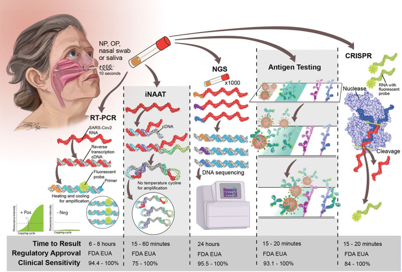
Most commonly, the diagnosis is made using a nasopharyngeal swab, but nasal swabs and saliva samples are becoming more common. Samples are preserved and transported in a viral stabilizing medium. The major laboratory-based methods currently available for the detection of SARS-CoV2 include real-time reverse-transcription polymerase chain reaction (RT-PCR), loop or isothermal nucleic acid amplification testing, next-generation sequencing, antigen-based testing, and clustered regularly interspaced short palindromic repeat–based technologies (Figure). RT-PCR is the most widely used technique for diagnosis of COVID-19. It is a specialized, quantitative molecular technique available in most major laboratories and the current gold standard for SARS-CoV2 infection diagnosis. RT-PCR involves 2 major steps: viral extraction and amplification. Initially, no SARS-CoV2–specific primers existed. Once the nucleotide sequence of SARS-CoV2 was determined, then primers were designed that bind target regions of the genome (eg, nucleocapsid and envelope regions). In RT-PCR, viral RNA is first extracted from the medium and then reverse transcribed to cDNA. Using SARS-CoV2–specific primers, a DNA polymerase amplifies cDNA through rapid heating and cooling (ie, thermocycling). In RT-PCR, a fluorescent nucleotide probe can then bind its target sequence region, and a fluorophore is emitted. The process is repeated ≈35 to 45 times, and once the accumulation of fluorescence signal passes a specified level, the cycle threshold, the test is considered positive. The highly manual and technical aspects of RT-PCR limit testing batches to ≈100 to 400 samples, which require 6 to 8 hours to complete. Recent technological innovations in RT-PCR, including the elimination of a dedicated extraction step, have reduced testing time to as little as 45 minutes.
Figure.

COVID-19 testing. A nasopharyngeal (NP), oropharyngeal (OP), or nasal swab or saliva is collected, and the sample is placed in a viral medium to provide stability. The sample is then processed by molecular and antigen-based technologies that detect the presence of severe acute respiratory syndrome coronavirus 2 (SARS-CoV2). These include molecular techniques that directly detect the SARS-CoV2 nucleic acid sequence (reverse-transcription polymerase chain reaction [RT-PCR], isothermal nucleic acid amplification testing [iNAAT], next-generation sequencing [NGS], and clustered regularly interspaced short palindromic repeat [CRISPR]) or other technologies that detect viral antigens. The clinical sensitivities of the tests are provided for reference from the US Food and Drug Administration Emergency Use Authorization (FDA EUA) filing and vary according to manufacturer within each test category. The specificity of testing is generally >95%. COVID-19 indicates coronavirus disease 2019.
Loop-mediated isothermal amplification or standard isothermal nucleic acid amplification testing is an alternative to RT-PCR that uses multiple primers that bind to complementary regions of the SARS-CoV2 genome and synthesize cDNA at a fixed temperature. Because no temperature cycling is required, the test can provide results within 15 to 20 minutes.1 The sensitivity of isothermal nucleic acid amplification testing came under scrutiny recently with the ID Now assay (Abbott Laboratories, Abbott Park IL), which approximates 75%2 and is more prone to error because it is a point-of-care device with a wide range of end-user technical expertise. The likelihood of getting a false-negative test for all technologies varies according to the time between symptom onset and testing because of lower viral shedding early after infection.3 Large-scale testing of SARS-CoV2 will likely require leveraging of next-generation sequencing technologies. Similar to RT-PCR, RNA is converted to cDNA. Each sample can be uniquely barcoded with a nucleotide sequence that allows sequencing of thousands of patient samples simultaneously. Sequence data are then processed through a bioinformatics analysis to distinguish individual samples and to detect the SARS-CoV2 genome. The sensitivity parallels that of RT-PCR. Next-generation sequencing will be helpful for wide-scale disease detection efforts that are not time sensitive. Specificity of all these nucleic acid amplification techniques is >95% because the primers are specific to the SARS-CoV2 genome.
In contrast, antigen-based testing detects the presence of viral proteins. When the patient sample is added to the testing device, specific SARS-CoV2 proteins present in the sample will bind antibody conjugated to a detector. These antigen-antibody complexes then bind to antibodies coated on a solid surface, and a signal is observed. It is a simpler and less expensive technology compared with nucleic acid technologies. However, antigen-based detection is generally less sensitive, and this may be especially true in populations with lower expected viral loads such as asymptomatic patients undergoing preprocedural testing. Last, modifications of clustered regularly interspaced short palindromic repeat–based technology have been developed to detect SARS-CoV2. A nuclease is programmed to detect a unique SARS-CoV2 nucleotide sequence. After the SARS-CoV2 genome is amplified, the nuclease can bind the target sequence, leading to enzyme activation and cleavage of the genome and a neighboring fluorescent probe. This releases a signal indicating the presence of SARS-CoV2.4
Given the wide array of tests available for the detection of SARS-CoV2, navigating the testing landscape can be daunting for cardiovascular practitioners. The selection of testing will often be driven by the presence of clinical symptoms, contact with affected individuals, local COVID-19 disease prevalence, and testing availability. Patients with clinically suspected COVID-19 can be tested with the most rapid form of testing available locally, typically isothermal nucleic acid amplification testing, antigen-testing, and clustered regularly interspaced short palindromic repeat–based testing. Given the high pretest probability, cardiovascular team members should be protected with full personal protective equipment regardless of results to reduce disease acquisition given the imperfect sensitivity of these methodologies. Urgent/emergent cardiovascular procedures should proceed to reduce cardiovascular morbidity and mortality, but elective procedures can be delayed until the patient convalesces from the infection. For asymptomatic individuals, rapid point-of-care testing, if available, should improve clinical workflows without introducing an unnecessary patient burden and minimizing turn-around time. This may be particularly useful for high-volume noninvasive procedures such as echocardiography. Ultimately, if the local disease prevalence is low (<5%) and the patient is asymptomatic, then the posttest probability is so low that false-negative results are unlikely to have a significant effect on the posttest likelihood of disease. Therefore, even tests with a low sensitivity of ≈75% are likely to be sufficient for screening of asymptomatic patients.5
It is clear that testing is critical to ensure the safety of cardiovascular practitioners and patients and in our success to be able to get back to providing “normal” cardiovascular care.
Acknowledgments
The authors acknowledge Devon Stuart for assistance with medical illustration and Dr Warren Levy who, in part, inspired them to write this piece, working relentlessly to navigate logistics to provide SARS-CoV2 testing for the thousands of patients of his Virginia Heart practices needing noninvasive testing.
Sources of Funding
The work in this study is supported by the National Institutes of Health Career Development Award K23 1K23HL143179-01A1 awarded to Dr Shah. Dr deFilippi receives funding from the National Center for Advancing Translational Science of the National Institutes of Health Award UL1TR003015.
Disclosures
Dr Shah reports personal fees from Procyrion and Ortho Clinical Diagnostics and research grants American Heart Association/Enduring Hearts, Abbott, Merck, and Bayer. Dr deFilippi reports consulting fees from Abbott Diagnostics, FujiRebio, Ortho Diagnostics, Quidel, Roche Diagnostics, and Siemens Healthineers. Dr Mullins reports no conflicts.
Footnotes
The opinions expressed in this article are not necessarily those of the editors or of the American Heart Association.
References
- 1.Bulterys PL, Garamani N, Stevens B, Sahoo MK, Huang C, Hogan CA, Zehnder J, Pinsky BA. Comparison of a laboratory-developed test targeting the envelope gene with three nucleic acid amplification tests for detection of SARS-CoV-2. J Clin Virol. 2020;129:104427 doi: 10.1016/j.jcv.2020.104427 [DOI] [PMC free article] [PubMed] [Google Scholar]
- 2.Harrington A, Cox B, Snowdon J, Bakst J, Ley E, Grajales P, Maggiore J, Kahn S. Comparison of Abbott ID Now and Abbott m2000 methods for the detection of SARS-CoV-2 from nasopharyngeal and nasal swabs from symptomatic patients. J Clin Microbiol. 2020;58:e00798-20 doi: 10.1128/JCM.00798-20 [DOI] [PMC free article] [PubMed] [Google Scholar]
- 3.Kucirka LM, Lauer SA, Laeyendecker O, Boon D, Lessler J. Variation in false-negative rate of reverse transcriptase polymerase chain reaction-based SARS-CoV-2 tests by time since exposure. Ann Intern Med. 2020;173:262–267. doi: 10.7326/M20-1495 [DOI] [PMC free article] [PubMed] [Google Scholar]
- 4.Kellner MJ, Koob JG, Gootenberg JS, Abudayyeh OO, Zhang F. SHERLOCK: nucleic acid detection with CRISPR nucleases. Nat Protoc. 2019;14:2986–3012. doi: 10.1038/s41596-019-0210-2 [DOI] [PMC free article] [PubMed] [Google Scholar]
- 5.Woloshin S, Patel N, Kesselheim AS. False negative tests for SARS-CoV-2 infection: challenges and implications. N Engl J Med. 2020;383:e38 doi: 10.1056/NEJMp2015897 [DOI] [PubMed] [Google Scholar]


