Abstract
Evergreen conifers in boreal forests can survive extremely cold (freezing) temperatures during long dark winter and fully recover during summer. A phenomenon called “sustained quenching” putatively provides photoprotection and enables their survival, but its precise molecular and physiological mechanisms are not understood. To unveil them, here we have analyzed seasonal adjustment of the photosynthetic machinery of Scots pine (Pinus sylvestris) trees by monitoring multi-year changes in weather, chlorophyll fluorescence, chloroplast ultrastructure, and changes in pigment-protein composition. Analysis of Photosystem II and Photosystem I performance parameters indicate that highly dynamic structural and functional seasonal rearrangements of the photosynthetic apparatus occur. Although several mechanisms might contribute to ‘sustained quenching’ of winter/early spring pine needles, time-resolved fluorescence analysis shows that extreme down-regulation of photosystem II activity along with direct energy transfer from photosystem II to photosystem I play a major role. This mechanism is enabled by extensive thylakoid destacking allowing for the mixing of PSII with PSI complexes. These two linked phenomena play crucial roles in winter acclimation and protection.
Subject terms: Biophysics, Forest ecology, Plant sciences, Non-photochemical quenching
Evergreen conifers rely on ‘sustained quenching’ to protect their photosynthetic machinery during long, cold winters. Here, Bag et al. show that direct energy transfer (spillover) from photosystem II to photosystem I triggered by loss of grana stacking in chloroplast is the major component of sustained quenching in Scots pine.
Introduction
Photosynthesis is the basis for most life on earth, and the ability to sustain growth by harvesting sunlight confers such an enormous evolutionary advantage that photosynthetic organisms have developed numerous adaptations that enable them to photosynthesize in diverse environments. These include vast boreal forests, which cover ~70% of all coniferous forests of the world (mainly in the northern hemisphere)1, which have lower species diversity than many other terrestrial ecosystems and are often dominated by evergreen conifers, like pine and spruce2. Broadleaf deciduous trees and shrubs are also present, and sometimes deciduous conifers such as Larch (Larix) species, but there is no doubt that evergreen conifers are particularly well adapted to harsh boreal conditions, including short growing seasons and cold, snowy winters. Thus, evergreen conifers acclimations to boreal regions must presumably include regulatory processes that protect the photosynthetic apparatus in their needles during the harsh winter and early spring. Knowledge of photoprotective mechanisms in various kinds of photosynthetic organisms has increased considerably in recent decades. Several control systems have been identified that allow the photosynthesis machinery to harmlessly dissipate excess excitation energy3–7. In 2003, Öquist and Huner8 published a seminal review of photosynthesis in evergreen plants, which pointed out that overwintering conifer needles enter a state of ‘sustained quenching’ during winter. They found strong evidence for major alterations in the organization and composition of the photosystem II (PS II) antenna but also concluded that photosystem I (PS I) may play an important role, via nonphotochemical quenching of absorbed light or via quenching absorbed light photochemically through cyclic electron transport. Recently it was also shown that alternative electron transport might add up to this as well9. Up to date, there exists no clear mechanism rationalizing winter quenching in conifers although there are several proposed hypotheses8,10. Several protection mechanisms may also occur in parallel and (if so) they are likely activated largely well before the extreme stress occurs11,12. But the major conundrum arises in early spring, when temperatures are typically still low, so plants’ biochemical and metabolic activities are strongly limited, but solar radiation is already high8. Once induced, the mechanisms need to be sustained, i.e., locked-in over the long winter season and protect the photosynthetic machinery. When conditions improve late in the spring, photosynthetic apparatus is restored and attains its active growth state in summer.
In previous explorations of these phenomena, a marked drop in Photosystem II photochemistry (measured as Fv/Fm) has been recorded in several overwintering conifers, and some indications of the quenching mechanisms involved have been suggested. These reportedly include destacking of thylakoid membranes and associated changes in chloroplast ultrastructure13, in accordance with the drop in PSII maximal fluorescence8. In higher plants, thylakoid stacking and heterogeneity play crucial roles in the localizations of PSII and PSI in grana (tightly appressed thylakoid layers) and stroma lamellae, respectively. In conifers, although an extreme drop in maximal fluorescence (Fm) in winter/early spring has been observed, its mechanistic relationship to thylakoid structural changes has not been explained. It was recently shown that phosphorylation of PsbS and Lhcb1 might be a prerequisite for this phenomenon in spruce14, triple phosphorylation of Lhcb1 was linked with massive destacking of thylakoids but its physiological relevance was yet undefined. In the study reported here we monitored steady-state chlorophyll fluorescence, ultrafast time-resolved fluorescence, and chloroplast ultrastructure in Scots pine needles from autumn to summer in three successive years. Our data strongly indicate that chlorophyll fluorescence quenching and thylakoid destacking are strongly linked, mutually dependent and crucial for the survival of evergreen conifers in the extreme northern boreal winter and early spring, when temperatures are low but solar radiation levels may be high.
Results
Seasonal changes in Photosystem II- and Photosystem I-related functional activities
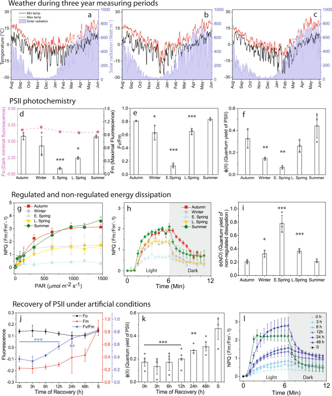
To monitor photosynthetic performance, we recorded several PSII and PSI parameters during three consecutive growth seasons (2015–2016, 2016–2017, and 2017–2018) along with concomitant changes in daily air temperature and solar radiation (Fig. 1a–c). For simplicity, in the main figures we only present (here onwards) data from 2017 to 2018, which we divided into five distinct seasons, based on weather parameters: Summer (S, June–Aug), autumn (A, Sept–mid Nov), winter (W, mid Nov–mid Feb), early spring (ES, mid Feb–mid Apr), and late spring (LS, mid Apr–June). Data from the other two growth seasons are provided in Supplementary Figs. 1–3.
Fig. 1. Seasonal dynamics of weather and photochemical performance of PSII measured by chlorophyll fluorescence in Scots pine needles.
Changes in temperature (°C) (Left Y-axis) and solar radiation (watt m−2) (Right Y-axis) during 2015–2016 (a), 2016–2017 (b), 2017–2018 (c) measuring seasons. Seasonal dynamics of PSII photochemistry: (d) Changes in maximal (Fm) and basic (Fo) fluorescence. (e) Maximal quantum efficiency of PSII measured as Fv/Fm (f) Effective quantum yield of PSII (Φ(II)). Energy dissipation measured as regulated and non-regulated non-photochemical quenching: (g) Changes in NPQ with increasing PAR. (h) Induction of NPQ with constant actinic light. (i) Quantum yield of non-regulated non-photochemical quenching. Recovery of pine needles under artificial conditions at 80 µmol photons m−2 s−1 of light for 48 h with 18/6 photoperiod: (j) Changes in Fo. Fm and Fv/Fm. (k) Changes in Φ(II). (l) Induction of NPQ with constant actinic light. Quantum yields were calculated at actinic light illumination of 300 μmol m−2 s−1 in the light response curve and NPQ induction was measured either at 300/800 μmol m−2 s−1 of actinic light illumination. All measurements were taken after 30 min of dark adaptation at 4 °C from winter to late spring and room temperature in summer. Error bars indicate the mean ± SD (n = 3) for (d–g), and (i); ± SD (n = 4) for (j, k). Error bars indicate the mean ± SE (n = 3) for (h, l). The statistically significant mean differences (t-test) are marked by asterisks indicating the following confidence intervals: *≤95%; **≤99%.; ***≤99.9%. Exact p values are provided in the source data file.
We found characteristic seasonal patterns in maximum PSII fluorescence (Fm) and maximum quantum efficiency of PSII (expressed as Fv/Fm), in accordance with earlier reports8,10. Fv/Fm was highest in S, fell with reductions in ambient temperatures during A and W (Fig. 1d, e), and was lowest in ES (63% lower than in S), when low temperatures coincided with rises in solar irradiance (Fig. 1a–c; see also Supplementary Fig. 2a). During LS, Fv/Fm gradually increased with increasing temperatures and peaked in S. The seasonal changes recorded in Fv/Fm were found to be mostly affected by changes in Fm rather than Fo (basic intrinsic fluorescence) (Fig. 1d). For a deeper understanding of PSII performance, the fraction of absorbed light energy utilized by PSII photochemistry15 (ΦPSII) was measured (Fig. 1f). During W ΦPSII decreased significantly and reached minimal values (19% of S values) in ES (Supplementary Fig. 2b).
Furthermore, we tried to quantify the amount of absorbed light energy thermally dissipated by non-photochemical quenching (NPQ)3. The component of NPQ that play a crucial role under fluctuating light conditions is the fast component (ΔpH-PsbS dependent-qE3 or zeaxanthin dependent-qZ4), which increased with increasing light intensities in all the seasons. In S and A, the fast component did not reach a stationary phase even at 1500 µmol m−2 s−1 illumination, while in W and LS samples a stationary phase was reached at 500 µmol m−2 s−1 and in ES already at 300 µmol m−2 s−1 (Fig. 1g). Most importantly, in ES inducible steady-state NPQ was much smaller and overall slower due to the smaller amplitude of the fast component (~50% less than in S) in ES (Fig. 1h). This is due to the fact that ES needles have already developed static NPQ. Instead, the quantum yield of non-regulated and/or constitutive loss (ΦNO) of energy was high during ES (Fig. 1i) (Supplementary Fig. 2d). This strongly suggests that this static NPQ is the fraction of absorbed light energy neither going to drive photochemistry (ΦPSII) nor thermally dissipated by rapid regulated NPQ processes (qE/qZ).
To confirm that this static quenching is a manifestation of a “sustained mode of quenching” we artificially relaxed the ES needles (hereafter, ER) for 48 h in low light (80 µmol m−2 s−1) with 18/6 h photoperiod. During this recovery, we observed no significant changes in Fv/Fm for 6–8 h (less than a 10% increase), a modest increase (30%) after 24 h (Fig. 1j), and almost complete (95%) recovery to S levels after 48 h. Fo did not change significantly throughout the recovery period (Fig. 1j), but changes in Fm followed the same recovery dynamics as Fv/Fm (Fig. 1j), although maximal fluorescence (Fm) was much higher in S than in 48 h ER samples. ΦPSII did not change significantly until 12 h of recovery but recovered to 50% and 90% of S levels after 24 and 48 h, respectively (Fig. 1k). qE/qZ-dependent fast NPQ kinetics followed similar patterns to ΦPSII (Fig. 1l). It should be noted that light-dependent induction of the fast component of NPQ (qE/qZ) was slower in the ER samples than in S samples, even after 48 h of recovery, which appears to be mainly due to photoinhibition.
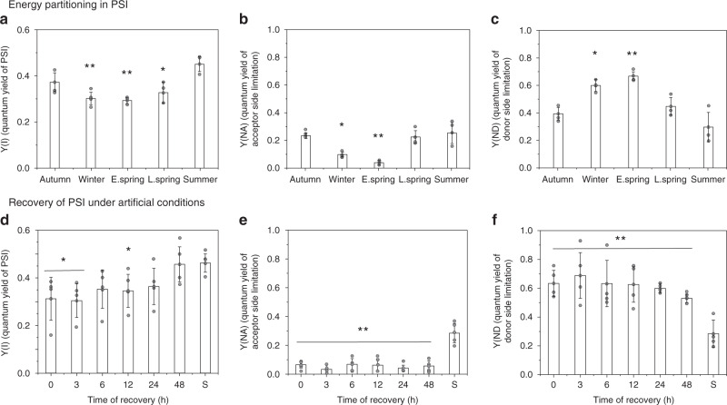
Seasonal changes in the partitioning of absorbed light energy within PSI showed that Y(I) was highest in S, declined in A and W, and reached minimal values in ES (Fig. 2a). Although it decreased, Y(I) was much less affected than the PSII activity (ΦPSII) during the cold periods (Fig. 1f) (Supplementary Fig. 3a). Y(NA) followed the same seasonal pattern, with minimum values registered in ES and a steady recovery until S (Fig. 2b) (Supplementary Fig. 3b). In contrast to Y(NA), Y(ND) considerably increased during the cold periods and peaked (at 69% higher than S values) in ES, then gradually declined to minimum values in S (Fig. 2c, Supplementary Fig. 3c). This suggests that during early spring most of the absorbed light energy is dissipated by oxidized P700 (P700+) to prevent photoinhibition caused by over reduction of the acceptor side as iron-sulfur clusters get damaged16. During the first 3 h of the recovery period, ΦPSI did not significantly change, but it gradually returned to S levels over 48 h (Fig. 2d). Y(NA) did not change much during recovery (Fig. 2e). Y(ND) increased slightly during the first 3 h, but then gradually decreased, and ER samples did not quite reach S values. Based on these results, we conclude that the restoration of PSI light use efficiency takes longer than 48 h (Fig. 2f).
Fig. 2. Seasonal changes of PSI photochemistry in Scots pine needles.
Energy partitioning in PSI considering Y(I) + Y(ND) + Y(NA) = 1, where Y(I) (a), Y(NA) (b), Y(ND) (c) are the photochemical quantum yield of PSI (when P700 is reduced and A is oxidized), energy dissipation in PSI (a measure of acceptor side limitation, when P700 and A both are reduced) and energy dissipation in PSI (a measure of donor side limitation, when P700 and A both are oxidized), respectively. Recovery of PSI photochemistry under artificial conditions: (d) Y(I), (e) Y(NA), (f) Y(ND) of PSI during the recovery period. Quantum yields were calculated from 300 μmol m−2 s−1 light illumination period of a light response curve. All measurements were taken after 30 min of dark adaptation at 4 °C from winter to late spring and room temperature in summer. Error bars indicate the mean ± SD (n = 3) for (a–c); ± SD (n = 4) for (d–f). The statistically significant mean differences (t-test) are marked by asterisks indicating the following confidence intervals: * ≤95%; ** ≤99%.; *** ≤99.9%. Exact p values are provided in the source data file.
Sustained non-photochemical quenching dominates in early spring needles
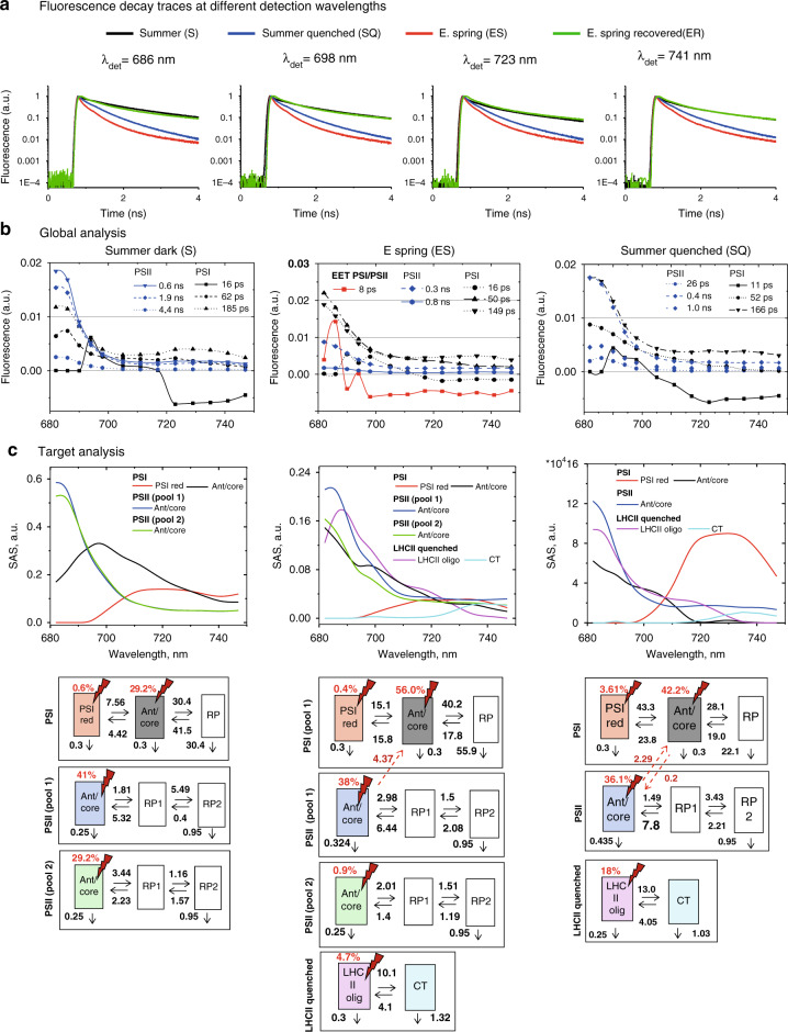
To elucidate the mechanism of “sustained quenching” observed in pine during the early spring periods, ultrafast time-resolved fluorescence measurements on intact pine needles were performed using the setup and approach described previously17,18. In addition, a special chamber was designed to keep the rotational cuvette with the sample at −20 °C (Supplementary Fig. 4). This chamber was used in order to maintain the “native” sustained quenched state in pine needles. Fluorescence decay traces were collected from pine needles in the same states as reported in the previous subsection: S, ES, ER: Summer needles (S) do not contain any “sustained quenching”, while E.spring needles (ES) contain the highest levels of “sustained quenching”. To resolve the mechanisms of this quenching, fluorescence decay traces of S needles in the original state (dark-adapted, Fig. 3a, Summer dark) were compared to the needles collected in ES when sustained quenching was present (Fig. 3a, Early spring) and after the quenching in the needles had been relaxed at room temperature (ER) (Fig. 3a, E. spring recovered). To understand whether sustained quenching observed in ES occurs via a similar quenching mechanism to light-induced quenching, S needles exposed to HL for 30 min (SQ) were also measured (Fig. 3a, Summer quenched). As shown by the direct comparison of the decay curves (Fig. 3a), the fluorescence in SQ needles is much shorter-lived as compared to that of S needles. However, the fluorescence decays of ES needles are still pronouncedly shorter-lived than that of the SQ samples, thus characterizing the “sustained quenching” in the ES samples as the most pronounced quenching at all detection wavelengths and under all conditions. In contrast, ER and S samples showed very similar fluorescence decays, indicating that the ES sample recovered quite (although not completely, vide infra) well from sustained quenching within 48 h of recovery treatment.
Fig. 3. Time-resolved fluorescence of intact pine needles measured using TCSPC.
(a) Fluorescence decay traces measured at −20 °C and at four characteristic wavelengths: 686 nm (mainly PSII, LHCII contributions), 698 nm (PSII, PSI contributions), 723 nm (mainly PSI contribution), and 741 nm (mainly PSI contribution). (b) Global analysis of pine needles in three states: Summer dark (S, dark-adapted summer needles, left row), ES (E.spring needles with “sustained quenching” present, middle row), Summer quenched (SQ, right row) (c) Kinetic target analysis of pine needles in the three states. See the main text for the explanations. The kinetic target analysis (SAS top, a kinetic model with rate constants in ns-1, bottom) shows the results of the detailed target modeling of the fluorescence kinetics of pine needles. The rate constants (ns−1) and Species-associated emission spectra (SAS) resulted were determined from global target analysis. Species-associated emission spectra (SAS) resulted from the fit of the target kinetic model in the corresponding state. Note that fluorescence decay measurements below 680 nm to detect the decreasing short-wavelength part of the spectra were not possible due to the extremely high scattering of the pine needles. This has no effect however on the ability to distinguish the various lifetime components kinetically and spectrally.
Global analysis of the fluorescence kinetics of pine needles
To get the first hints on the mechanism(s) underlying sustained quenching in early spring global analysis19 was performed on all datasets: ES, S, SQ (Fig. 3b), and ER (Supplementary Figs. 5–8) needles. Six decay-associated spectra (DAS) were required to fit the fluorescence kinetics in all four cases. Three DAS were tentatively assigned to PSI: 11–16 ps, 50–60 ps, 150–180 ps (Fig. 3b, Supplementary Figs. 5, 6). Their spectra and lifetimes are reminiscent of previously reported PSI-related DAS in other plant species in vivo17,18,20. The fastest component represents energy equilibration between Chlred and Chlblue21,22. The second DAS in S needles peaks around 685–690 nm with a broad emission in the 700–720 nm region. Therefore, it mainly represents PSI core decay in combination with the 710–720 Chls of Lhcas. The third DAS (185 ps) peaks at 680 nm with some contribution at 730 nm, and therefore, it should be primarily assigned to LHCII and some Chlred in PSI. In quenched states (SQ and ES, in particular) these two DAS have substantially higher contribution around 680 nm and, as a result, the reconstructed steady-state PSI spectra have higher emission around 680–685 nm as compared to the unquenched states (S and ER see Supplementary Fig. 5). It indicates that upon quenching PSII and/or LHCII kinetics also contribute to these PSI-related components. In S and ER states, the remaining three DAS peak at 682 nm and therefore, are assigned to PSII (Fig. 3b and Supplementary Fig. 9). Their lifetimes are 0.5–0.6 ns, 1.5–2.0 ns, and 4 ns, similar to what was resolved previously for PSII in the closed state17,18. However, in ES two considerably shorter-lived PSII-related DAS were observed with lifetimes of 0.3 ns and 0.8 ns. This demonstrates that PSII kinetics is indeed quenched strongly in ES needles. Additionally, we resolved a DAS with 8 ps lifetime in the ES sample. The “oscillations” in the spectra giving rise to the sharp bands are due to the mixing of the contributing components, representing excitation energy transfer (EET) from Chl a pools emitting at ~680 nm to those emitting at 690 nm and 700–730 nm, respectively. This suggests that there occurs efficient EET between PSII and/or LHCII and PSI in the ES needles. However, these processes could be unambiguously resolved only by target modeling.
In a nutshell, global analysis results suggest that sustained quenching in the ES state involves both PSII/LHCII and PSI through a direct energy transfer. This mechanism provides much stronger quenching than the high-light-induced quenching in the summer needles (SQ).
Target modeling of the fluorescence kinetics of unquenched pine needles
To resolve the mechanism(s) responsible for the “sustained quenching” in pine needles in detail we performed target analysis23 of all the kinetic data. In target analysis, a specific kinetic model is tested using a linear time-invariant compartmental model. Each compartment in Fig. 3 is presented by a square box. Each box belongs to any of the three different components: PSI, PSII, LHCII quenched. Fluorescence emitting compartments are presented by colored boxes. The red flashes with numbers directing to that compartments represent the so-called excitation vector, providing the percentage of excitations directed to each compartment and should roughly illustrate the relative number of Chl a (i.e., relative Chl a antenna size) in it. Non-colored boxes represent non-emitting compartments, like radical pairs (RP) in RCs and CT states, which are contributing to the overall observable kinetics and thus have to be included. Target modeling is required to describe the real time-dependent concentrations of each compartment, rates between them (arrows in Fig. 3, ns−1), and spectra (species associated spectra (SAS)) of the interacting components. Note, that the global analysis, presented in the previous subsection, is equivalent to a number of noninteracting, parallelly decaying compartments and gives only the first indications of the processes involved. It is a mere mathematical analysis, not considering any physical description or information on the studied system. By testing kinetic models that had been validated previously on isolated subcomplexes24–26 it was possible to separate the PSI from the PSII kinetics. Therefore, our assignment of PSI and PSII is based on a combination of the used kinetic model, resolved rates as well as SAS. The target model and the excitation vector presented in Fig. 3c resulted in the best fitting quality (Supplementary Fig. 7). In S needles (Fig. 3c, Summer dark) the PSI kinetics was described with two emitting compartments (PSI red, Ant/core) and one non-emitting radical pair (RP, Fig. 3c). The resolved rates and SAS are strongly reminiscent of the ones reported for the PSI-LHCI complex26. Kinetics from the PSII antenna/RC complex was described by one emitting compartment (Ant/core) and two non-emitting radical pairs (RP1 and RP2, Fig. 3c) as reported previously17,24,25,27. According to their SAS and rates, the emitting species represents the equilibrated excited state of the PSII-LHCII super-complex. In our analysis, to achieve a very good fit, two pools of PSII complexes with different decay rates were required (pool 1, pool 2, Fig. 3c, see the detailed comparison of the residuals plots in Supplementary Fig. 7) for the S sample. The two pools differ in the rate constants of charge separation in a manner that is typical for PSII particles with different antenna sizes (see18,19 for discussion and explanation), which result in different average lifetimes (Supplementary Table 3). In the S sample, PSI with its antenna accounts for 30% of the total absorption cross-section at the excitation wavelength (662 nm), while the two PSII pools account for 40% and 30% of the total absorption cross-section. No internal quenching related to any NPQ process of these PSII compartments was observed.
The target model for the ER needles is rather similar to that of S needles (Supplementary Fig. 5). The main difference is that the smaller PSII pool (with a very small absorption cross-section of ca. 10%) shows an unusually small charge separation rate. The most likely explanation of this phenomenon is a small percentage of PSII RCs has been photoinhibited during the 48 h relaxation process from the sustained quenching state, which agrees with NPQ measurements.
Direct energetic transfer between PSII and PSI is the dominating component of sustained quenching
As for the S samples, also the description of the ES sample (Fig. 3c) required a three-compartmental target model for PSI and the presence of two PSII pools, each described by a three-compartmental model. The two PSII pools differ in the rate constants of charge separation in a similar manner as in S samples. The kinetics are also very similar to that of the S samples, however with a slightly increased charge separation rate. This can be interpreted as a consequence of the partial reduction in the physical antenna size. This goes in line with an increase in the Chl a/b ratio in ES as compared to S samples (Supplementary Fig. 9, Supplementary Table 2). Resolved antenna SAS (Ant/Core compartment of PSI, Fig. 3) of PSI in ES samples contains stronger emission in the 680 nm region, as compared to S needles. That is why we suggest that some LHCII strongly coupled to PSI contribute to it, like recently reported20.
The striking feature is the presence of a very strong contribution of a direct energy transfer from PSII to PSI. Without allowing for this direct energy transfer, the data could not at all be fitted adequately (Supplementary Figs. 6, 7). This process has a transfer rate of 4.4 ns−1. A small compartment representing quenched and functionally detached LHCII was also needed for a satisfactory description of the ES samples (See more details on this component in the next section). Overall, the target analysis shows that direct energy transfer from PSII to PSI complexes provides the dominant quenching mechanism of PSII complexes in ES needles causing the pronounced “sustained quenching”.
Comparison of sustained quenching and light-induced quenching
To model the fluorescence kinetics of SQ needles again three-compartmental models for PSI and PSII were required. However, unlike in previous cases, one single PSII pool was sufficient to describe the PSII kinetics.
Besides PSII and PSI, one additional component was needed to satisfactorily describe the fluorescence kinetics in the SQ samples. The rates, spectra, and average lifetime (0.4 ns, Supplementary table 3) of these compartments are strongly reminiscent of what was reported previously for highly quenched LHCII aggregates28, and the quenched LHCII complexes under NPQ conditions in wild type Arabidopsis17 and in Arabidopsis mutants depleted of the reaction centers29. We, therefore, assign this highly quenched component to the quenched LHCII antenna energetically disconnected from both PSII and PSI. However, the addition of this quenched LHCII compartment was not sufficient to fully describe the fluorescence kinetics (Supplementary Fig. 7). The species-associated spectra (SAS) and kinetics of PSII still contained a substantial amount of 690–700 and 730 nm emission, entirely uncharacteristic for PSII super-complexes, but characteristic of PSI complexes22,26. Interestingly, also the SAS of the PSI antenna/core complex contains a 680 nm emission, which is characteristic of PSII. This strongly suggests—as can be directly deduced from the spectral properties—the presence of direct energetic contact between PSI and PSII (Fig. 3c). Introducing such a connection in the target model indeed resulted in a considerably better fit (χ2 decreased from 1.198 to 1.093, Supplementary Fig. 6). Also, the PSII and PSI kinetics became well-separated in such a model. The resolved direct energy transfer rate from PSII to PSI is quite high (2.3 ns−1), implying that a part of energy absorbed by PSII is actually funneled to PSI. However, this rate of direct energy transfer in SQ needles is only about half of that in ES needles, suggesting that unlike in cold-induced sustained quenching, upon light illumination, spillover is not the major process. The contribution of the quenched antenna, on the other hand, plays a major role.
Efficiency of direct energy transfer quenching in different conditions
By evaluating the kinetic information provided in Fig. 3c one can get a quantitative estimation of the degrees of light use efficiencies (for photosynthesis) on the one hand, and for photoprotection/quenching on the other hand, present in both PSII and PSI in the different situations (c.f. Supplementary Fig. 8, Supplementary Table 4). In S needles 70% of the total absorbed energy drives PSII charge separation and only 30% PSI charge separation. In the SQ needles the percentage of energy flowing into the PSII reaction center drops to 7.1%, while it increases to 67% for PSI. Upon strong light illumination, most of the PSII reaction centers will be closed, and a fraction of that total energy flowing through the RC can produce harmful reactive species like ROS30,31. Since the percentage of energy flow into PSII decreases 10 times (70% in S and 7.1% in SQ) in SQ as compared to S, this means that the potential of oxidative damage is reduced by a factor of 10 by the quenching process. This factor is increased by a further factor of approx. 2 due to the large pool of functionally detached and quenched LHCII. So, in SQ needles the overall quenching provides a protection factor of 20 for PSII compared to S needles. In the sustained quenched ES samples this effect is much more extreme: Only 1.5% of the total absorbed energy in the system flows through the PSII reaction center potentially causing oxidative damage (assuming all other factors are equal). Thus, in total, “sustained quenching” in ES samples provides a protection factor of approx. 40 to PSII (double of SQ) compared to S needles. This huge protection factor explains well why “sustained quenching” is so effective enabling pine needles to survive the harsh winter and early spring conditions.
Massive destacking of grana membranes in the winter
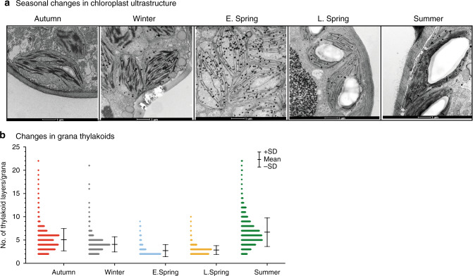
In accordance with early reports32,33, we observed strong seasonal changes in chloroplast ultrastructure, including loss of grana stacks during early spring (Fig. 4a). Ultrastructural changes derived from morphometric analysis of electron micrographs (Fig. 4a) indicate that average numbers of grana per chloroplast steadily decreased from autumn to winter and reached minimal values in early spring, corresponding to just 68% of S values (Supplementary Table 1). More strikingly, the number of appressed thylakoid layers/granum dramatically declined from A (4.97) to ES (2.72) and rose back in S (6.50) (Supplementary Table 1). These changes corroborate the shift in grana stacking illustrated in Fig. 4b. In S and A, 3–6 layered grana stacks accounted for 60–70% of total stacks. In contrast in ES samples, 70–75% and 15–20% of the grana had only two and three layers, respectively (Fig. 4b). These changes in ES samples were accompanied by a transient doubling of the number of lipid globules (plastoglobuli) per cell (Supplementary Table 1) when the chloroplast structure deviated most strongly from the S state.
Fig. 4. Transmission electron microphotographs depicting seasonal variations in chloroplast ultrastructure in pine needles.
a Chloroplast structure in Autumn, Winter, E. Spring, L. Spring, and Summer. b Histograms of frequency distributions of numbers of thylakoids per granum during the five distinct seasonal periods. The histograms were calculated from 80–100 electron micrographs per season, representing 2–3 chloroplasts per image [Error bars indicate mean ± SD calculated from n = 1124 (Autumn), 1235 (Winter), 521 (Early Spring), 578 (Late Spring) and 1048 (Summer) of grana stacks].
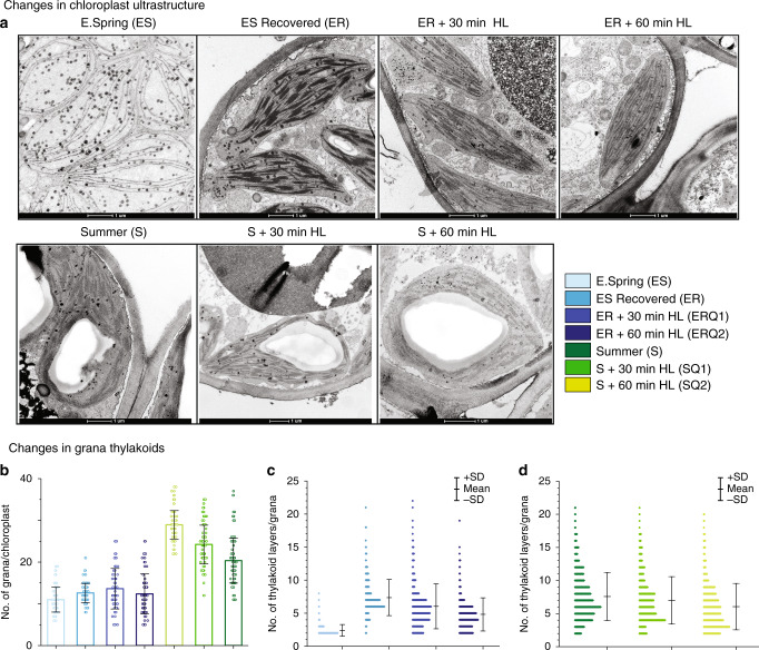
To obtain a deeper understanding of the thylakoid plasticity in ES needles we also subjected needles collected at specific early spring and summer dates to EM analysis after artificially induced recovery under different light conditions (Fig. 5a). When both S and ER samples were exposed to HL for 30 (SQ1 for S samples, ERQ1 for ER samples) and 60 min (SQ2 for S samples, ERQ2 for ER samples) similar destacking of grana (as of ES samples) have been observed (Fig. 5b). These changes in grana structures recorded in ER and S samples (Fig. 5c, d) following short term HL exposure strongly corroborate the high dynamic plasticity of the thylakoid membrane, which appears to be essential for acclimation to the harsh boreal winters.
Fig. 5. Artificial induction of changes in chloroplast ultrastructure of pine needles.
a Changes in chloroplast ultrastructure in E. spring (ES), E. spring samples recovered (ER) at 18oC for 48 h with a photoperiod of 18 h at 80 µmol m−2 s−1, ER samples treated with 800 µmol m−2 s−1 high light for 30 min (ERQ1), for 60 min (ERQ2). Summer (S), Summer samples treated with 1200 µmol m−2 s−1 high light for 30 min (SQ1), for 60 min (SQ2). b The number of grana per chloroplasts (Error bars indicate mean ± SD (n = 75); c Histograms of frequency distributions of numbers of thylakoids per granum in different E. spring treated [n = 220 (ES), 250 (ER), 272 (ERQ1), 246 (ERQ2)]. d Summer treated [n = 576 (S), 498 (SQ1), 415 (SQ2)] samples. Error bars indicate the mean ± SD obtained from the analysis of grana stacks.
Changes in pigment and protein composition of ES, ER, SQ, and S samples
We also analyzed pigments of the pine needles used for spectroscopic measurements and found substantial differences in pigment contents between S and ES needles (Supplementary Table 2). In S and ES samples the Chl a/b ratios were 2.8 and 3.4, respectively, indicating that S needles had higher relative amounts of Lhcbs and other Chl b-binding proteins. The Chl/carotenoid ratio was also substantially lower in ES samples than in S samples, indicating that they had higher amounts of carotenoids (mainly lutein and violaxanthin/zeaxanthin). We observed no significant difference in total Chl pigment contents between S and SQ samples.
For quantifying the core and antenna proteins, isolated thylakoids were subjected to SDS-PAGE and were immunoblotted against specific antibodies (Supplementary Fig. 9). This revealed that ES samples contain a lower abundance of core (PsaD, PsbD) and antenna (Lhcb2, Lhca4) proteins than in S, in line with earlier reports34 (Supplementary Fig. 9). ER samples contained lower amounts of PsbD and Lhcb2 compared to S, which might reflect on the minor amount of photoinhibition seen in fluorescence data.
Discussion
Distinct changes in chloroplast ultrastructure, including loss of grana thylakoids and disorganization resulting in mostly single-/bi-stromal thylakoid membranes during the cold winter season, followed by recovery during the late spring/summer season have been observed in various conifers8,13,33. We observed similar strong seasonal variations in the chloroplast architecture of pine chloroplasts (Fig. 4, Supplementary Table 1), and together with the fluorescence data presented above we are able to decipher the major molecular mechanism that connects these changes to the pronounced ‘sustained quenching’ described in conifers in the winter.
The seasonal reductions in the maximal quantum efficiency of PSII (Fig. 1e) and light energy utilization during the winter (Fig. 1f) were associated with a substantial increase in excitation pressure, indicating overall reduction of the photosynthetic electron transport chain35. In early spring, conifers harvest light energy but the use of the excitation energy for photosynthesis is severely restricted, resulting in potential photoinhibition. Hence, all overwintering conifers must develop highly efficient mechanisms for photoprotection of the photosynthetic apparatus. The major photoprotective mechanism for de-excitation of excess light energy in green plants and algae is the rapidly/ reversible ΔpH- dependent non-photochemical quenching (qE) that occurs in the pigment bed of LHCII proteins3,17,36. However, several alternative and/or supplementary photoprotective mechanisms for effective thermal deactivation of excess light energy have also been proposed5,12,13,37–39.
At first glance, the sharp decline in ΦPSII during early spring (Fig. 1f) might suggest that excess irradiance is dissipated via qE, which has been established as one of the most effective mechanisms for coping with the excess energy flux3. However, fast inducible qE was extremely low (Fig. 1g, h) in ES samples and was not restored to S levels even after 48 h of recovery treatment. (Fig. 1l). This is not unexpected since, in contrast to the fast energy dissipation (qE) depending on ΔpH and PsbS-protein3 the energy dissipation during winter (sustained quenching) occurs under strong down-regulation, even perhaps in the absence of ΔpH8,10,40. However, we can assign this relatively small qE contribution to quench to the small component (accounting for 4.7% of the absorption cross-section) of detached and quenched LHCII. This fraction was much smaller than in light-quenched samples (18%), and absent in both the dark-adapted S samples and recovered ES samples. Incidentally, a so-called “cold hard band” has been reported in low-temperature fluorescence spectra and kinetics of cold-acclimated evergreens40,41. Features of this band are reminiscent of quenched LHCII aggregates at low temperature42, hence it is probably correlated with the fraction of detached and quenched LHCII observed in “sustained quenching” conditions. This led us to conclude that this quenching seen in conifers is not only “Sustained NPQ” but also has a different mechanism.
Detailed quantitative energy partitioning analysis of total absorbed light energy by PSII demonstrates a strong increase (4 fold) of the fraction of constitutive thermal dissipation (ΦNO)43,44 (Fig. 1i). Usually, this thermal dissipation (ΦNO) contribution is not explained by any clear mechanisms. However, in the case of ES samples, it is strongly suggested by the data that the high levels of ΦNO do reflect the ΔpH-independent “sustained” energy quenching8,10,40. Our lifetime data provide an explanation for the origin of this component: A very high rate of direct energy transfer from PSII to PSI, provides this delta-pH independent (non-qE) quenching of PSII. Other photoprotective mechanisms proposed, such as, role of photo-inactivated PSII complexes may also effectively dissipate excess excitation energy as heat45, and since the photoinhibitory quenching (qI), dependent on inactivation and/or degradation of D1, relaxes within hours (or longer), the process could also be considered a form of “sustained quenching”. Moreover, the involvement of RC quenching, based on enhanced S2QA- and S2QB- charge recombination, favoring non-radiative PSII RC dissipation of excess light has been suggested to supplement or even replace the fast component of non-photochemical quenching during winter, and thus play a significant role in overwintering conifers37,38. However, we found no indication of photoinhibited PSII centers in the unrelaxed ES samples that displayed sustained quenching. A component with these characteristics was not observed in the ES samples. However, even if it would be present it could not explain the strong quenching effect since this component has a longer average lifetime and higher fluorescence yield than the intact PSII units. In addition, we found no experimental evidence for PSII RC quenching either. All rate constants obtained from target modeling of the PSII antenna/RC pools in both unquenched and quenched states are quite normal. Thus, neither photoinhibition nor RC quenching alone could explain the strong “sustained quenching”. Moreover, changes in Y(I) suggest a lowered activity of PSI, even though PSI activity was less affected than PSII. In the absence of carbon fixation one would expect higher acceptor side limitation, but contrastingly we found Y(NA) less affected and donor side limitation [Y(ND)] was very high (compared to S). This makes sense if PSII directly transfers energy to PSI, making PSI an energy sink for PSII by reducing linear electron flow and in turn, causing PSI donor side limitation (oxidized P700) to increase.
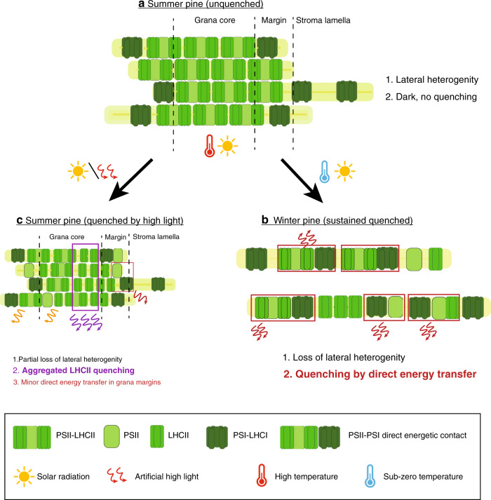
The detailed structural and functional data presented here enable inference of a coherent quenching/photoprotection mechanism involving structural rearrangements of the thylakoids. The model explains Scots pine’s acclimation to the combination of harsh freezing temperatures and high solar radiation that occurs during early spring in northern boreal forests (Fig. 6). Acclimation to early spring conditions (Fig. 6b) results in massive loss of grana stacks (destacking) and formation of uniform membrane structures of stromal thylakoids and phosphorylation of Lhcb1 and/or PsbS, that has been identified in Norway spruce, could be a trigger for the process14. This allows for a redistribution and randomization of PSII and PSI. The reduction in the spatial distance between PSII and PSI complexes increases the probability of direct energy transfer from PSII to PSI46–48. This protective mechanism severely restricts linear electron flow since it strongly quenches PSII activity. It also has strong similarities to the potent quenching observed in other extreme environmental conditions when PSII requires strong protection. Examples include drying-induced quenching in lichen49 and heat-induced quenching in Symbiodinium cells of coral50. In both cases, destacking and reorganization of the thylakoid membrane occur, resulting in proximity of PSII to PSI complexes with direct energy transfer.
Fig. 6. Molecular model for acclimation of photosynthetic machinery under changing natural environmental conditions.
Changes in Summer unquenched (a); Winter quenched (b); Summer quenched (c) in Scots pine. The larger text indicates the process is dominating under that condition. Text colors correspond to the legend colors of the figure.
In conclusion, during early spring only a very small fraction of the absorbed light energy in PSII antennae is utilized for PSII photochemistry (ΦPSII) and the fast inducible NPQ (qE) is low since it not only requires a high ΔpH3,6,17 but there is a strong static quenching present already. On the other hand protection from extreme oxidative stress and rearrangement of thylakoid complexes are likely coupled to the high zeaxanthin content51–53 (Supplementary table 2), and probably to pronounced phosphorylation of antenna and core proteins present under these conditions54. In this situation, the excess energy is dissipated primarily through direct energy transfer from PSII to PSI, which appears in steady-state (PAM) fluorescence measurements as a non-regulated constitutive energy quenching. An additional, albeit smaller, amount of quenching and photoprotection is provided under sustained quenching conditions by the qE mechanism in functionally detached LHCII. It is possible that this fraction is linked to large aggregates containing LHCII, PsbS, and other small proteins, previously detected in cold-adapted pine needles55. Thus, conifers appear to have a powerful protective mechanism that resembles those found in other photosynthetic organisms of relatively harsh environments49,50. This protective mechanism plays a crucial role in the survival of pine from harsh boreal winters.
Methods
Weather
Weather data were recorded from October 2015 to May 2016, from November 2016 to June 2017, and from October 2017 to June 2018 on an hourly basis at the weather station of the Sweden’s meteorological and hydrological institute, Umeå stations. (available on https://www.smhi.se).
Plant material and sampling
Fully developed needles were collected frequently, from Oct 2015 to May 2016, from Nov 2016 to June 2017 and Oct 2017 to June 2018, inclusive, at midday (8.00–10.00) from fully exposed south-facing branches of five Scots pine (Pinus sylvestris L.) trees growing on Umeå University campus (63° 49’ N, 20° 18’ E). The collected needles were immediately stored in darkness, at 0–5 °C, in a laboratory at Umeå Plant Science Centre, and subjected to measurements (described in the following sections) shortly after sampling to minimize any further changes in the photosynthetic machinery. For recovery treatment samples from winter-stressed branches were collected in mid-March and subjected to recovery treatment, involving exposure to very low light (80 µmol photons m−2s−1) at 20 °C55. Further details are provided (together with other details regarding the sampling dates and procedures) in Supplementary Fig. 1.
Modulated chlorophyll fluorescence and P700 + measurements
Chlorophyll fluorescence was measured using a Dual-PAM-100 instrument (Heinz Walz GmbH, Effeltrich, Germany) at room temperature (20 °C) after 30 min dark adaptation at low temperature [below 5 °C, winter samples (samples collected during November to May)] (No changes occurred in sustained quenched (E.Spring) samples during the measuring period) or room temperature [(20 °C, summer samples (samples collected during June to October)]. Maximum photochemical efficiency of PSII was calculated as Fv/Fm = (Fm − Fo)/Fm. To perform quantum yield measurements, we chose to record a light response curve where we measured Fv/Fm first, followed by Pm (maximal P700 oxidation), and then changing the actinic light intensity stepwise from 3 µmol photons m−2s−1 to 1469 µmol photons m−2s−1. During the recording of light response curves, samples were exposed to each light intensity for 3 min followed by a saturating pulse (SP) of 4000 µmol photons m−2s−1 was superimposed in the presence of actinic light to get the maximum chlorophyll fluorescence in light-adapted state (Fm’). All the quantum yields were calculated from the saturating pulse applied after 300 μmol m−2 s−1 of the light illumination period of the light response curve. Partitioning of absorbed light energy in PSII was estimated as ΦPSII + ΦNPQ + ΦNO = 143,44. Where, ΦPSII is the effective quantum yield of PSII, ΦNPQ is the quantum yield of energy dissipated as regulated NPQ mainly by heat, and ΦNO is the fraction of energy dissipated by other non-regulatory pathways. The effective photochemical quantum yield of PSI, Y(I), was calculated as Y(I) = 1−(Y(ND) + Y(NA))56. Here, Y(I) is the effective quantum yield of PSI (ΦPSI) when reaction centers (RCs) are open with reduced donor side (P700) and oxidized acceptor side (A); Y(NA) is the energy dissipation due to acceptor side limitation, reflecting the redox state of the acceptor side when RCs are closed with P700 reduced and both acceptors reduced; and Y(ND) is the energy dissipation due to donor side limitation, reflecting the redox state with both P700 and A oxidized. Steady-state NPQ induction was measured with SP applied under constant actinic light illumination of either 300 (seasonal measurements)/800 (recovery experiment) μmol m−2 s−1 with saturating pulse applied every 30 s.
Ultrafast fluorescence measurements
Intact needles were subjected to ultrafast time-correlated single-photon counting measurements at −20 °C in a rotating cuvette, as previously described17,18. Pine needles were immersed in 60% PEG solution (pH 7.5), which was used as an anti-freeze. Tests confirmed that this solution did not significantly affect either the Fv/Fm values or fluorescence kinetics. These conditions allowed maintenance of the physiological and quenching states for a long time during the measurements and prevention of radiation damage or further quenching induction by the very weak measuring light. Needles sampled in summer (S needles) were measured in both unquenched and fully light-quenched states. For measurement in the unquenched state, they were dark-adapted for 30 min, then quickly frozen to −20 °C in the rotation cuvette. For measurement in the light-quenched state, the needles were illuminated by red light (intensity 1000 µmol m−2sec−1) for 30 min. at room temperature. During this time, fluorescence intensity was monitored and after 30 min a steady-state level was reached. The needles were then frozen quickly under illumination in the rotation cuvette to −20 °C. Early spring samples (ES samples) were measured either in the original “sustained quenching state” by transferring them directly at −20 °C into the cooled rotation cuvette (Supplementary Fig. 4), or in a “recovery state” induced by exposing them to very low light (80 µmol m−2s−1) at 20 °C, as described earlier, then freezing them (as in the measurements of the summer needles in unquenched state). After each fluorescence lifetime measurement, the samples were fast-frozen in liquid nitrogen and stored at −80 °C for further pigment analysis. The laser settings for the measurements were 40–60 µW power, 4 MHz repetition rate, and 1 mm diameter beam size. Excitation wavelength was set to 662 nm to excite Qy band of Chl a in order to resolve fluorescence kinetics by sufficiently advanced kinetic target models established previously25,28,30 without their further overcomplication. Soret band excitation or Chl b excitation would result in substantially more complex fluorescence kinetics than the one observed here due to the contribution of Soret→Qy deactivation and Chl b → Chl a energy transfer pathways, which have multiexponential character57–60. Furthermore, usage of excitation wavelengths further from Boltzmann equilibrium at 665–670 nm, would result in the necessity to include complex equilibration kinetics61.
Pigment and protein analysis
Pigments were extracted from frozen pine needles with 80% acetone. Chl a/b and Chl/carotenoid ratios were calculated by fitting the extracts’ absorption spectra with the spectra of the individual pigments, and the relative amounts of carotenoids were determined by HPLC62. First acetone extracts (~200 μL, Qy OD 0.3–0.8/cm) of the needles were loaded on a SephereClone 5 u ODS1 column (Phenomenex) with a 1.5 mL/min flow rate. Buffer A was composition was acetonitrile/methanol/0.1 M Tris-HCl pH 7.6 in 72:8:10 ratio. Buffer composition was methanol/hexane in 4:1 ratio. The amount of buffer B was linearly increased to 50% in 10 min, held at this value for 0.5 min, and then increased linearly to 100% in 4 min. Thylakoid membranes for SDS-PAGE protein composition analysis were isolated from fully developed Pinus sylvestris needles using three different buffers. First, needles were homogenized in grinding buffer (50 mM Hepes–KOH, pH 7.5; 330 mM sorbitol; 5 mM MgCl2; 10 mM NaF; 10% (w/v) polyethylene glycol, 6000 kDa; 0.075% (w/v) bovine serum albumin; 0.065% (w/v) Na-ascorbate) in a blender (Multiblender Coline, Clas Ohlson), followed by filtering through two layers of Miracloth (VWR, MiliporeSigma, #EM475855-1R). The filtered homogenate was centrifuged at 6000 g for 6 min. The pellet was resuspended in a medium containing: 50 mM Hepes–KOH, pH 7.5; 5 mM MgCl2; 10 mM NaF and centrifuged as above. The pelleted thylakoid membranes were resuspended again in storage buffer (50 mM Hepes–KOH, pH 7.5; 5 mM MgCl2; 100 mM sorbitol; 10 mM NaF), frozen in small aliquots and stored in −80 °C for further use63. Thylakoid membranes containing equal amounts of protein were subjected to SDS-PAGE separation64 using Criterion TGX Any kD™ precasted gels (BioRad, #5678124) and electrophoretically transferred to a PVDF membrane (BioRad, #1704156) using Trans-Blot® Turbo™ Transfer System (BioRad, #1704150). Membranes were probed with specific primary antibodies against: PsbD (1:5000 dilution), PsaD (1:10000 dilution), Lhcb2 (1:15000 dilution) and Lhca4 (1:15000 dilution). After incubation with horseradish peroxidase-conjugated secondary antibody (Goat anti-Rabbit IgG (H&L), #AS09 602, all antibodies from Agrisera AB, Vännäs, Sweden) the antibody complexes were visualized by incubation of the blots in Agrisera ECL chemiluminescent detection set (#AS16 ECL-S-N) (as per manufacturer’s instructions) in Azure c600 developing device (Azure Bioscience, USA)
Transmission electron microscopy
Thin slices (< 0.5 mm) from the middle region of pine needles were cut in tap water and fixed in 4% paraformaldehyde, 2.5% glutaraldehyde (TAAB Laboratories, Aldermaston, England) in 0.1 M or 0.05 M (May and June) sodium cacodylate buffer, pH 7.4 (TAAB Laboratories, Aldermaston, England). Thoroughly washed samples were post-fixed in 1% osmium tetroxide (TAAB Laboratories, Aldermaston, England). The fixed material was dehydrated in ethanol series with increasing concentrations and propylene oxide and finally embedded in Spurr resin (TAAB Laboratories, Aldermaston, England). Ultrathin sections (70 nm) were post contrasted in uranyl acetate and Reynolds lead citrate and further examined with Talos 120 C electron microscope (FEI, Eindhoven, The Netherlands) operating at 120 kV. Micrographs were acquired with a Ceta 16 M CCD camera (FEI, Eindhoven, The Netherlands) using TEM Image & Analysis software ver. 4.14 (FEI, Eindhoven, The Netherlands). The chloroplast ultrastructure was analyzed from the electron micrographs by measuring the average number of chloroplasts per cell, average number of grana per chloroplasts and average number of appressed thylakoids per grana stack (Ng)65,66.
Statistical analysis
The significance of between-mean differences was assessed by t-tests and p values were recorded. One (*), two (**), and three (***) asterisks in the presented figures indicate *≤95%; **≤99%; ***≤99.9% confidence levels, respectively, for fluorescent and electron microscopic measurements.
Reporting summary
Further information on research design is available in the Nature Research Reporting Summary linked to this article.
Supplementary information
Acknowledgements
This work was supported by SE2B Horizon 2020 under Grant agreement no. 675006 (SE2B) to S.J. (Umeå University) and R.C. (Vrije Universiteit Amsterdam), the Swedish Research council (VR), and the Kempe Foundation to S.J. and the Netherlands Organization for Scientific Research (NWO-Vici grant) to R.C. We would like to thank Sara Henriksson of the Umeå Core Facility for Electron Microscopy (UCEM) for help with the sample preperation. We would also like to thank Prof. Eva-Mari Aro for helpful discussions.
Source data
Author contributions
S.J. and A.R.H. conceived the idea; P.B., A.G.I., A.R.H., and S.J. designed the experiments; P.B., Z.Z., S.P., and T.S. performed the chlorophyll fluorescence and P700 measurements; P.B. and A.G.I. analyzed chlorophyll fluorescence data; P.B. performed TEM studies and protein quantification; V.C. performed ultrafast time-resolved fluorescence measurements; V.C. and A.R.H. analyzed the time-resolved fluorescence data; P.B., V.C., A.G.I., A.R.H., R.C., and S.J. analyzed and discussed all results; P.B., A.G.I., V.C., A.R.H., and S.J. wrote the paper.
Funding
Open Access funding provided by University Of Umea.
Data availability
Source and raw data for Figs. 1, 2, 4, and 5 are provided in the source data file. Source data for Fig. 3 is provided in the manuscript. Source data for Supplementary Figs. 2, 3, 4, 5, 6, 7, 8, and 9 are provided in the source data file. Representative electron micrographs in Figs. 4 and 5 are provided in the manuscript, all other electron micrographs which were analyzed to obtain the graphs in Figs. 4 and 5 can be provided upon reasonable request. All Source data are provided with this paper.
Competing interests
The authors declare no competing interests.
Footnotes
Peer review information Nature Communications thanks the anonymous reviewers for their contribution to the peer review of this work. Peer review reports are available.
Publisher’s note Springer Nature remains neutral with regard to jurisdictional claims in published maps and institutional affiliations.
These authors contributed equally: Pushan Bag, Volha Chukhutsina.
Change history
3/8/2021
A Correction to this paper has been published: 10.1038/s41467-021-22013-6
Contributor Information
Alfred R. Holzwarth, Email: a.holzwarth@vu.nl
Stefan Jansson, Email: stefan.jansson@umu.se.
Supplementary information
Supplementary information is available for this paper at 10.1038/s41467-020-20137-9.
References
- 1.Gowthrop D. Vanishing Halo. Greystone Books; 2009. https://books.google.se/books?id=t-SDCgAAQBAJ.
- 2.Nystedt B, et al. The Norway spruce genome sequence and conifer genome evolution. Nature. 2013;497:579–584. doi: 10.1038/nature12211. [DOI] [PubMed] [Google Scholar]
- 3.Li X-P, et al. A pigment-binding protein essential for regulation of photosynthetic light harvesting. Nature. 2000;403:391–395. doi: 10.1038/35000131. [DOI] [PubMed] [Google Scholar]
- 4.Jahns P, Holzwarth AR. The role of the xanthophyll cycle and of lutein in photoprotection of photosystem II. Biochim Biophys. Acta-Bioenerg. 2012;1817:182–193. doi: 10.1016/j.bbabio.2011.04.012. [DOI] [PubMed] [Google Scholar]
- 5.Malnoë A. Photoinhibition or photoprotection of photosynthesis? Update on the (newly termed) sustained quenching component qH. Environ. Exp. Bot. 2018;154:123–133. [Google Scholar]
- 6.Niyogi KK. Photoprotection revisited: genetic and molecular approaches. Annu Rev. Plant Biol. 1999;50:333–359. doi: 10.1146/annurev.arplant.50.1.333. [DOI] [PubMed] [Google Scholar]
- 7.Horton P, Ruban AV, Walters RG. Regulation of light harvesting in green plants. Annu Rev. Plant Biol. 1996;47:655–684. doi: 10.1146/annurev.arplant.47.1.655. [DOI] [PubMed] [Google Scholar]
- 8.Öquist G, Huner NPA. Photosynthesis of overwintering evergreen plants. Annu Rev. Plant Biol. 2003;54:329–355. doi: 10.1146/annurev.arplant.54.072402.115741. [DOI] [PubMed] [Google Scholar]
- 9.Yang Q, et al. Two dominant boreal conifers use contrasting mechanisms to reactivate photosynthesis in the spring. Nat. Commun. 2020;11:1–12. doi: 10.1038/s41467-019-13954-0. [DOI] [PMC free article] [PubMed] [Google Scholar]
- 10.Verhoeven A. Sustained energy dissipation in winter evergreens. N. Phytol. 2014;201:57–65. [Google Scholar]
- 11.Harsch MA, Zhou Y, HilleRisLambers J, Kot M. Keeping pace with climate change: stage-structured moving-habitat models. Am. Nat. 2014;184:25–37. doi: 10.1086/676590. [DOI] [PubMed] [Google Scholar]
- 12.Demmig‐Adams B, Adams WW., III Photoprotection in an ecological context: the remarkable complexity of thermal energy dissipation. N. Phytol. 2006;172:11–21. doi: 10.1111/j.1469-8137.2006.01835.x. [DOI] [PubMed] [Google Scholar]
- 13.Krol M, et al. Low growth temperature inhibition of photosynthesis in cotyledons of jack pine seedlings (Pinus banksiana) is due to impaired chloroplast development. Can. J. Bot. 2002;80:1042–1051. [Google Scholar]
- 14.Grebe S, et al. Specific thylakoid protein phosphorylations are prerequisites for overwintering of Norway spruce (Picea abies) photosynthesis. Proc. Natl Acad. Sci. USA. 2020;117:17499–17509. doi: 10.1073/pnas.2004165117. [DOI] [PMC free article] [PubMed] [Google Scholar]
- 15.Genty B, Briantais J-M, Baker NR. The relationship between the quantum yield of photosynthetic electron transport and quenching of chlorophyll fluorescence. Biochim Biophys. Acta. 1989;990:87–92. [Google Scholar]
- 16.Tiwari A, et al. Photodamage of iron–sulphur clusters in photosystem I induces non-photochemical energy dissipation. Nat. Plants. 2016;2:1–9. doi: 10.1038/nplants.2016.35. [DOI] [PubMed] [Google Scholar]
- 17.Holzwarth AR, Miloslavina Y, Nilkens M, Jahns P. Identification of two quenching sites active in the regulation of photosynthetic light-harvesting studied by time-resolved fluorescence. Chem. Phys. Lett. 2009;483:262–267. [Google Scholar]
- 18.Chukhutsina VU, Holzwarth AR, Croce R. Time-resolved fluorescence measurements on leaves: principles and recent developments. Photosynth Res. 2019;140:355–369. doi: 10.1007/s11120-018-0607-8. [DOI] [PMC free article] [PubMed] [Google Scholar]
- 19.Van Stokkum IHM, Larsen DS, Van, Grondelle R. Global and target analysis of time-resolved spectra. Biochim Biophys. Acta-Bioenerg. 2004;1657:82–104. doi: 10.1016/j.bbabio.2004.04.011. [DOI] [PubMed] [Google Scholar]
- 20.Chukhutsina V. U., Liu Xin, Xu Pengqi CR. The major light-harvesting complex (LHCII) is an antenna of photosystem I in dark-adapted plants. Nat Plants. Published online 2020. 10.1038/s41477-020-0693-4. [DOI] [PubMed]
- 21.Croce R, Dorra D, Holzwarth AR, Jennings RC. Fluorescence decay and spectral evolution in intact photosystem I of higher plants. Biochemistry. 2000;39:6341–6348. doi: 10.1021/bi992659r. [DOI] [PubMed] [Google Scholar]
- 22.Wientjes E, Van Stokkum IHM, Van Amerongen H, Croce R. Excitation-energy transfer dynamics of higher plant photosystem I light-harvesting complexes. Biophys. J. 2011;100:1372–1380. doi: 10.1016/j.bpj.2011.01.030. [DOI] [PMC free article] [PubMed] [Google Scholar]
- 23.Holzwarth A. R. Data analysis of time-resolved measurements. In: Biophysical Techniques in Photosynthesis. Springer; 1996:75–92.
- 24.Holzwarth AR, Müller MG, Niklas J, Lubitz W. Ultrafast transient absorption studies on photosystem I reaction centers from Chlamydomonas reinhardtii. 2: mutations near the P700 reaction center chlorophylls provide new insight into the nature of the primary electron donor. Biophys. J. 2006;90:552–565. doi: 10.1529/biophysj.105.059824. [DOI] [PMC free article] [PubMed] [Google Scholar]
- 25.Miloslavina Y, et al. Charge separation kinetics in intact photosystem II core particles is trap-limited. A picosecond fluorescence study. Biochemistry. 2006;45:2436–2442. doi: 10.1021/bi052248c. [DOI] [PubMed] [Google Scholar]
- 26.Slavov C, Ballottari M, Morosinotto T, Bassi R, Holzwarth AR. Trap-limited charge separation kinetics in higher plant photosystem I complexes. Biophys. J. 2008;94:3601–3612. doi: 10.1529/biophysj.107.117101. [DOI] [PMC free article] [PubMed] [Google Scholar]
- 27.Szczepaniak M, et al. Charge separation, stabilization, and protein relaxation in photosystem II core particles with closed reaction center. Biophys. J. 2009;96:621–631. doi: 10.1016/j.bpj.2008.09.036. [DOI] [PMC free article] [PubMed] [Google Scholar]
- 28.Miloslavina Y, et al. Far‐red fluorescence: A direct spectroscopic marker for LHCII oligomer formation in non‐photochemical quenching. FEBS Lett. 2008;582:3625–3631. doi: 10.1016/j.febslet.2008.09.044. [DOI] [PubMed] [Google Scholar]
- 29.Ware MA, Giovagnetti V, Belgio E, Ruban AV. PsbS protein modulates non-photochemical chlorophyll fluorescence quenching in membranes depleted of photosystems. J. Photochem Photobiol. B Biol. 2015;152:301–307. doi: 10.1016/j.jphotobiol.2015.07.016. [DOI] [PubMed] [Google Scholar]
- 30.Lambrev PH, Miloslavina Y, Jahns P, Holzwarth AR. On the relationship between non-photochemical quenching and photoprotection of photosystem II. Biochim. Biophys. Acta-Bioenerg. 2012;1817:760–769. doi: 10.1016/j.bbabio.2012.02.002. [DOI] [PubMed] [Google Scholar]
- 31.Khorobrykh S, Havurinne V, Mattila H, Tyystjärvi E. Oxygen and ROS in Photosynthesis. Plants. 2020;9:91. doi: 10.3390/plants9010091. [DOI] [PMC free article] [PubMed] [Google Scholar]
- 32.Parker J, Philpott DE. An electron microscopic study of chloroplast condition in summer and winter in Pinus strobus. Protoplasma. 1961;53:575–583. [Google Scholar]
- 33.Martin B, Öquist G. Seasonal and experimentally induced changes in the ultrastructure of chloroplasts of Pinus silvestris. Physiol. Plant. 1979;46:42–49. [Google Scholar]
- 34.Verhoeven A, Osmolak A, Morales P, Crow J. Seasonal changes in abundance and phosphorylation status of photosynthetic proteins in eastern white pine and balsam fir. Tree Physiol. 2009;29:361–374. doi: 10.1093/treephys/tpn031. [DOI] [PubMed] [Google Scholar]
- 35.Huner N, et al. Chloroplast redox imbalance governs phenotypic plasticity: the “grand design of photosynthesis” revisited. Front Plant Sci. 2012;3:255. doi: 10.3389/fpls.2012.00255. [DOI] [PMC free article] [PubMed] [Google Scholar]
- 36.Nilkens M, et al. Identification of a slowly inducible zeaxanthin-dependent component of non-photochemical quenching of chlorophyll fluorescence generated under steady-state conditions in Arabidopsis. Biochim Biophys. Acta-Bioenerg. 2010;1797:466–475. doi: 10.1016/j.bbabio.2010.01.001. [DOI] [PubMed] [Google Scholar]
- 37.Ivanov A, et al. Seasonal responses of photosynthetic electron transport in Scots pine (Pinus sylvestris L.) studied by thermoluminescence. Planta. 2002;215:457–465. doi: 10.1007/s00425-002-0765-x. [DOI] [PubMed] [Google Scholar]
- 38.Sveshnikov D, et al. Excitation energy partitioning and quenching during cold acclimation in Scots pine. Tree Physiol. 2006;26:325–336. doi: 10.1093/treephys/26.3.325. [DOI] [PubMed] [Google Scholar]
- 39.Tikkanen M, Aro E-M. Integrative regulatory network of plant thylakoid energy transduction. Trends Plant Sci. 2014;19:10–17. doi: 10.1016/j.tplants.2013.09.003. [DOI] [PubMed] [Google Scholar]
- 40.Gilmore AM, Ball MC. Protection and storage of chlorophyll in overwintering evergreens. Proc. Natl Acad. Sci. USA. 2000;97:11098–11101. doi: 10.1073/pnas.150237697. [DOI] [PMC free article] [PubMed] [Google Scholar]
- 41.Gilmore AM, Matsubara S, Ball MC, Barker DH, Itoh S. Excitation energy flow at 77 K in the photosynthetic apparatus of overwintering evergreens. Plant Cell Environ. 2003;26:1021–1034. [Google Scholar]
- 42.Ostroumov E. E., Götze J. P., Reus M., Lambrev P. H., Holzwarth A. R. Characterization of fluorescent chlorophyll charge-transfer states as intermediates in the excited state quenching of light-harvesting complex II. Photosynth Res. 144, 1–23 (2020). [DOI] [PubMed]
- 43.Kramer DM, Johnson G, Kiirats O, Edwards GE. New fluorescence parameters for the determination of Q A redox state and excitation energy fluxes. Photosynth Res. 2004;79:209. doi: 10.1023/B:PRES.0000015391.99477.0d. [DOI] [PubMed] [Google Scholar]
- 44.Hendrickson L, Furbank RT, Chow WS. A simple alternative approach to assessing the fate of absorbed light energy using chlorophyll fluorescence. Photosynth Res. 2004;82:73. doi: 10.1023/B:PRES.0000040446.87305.f4. [DOI] [PubMed] [Google Scholar]
- 45.Krause GH. Photoinhibition of photosynthesis. An evaluation of damaging and protective mechanisms. Physiol. Plant. 1988;74:566–574. [Google Scholar]
- 46.Barber J. An explanation for the relationship between salt‐induced thylakoid stacking and the chlorophyll fluorescence changes associated with changes in spillover of energy from photosystem II to photosystem I. FEBS Lett. 1980;118:1–10. [Google Scholar]
- 47.Barber J. Influence of surface charges on thylakoid structure and function. Annu Rev. Plant Physiol. 1982;33:261–295. [Google Scholar]
- 48.Barber J. Photosynthetic energy conversion: natural and artificial. Chem. Soc. Rev. 2009;38:185–196. doi: 10.1039/b802262n. [DOI] [PubMed] [Google Scholar]
- 49.Slavov C, Reus M, Holzwarth AR. Two different mechanisms cooperate in the desiccation-induced excited state quenching in Parmelia lichen. J. Phys. Chem. B. 2013;117:11326–11336. doi: 10.1021/jp402881f. [DOI] [PubMed] [Google Scholar]
- 50.Slavov C, et al. “Super-quenching” state protects Symbiodinium from thermal stress—implications for coral bleaching. Biochim Biophys. Acta-Bioenerg. 2016;1857:840–847. doi: 10.1016/j.bbabio.2016.02.002. [DOI] [PubMed] [Google Scholar]
- 51.Vajravel S, et al. Zeaxanthin and echinenone modify the structure of photosystem I trimer in Synechocystis sp. PCC 6803. Biochim Biophys. Acta-Bioenerg. 2017;1858:510–518. doi: 10.1016/j.bbabio.2017.05.001. [DOI] [PubMed] [Google Scholar]
- 52.Tian L, Xu P, Chukhutsina VU, Holzwarth AR, Croce R. Zeaxanthin-dependent nonphotochemical quenching does not occur in photosystem I in the higher plant Arabidopsis thaliana. Proc. Natl Acad. Sci. USA. 2017;114:4828–4832. doi: 10.1073/pnas.1621051114. [DOI] [PMC free article] [PubMed] [Google Scholar]
- 53.Havaux M, Dall’Osto L, Bassi R. Zeaxanthin has enhanced antioxidant capacity with respect to all other xanthophylls in arabidopsis leaves and functions independent of binding to PSII antennae. Plant Physiol. 2007;145:1506–1520. doi: 10.1104/pp.107.108480. [DOI] [PMC free article] [PubMed] [Google Scholar]
- 54.Merry R, Jerrard J, Frebault J, Verhoeven A. A comparison of pine and spruce in recovery from winter stress; changes in recovery kinetics, and the abundance and phosphorylation status of photosynthetic proteins during winter. Tree Physiol. 2017;37:1239–1250. doi: 10.1093/treephys/tpx065. [DOI] [PubMed] [Google Scholar]
- 55.Ottander C, Campbell D, Öquist G. Seasonal changes in photosystem II organisation and pigment composition in Pinus sylvestris. Planta. 1995;197:176–183. [Google Scholar]
- 56.Schreiber U. Saturation pulse method for assessment of energy conversion in PSI. PAM Appl Notes. 2008;1:11–14. [Google Scholar]
- 57.Gradinaru CC, et al. The flow of excitation energy in LHCII monomers: implications for the structural model of the major plant antenna. Biophys. J. 1998;75:3064–3077. doi: 10.1016/S0006-3495(98)77747-1. [DOI] [PMC free article] [PubMed] [Google Scholar]
- 58.Gradinaru CC, et al. Ultrafast evolution of the excited states in the chlorophyll a/b complex CP29 from green plants studied by energy-selective pump−probe spectroscopy. Biochemistry. 1998;37:1143–1149. [Google Scholar]
- 59.Gradinaru C. C., van Stokkum I. H. M., van Grondelle R., van Amerongen H. Ultrafast Absorption Changes of the Lhcii Carotenoids upon Selective Excitation of the Chlorophylls BT - Photosynthesis: Mechanisms and Effects: Volume I–V: Proceedings of the XIth International Congress on Photosynthesis, Budapest, Hungary, August 17–22. In: Garab G., ed. Springer Netherlands; 1998:277–280. 10.1007/978-94-011-3953-3_64.
- 60.Connelly JP, Müller MG, Bassi R, Croce R, Holzwarth AR. Femtosecond transient absorption study of carotenoid to Chlorophyll energy transfer in the light-harvesting complex II of photosystem II. Biochemistry. 1997;36:281–287. doi: 10.1021/bi962467l. [DOI] [PubMed] [Google Scholar]
- 61.van Grondelle R, Bergström H, Sundström V, Gillbro T. Energy transfer within the bacteriochlorophyll antenna of purple bacteria at 77 K, studied by picosecond absorption recovery. Biochim Biophys. Acta - Bioenerg. 1987;894:313–326. [Google Scholar]
- 62.Xu P, Tian L, Kloz M, Croce R. Molecular insights into Zeaxanthin-dependent quenching in higher plants. Sci. Rep. 2015;5:1–10. doi: 10.1038/srep13679. [DOI] [PMC free article] [PubMed] [Google Scholar]
- 63.Grebe S, et al. The unique photosynthetic apparatus of Pinaceae: analysis of photosynthetic complexes in Picea abies. J. Exp. Bot. 2019;70:3211–3225. doi: 10.1093/jxb/erz127. [DOI] [PMC free article] [PubMed] [Google Scholar]
- 64.Damkjær JT, et al. The photosystem II light-harvesting protein Lhcb3 affects the macrostructure of photosystem II and the rate of state transitions in Arabidopsis. Plant Cell. 2009;21:3245–3256. doi: 10.1105/tpc.108.064006. [DOI] [PMC free article] [PubMed] [Google Scholar]
- 65.Ivanov AG, Ignatova NS, Christov AM. Comparative ultrastructural and fluorescence studies of grapevine (Vitis vinifera L.) chloroplasts isolated from stem and leaf tissues. Plant Sci. 1990;67:253–257. [Google Scholar]
- 66.Ivanov AG, Kitcheva MI, Christov AM, Popova LP. Effects of abscisic acid treatment on the thermostability of the photosynthetic apparatus in barley chloroplasts. Plant Physiol. 1992;98:1228–1232. doi: 10.1104/pp.98.4.1228. [DOI] [PMC free article] [PubMed] [Google Scholar]
Associated Data
This section collects any data citations, data availability statements, or supplementary materials included in this article.
Supplementary Materials
Data Availability Statement
Source and raw data for Figs. 1, 2, 4, and 5 are provided in the source data file. Source data for Fig. 3 is provided in the manuscript. Source data for Supplementary Figs. 2, 3, 4, 5, 6, 7, 8, and 9 are provided in the source data file. Representative electron micrographs in Figs. 4 and 5 are provided in the manuscript, all other electron micrographs which were analyzed to obtain the graphs in Figs. 4 and 5 can be provided upon reasonable request. All Source data are provided with this paper.


