Abstract
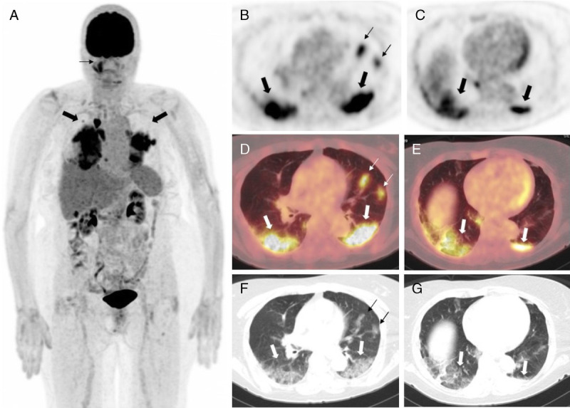
A 69-year-old woman with multiple myeloma came to our department for 18F-FDG PET/CT scan for routine surveillance. The patient denied any history of fever, cough, shortness of breath, or body aches. 18F-FDG PET/CT scan from vertex to knees was performed. PET/CT images revealed extensive peripheral ground-glass opacities showing intense FDG uptake (SUVmax 12) involving bilateral lower lobes. Possibility of an infective etiology including novel coronavirus (COVID-19) infection was raised. The patient’s oropharyngeal swab for COVID-19 by polymerase chain reaction amplification test came back positive for COVID-19 infection. The patient and her husband were advised home quarantine for 14 days.
Key Words: asymptomatic, COVID-19, PET/CT
FIGURE 1.

A, Maximum intensity projection image shows intense FDG uptake in bilateral lungs (thick black arrows). Incidentally noted is uptake in the right maxillary sinus (thin black arrows) suggestive of sinusitis. B and C, Axial PET images showing intense FDG uptake in the peripheral aspect of bilateral lower lobes (thick black arrows) and in the inferior lingular segment (thin black arrows). D and E, Fused PET/CT images show intense FDG uptake (SUVmax 12) in the peripheral aspect of bilateral lower lobes (thick white arrow). Patchy uptake also noted in the inferior lingular segment (thin white arrow). F and G, CT images (lung window) show extensive ground-glass opacities (GGOs) involving the subpleural bilateral lower lobes (thick white arrow), along with patchy GGOs in the inferior lingular segment (thin black arrow). The patient white blood cell count was within normal limits (5300/μL); C-reactive protein was elevated (4.7 mg/dL). The patient’s oropharyngeal swab for novel coronavirus (novel coronavirus [COVID-19], ePlex SARS-CoV-2 [severe acute respiratory syndrome–coronavirus 2] panel) by polymerase chain reaction amplification test came back positive for COVID-19 infection. Radiological imaging can help in the detection of such asymptomatic patients who obtain imaging for cancer surveillance. Many recently published articles described the incidentally detected CT findings in asymptomatic patients. In the early stages of the disease, CT scan of the chest can show patchy peripheral predominant GGOs; later as the disease progresses, these can coalesce to form larger areas of GGOs or consolidation areas.1–3 The imaging features are similar to those seen in MERS (Middle East respiratory syndrome)–CoV and SARS-CoV-2 viral pneumonia.4 FDG PET/CT plays an important role in detecting asymptomatic patients. Many authors have described the 18F-FDG PET/CT features in COVID-19 patients. In most patients, there was predominant bilateral peripheral GGOs with moderate FDG uptake with SUVmax 3 to 8.4–7 In our case, there were extensive GGOs involving peripheral bilateral lower lobes, which showed very intense FDG uptake with SUVmax 12. Patchy GGOs were also noted in bilateral upper lobes, which also showed intense FDG uptake. Although FDG PET/CT is not used for the diagnosis of COVID-19, it can play an important role in the detection of COVID-19 infection in asymptomatic patients.8 Radiologists should be aware of imaging findings of COVID-19 infection in asymptomatic patients. Early suspicion and testing can help in early detection and isolation of such patients, which can help prevent severe illness and stop the community transmission. Health care personnel doing procedures that can cause aerosols from the patients should use proper personal protective equipment including N95 masks, face shields, gown, and gloves as recommended.
Footnotes
Conflicts of interest and sources of funding: none declared.
REFERENCES
- 1.Kanne JP Little BP Chung JH, et al. Essentials for radiologists on COVID-19: an update—Radiology scientific expert panel. Radiology. 2020;296:E113–E114. [DOI] [PMC free article] [PubMed] [Google Scholar]
- 2.Shi H Han X Jiang N, et al. Radiological findings from 81 patients with COVID-19 pneumonia in Wuhan, China: a descriptive study. Lancet Infect Dis. 2020;20:425–434. [DOI] [PMC free article] [PubMed] [Google Scholar]
- 3.Lee EYP, Ng MY, Khong PL. COVID-19 pneumonia: what has CT taught us? Lancet Infect Dis. 2020;20:384–385. [DOI] [PMC free article] [PubMed] [Google Scholar]
- 4.Koo HJ Lim S Choe J, et al. Radiographic and CT features of viral pneumonia. Radiographics. 2018;38:719–739. [DOI] [PubMed] [Google Scholar]
- 5.Qin C Liu F Yen TC, et al. 18F-FDG PET/CT findings of COVID-19: a series of four highly suspected cases. Eur J Nucl Med Mol Imaging. 2020;47:1281–1286. [DOI] [PMC free article] [PubMed] [Google Scholar]
- 6.Zou S, Zhu X. FDG PET/CT of COVID-19. Radiology. 2020;296:E118. [DOI] [PMC free article] [PubMed] [Google Scholar]
- 7.Polverari G Arena V Ceci F, et al. 18F-fluorodeoxyglucose uptake in patient with asymptomatic severe acute respiratory syndrome coronavirus 2 (coronavirus disease 2019) referred to positron emission tomography/computed tomography for NSCLC restaging. J Thorac Oncol. 2020;15:1078–1080. [DOI] [PMC free article] [PubMed] [Google Scholar]
- 8.Doroudinia A, Tavakoli M. A case of coronavirus infection incidentally found on FDG PET/CT scan. Clin Nucl Med. 2020;45:e303–e304. [DOI] [PMC free article] [PubMed] [Google Scholar]


