Abstract
The near‐minimal bacterium Mesoplasma florum is an interesting model for synthetic genomics and systems biology due to its small genome (~ 800 kb), fast growth rate, and lack of pathogenic potential. However, fundamental aspects of its biology remain largely unexplored. Here, we report a broad yet remarkably detailed characterization of M. florum by combining a wide variety of experimental approaches. We investigated several physical and physiological parameters of this bacterium, including cell size, growth kinetics, and biomass composition of the cell. We also performed the first genome‐wide analysis of its transcriptome and proteome, notably revealing a conserved promoter motif, the organization of transcription units, and the transcription and protein expression levels of all protein‐coding sequences. We converted gene transcription and expression levels into absolute molecular abundances using biomass quantification results, generating an unprecedented view of the M. florum cellular composition and functions. These characterization efforts provide a strong experimental foundation for the development of a genome‐scale model for M. florum and will guide future genome engineering endeavors in this simple organism.
Keywords: Mesoplasma florum, Mollicutes, synthetic genomics, systems biology, whole‐cell characterization
Subject Categories: Microbiology, Virology & Host Pathogen Interaction
A deep characterization of the near‐minimal bacterium M. florum reveals important features of this emerging model organism for systems and synthetic biology.

Introduction
Since the first report of the in vitro synthesis of a complete gene (Agarwal et al, 1970), DNA synthesis and assembly techniques have improved considerably in terms of efficiency and capacity (Hughes & Ellington, 2017; Schindler et al, 2018). Large DNA molecules such as entire chromosomes can now be synthetized at reasonable cost, enabling the creation of synthetic or semi‐synthetic organisms, an emerging field known as synthetic genomics (Montague et al, 2012; van der Sloot & Tyers, 2017; Mitchell & Ellis, 2017; Schindler et al, 2018). Given proper design, synthetic organisms could play a very important role in addressing some of the most critical challenges of the 21st century such as the development of sustainable energy sources, the fight against antibiotic resistance, and the treatment of diseases such as cancer and diabetes (Khalil & Collins, 2010; Alper et al, 2010; Cambray et al, 2011).
Although the tools to build artificial chromosomes are now available, not even a handful of significatively modified synthetic genomes have been reported (Hutchison et al, 2016; Richardson et al, 2017; Fredens et al, 2019), and our ability to design complete genomes from scratch is extremely poor at best. Consequently, little is still truly understood about genome design principles. This is mainly explained by the overwhelming complexity of common model organisms, which outstrips our current analytical skills and inhibits our ability to rationally evaluate genome designs. Moreover, the number of possible artificial genome configurations can quickly become overwhelming, even for small genome bacteria. In that context, systems biology approaches such as genome‐scale metabolic models (GEMs) could soon become powerful tools to systematically evaluate genome designs and help select the most promising scenarios for total synthesis (preprint: Chalkley et al, 2019; Rees‐Garbutt et al, 2020). GEMs consist of mathematically structured knowledge frameworks describing the metabolism of organisms, offering phenotypic predictions capabilities useful in a wide‐range of applications from omics data integration to metabolic engineering (Oberhardt et al, 2009; Durot et al, 2009; Bordbar et al, 2014; O’Brien et al, 2015; Ebrahim et al, 2016; Kim et al, 2016; Gu et al, 2019). For example, the impact of multiple gene deletions or environmental stresses on metabolic fluxes and growth rate can be predicted, providing context‐specific hypotheses prior to experimental testing. To perform accurate predictions, GEMs must however be constrained and validated by experimental data such as the biomass composition of the cell (% of DNA, RNA, proteins, etc.) (Feist & Palsson, 2010; Lachance et al, 2019b). To date, more than 100 high‐quality GEMs have been reconstructed, including GEMs for many model organisms such as Escherichia coli, Saccharomyces cerevisiae, and Homo sapiens (Norsigian et al, 2020). GEMs have also been extended to include additional cellular processes such as proteome expression, thereby increasing their capabilities and breadth of applications (King et al, 2015; O’Brien & Palsson, 2015).
Because of their exceptionally small genomes (0.58–2.2 Mbp) (Sirand‐Pugnet et al, 2007), near‐minimal bacteria of the Mollicutes class have long been proposed as models to study the basic principles of life (Morowitz, 1984). These very small (0.2–0.6 µm) wall‐less bacteria do not constitute ancient or primitive forms of life but rather evolved from low G‐C content Gram positive bacteria through a process of massive gene loss (Pettersson & Johansson, 2002; Maniloff, 2002). This resulted in a drastic simplification of their metabolism, with many incomplete or missing metabolic pathways (Dybvig & Voelker, 1996; Pollack et al, 1997). The genomic simplicity of Mollicutes thus offers a unique opportunity to achieve an unprecedented characterization of cellular processes, reduces the number of artificial genome configurations to be tested using synthetic genomics approaches, and decreases the costs related to chromosome synthesis (Xavier et al, 2014; Lachance et al, 2019a). Among all Mollicutes, members of the Mycoplasma genus are the most extensively studied, with many species infecting various animals, including humans (Dybvig & Voelker, 1996; Maniloff, 2002). However, mycoplasmas recently gained particular attention with the development of whole‐genome chemical synthesis, assembly, and cloning in yeast (Gibson et al, 2008; Gibson & Benders, 2008; Benders et al, 2010). The total synthesis and cloning of the 1.08 Mb Mycoplasma mycoides subspecies capri GM12‐based genome followed by its transplantation into a recipient bacterium (Mycoplasma capricolum subspecies capricolum) notably led to the creation of the first cell controlled by an entirely synthetic chromosome, JCVI‐syn1.0 (Gibson et al, 2010; Sleator, 2010). This impressive tour de force recently culminated with the creation of the first artificial “working approximation” of a minimal cell, JCVI‐syn3.0 (Hutchison et al, 2016). This minimal bacterium harbors a reduced and synthetic version of the M. mycoides subspecies capri genome totalizing 531 kb and 473 genes (GenBank: CP014940.1), making it the smallest genome ever observed in any autonomously replicating cell (Hutchison et al, 2016; Glass et al, 2017). The JCVI‐syn3.0 strain however showed altered morphological traits and impaired growth rates compared with the M. mycoides parent strain (doubling time of ~ 2–3 h vs. ~ 1 h), which were restored by the incorporation of 19 additional genes (Breuer et al, 2019). The resulting strain, named JCVI‐syn3A, carried a genome of 543 kb and 493 genes (GenBank: CP016816.2).
First described in 1984 as Acholeplasma florum (McCoy et al, 1984), the near‐minimal bacterium Mesoplasma florum constitutes another member of the Mollicutes class particularly well suited for synthetic genomics and systems biology studies. While closely related to M. mycoides, M. florum however has a smaller genome, shows faster growth rates, and has no pathogenic potential (Sirand‐Pugnet et al, 2007; Gibson et al, 2010; Matteau et al, 2015; Baby et al, 2018). The genome of the L1 type strain, for example, comprises only 793 kb and 720 predicted genes (GenBank: AE017263.1), while the genome of the M. mycoides capri LC GM12 accounts for 1.09 Mb and 879 genes (GenBank: CP001621.1). These features greatly facilitate the manipulation of M. florum and its distribution throughout the scientific community. As for most Mollicutes, M. florum also uses an alternative genetic code (Mycoplasma/Spiroplasma code) that limits the exchange of genetic material from and to other microorganisms (Navas‐Castillo et al, 1992). Importantly, genetic manipulation tools have recently been developed specifically for this bacterium, including procedures for whole‐genome cloning in yeast and genome transplantation (Matteau et al, 2017; Baby et al, 2017). Gene conservation and essentiality analyses have also showed that 57 putatively essential M. florum genes have no homolog in the synthetic JCVI‐syn3.0 strain, suggesting that different minimal genome compositions and configurations probably exist, even within closely related species (Baby et al, 2018). In addition, these analyses enabled the formulation of different genome reduction scenarios for M. florum, providing starting points for genome minimization efforts (Baby et al, 2018). The comparison of the JCVI‐syn3.0 genome with other minimal genomes offers a unique opportunity to decipher genome design principles and some of the most fundamental principles of life, in the same way that the two first complete bacterial genome sequences provided insights about the minimal gene set required for cellular life (Mushegian & Koonin, 1996).
Here, we report the first integrative characterization of M. florum to advance fundamental knowledge on this emerging model. More specifically, we accurately measured several physical and physiological parameters of M. florum L1 growing in rich medium, including the cell diameter, buoyant density, dry mass, optimum growth temperature, growth rate, and growth kinetics. We also defined the macromolecular composition of the cell, identified and characterized more than 400 active promoters, and proceeded to the reconstruction of M. florum transcription units (TUs). Finally, we used transcriptomics and proteomics expression datasets to estimate RNA and protein species abundances, revealing the relative importance of the different cellular processes of a near‐minimal cell. Our work contributes to a detailed understanding of global cell functioning in a simple organism and provides an experimental foundation for the development of a systems and synthetic biology platform.
Results
Mesoplasma florum optimal growth temperature and growth kinetics
The doubling time and the optimal growth temperature represent fundamental parameters in the characterization of a bacterial strain. Moreover, the doubling time is a critical constraint in many cellular modeling approaches such as GEMs (Feist & Palsson, 2010; Lachance et al, 2019b). Accurate measurement of these parameters has however never been reported specifically for M. florum. The optimal growth temperature of the type strain M. florum L1 was therefore evaluated in ATCC 1161 medium by measuring the doubling time at different incubation temperatures typically used for Mollicutes (~ 30–38°C) (Brown et al, 2007). Doubling times were determined using colorimetric assays that measure the time needed for twofold culture dilutions to reach the same optical density at 560 nm (OD560 nm). Raw M. florum growth curves are presented in Appendix Fig S1. The smallest doubling time was observed at a temperature of 34°C (38 ± 5 min) while no growth was observed at a temperature higher than 36°C (Fig 1A), contrasting with pathogenic mycoplasmas such as M. mycoides, M. capricolum, and Mycoplasma pneumoniae. These results are consistent with previous observations concerning different M. florum strains (McCoy et al, 1984) and other members of the Mesoplasma genus (Tully et al, 1994).
Figure 1. Analysis of M. florum growth in ATCC 1161 medium.
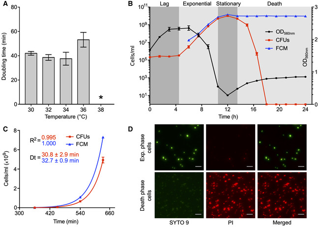
- M. florum doubling time at different incubation temperatures measured by colorimetric assays. The bars represent the mean and standard deviation values obtained from three technical replicates. The asterisk indicates the absence of significant growth, preventing the calculation of a doubling time.
- M. florum growth kinetics at 34°C. Growth was monitored for 24 h by measuring the optical density at 560 nm (black circles) as well as the cell concentrations using two different methods, colony‐forming units (CFUs, red squares) and flow cytometer (FCM, blue triangles). The four typical bacterial growth phases (lag, exponential, stationary, and death) are represented by gray shading. The dots and error bars indicate the mean and standard deviation values obtained from three independent biological replicates. CFU data points superimposed to the x‐axis represent values below the limit of detection (2 × 10⁻²).
- Exponential growth fit on CFU (red squares) and FCM (blue triangles) counts shown in B. Calculated doubling times (Dt) and correlation coefficients (R 2) are shown. The dots and error bars indicate the mean and standard deviation values obtained from three independent biological replicates.
- Representative images of SYTO 9 and propidium iodide (PI) double‐stained M. florum cells, harvested from an exponential or death‐phase culture, observed by widefield fluorescence microscopy. The brightness of each channel was adjusted equally between conditions. Scale bar: 5 µm.
We then used flow cytometry (FCM) and colony‐forming units (CFUs) to precisely measure the growth kinetics of M. florum incubated at the optimal growth temperature (34°C). We first validated that cell concentrations measured by FCM were well correlated with culture dilutions (Appendix Fig S2). By following cell concentrations over ~ 24 h, we could observe an overall pattern corresponding to the four typical bacterial growth phases (Fig 1B). The exponential phase coincided with a substantial drop in medium pH (from ~ 8.0 to 6.5) causing the phenol red present in the culture medium to change color from red to orange, corresponding to an important decrease in measured OD560 nm. Using exponential curve fitting on FCM and CFUs data, we determined a doubling time of 30.8 ± 2.9 min and 32.7 ± 0.9 min, respectively (Fig 1C). CFU and FCM cell concentrations were highly consistent with each other until late stationary phase, where they both reached a plateau at ~ 1 × 10¹⁰ cells/ml and started to diverge. The stationary phase was also marked by the lowest OD560 nm value observed for the entire experiment, corresponding to a yellow medium color and a medium pH around 6.0. This was followed by a gradual formation of cell aggregates in the culture, resulting in a notable increase in the measured OD560 nm. This phenomenon was accompanied by a rapid diminution of CFU counts, suggesting an important loss in cell viability reminiscent of the death phase (Fig 1B). We validated that the decrease in CFU counts was effectively due to an altered cell viability by SYTO 9 and propidium iodide (PI) dual staining fluorescence microscopy (Fig 1D). As expected, M. florum cells harvested at the death phase showed an intense PI signal and practically no SYTO 9 fluorescence, indicating a significantly compromised cell membrane integrity. Similar signals were observed for formaldehyde fixed and permeabilized cells (Appendix Fig S3), whereas exponential‐phase cells showed a strong SYTO 9 fluorescence and almost no PI signal, typical of healthy cells (Fig 1D).
Physical characteristics and macromolecular composition of the cell
To better define the physical constraints shaping the biology of M. florum, we sought to precisely measure its cell diameter since the only quantitative data available for this species relied on filtration studies (McCoy et al, 1984). Filtration constitutes an indirect approach that can be subjected to different sources of variation such as pore size heterogeneity and deformation of cellular morphology, especially for wall‐less bacteria. We analyzed exponential‐phase M. florum cells using two different techniques, transmission electron microscopy (TEM) and stimulated emission depletion (STED) microscopy. Cells were stained with PicoGreen and mCLING (Revelo et al, 2014), respectively, targeting the DNA and the cellular membrane, prior to STED microscopy examination. Representative images obtained from both techniques are shown in Fig 2A and B. Both TEM and STED microscopy showed predominantly ovoid cells, with a cell diameter ranging from approximately 300 to 600 nm and 500 to 1,000 nm, respectively (Fig 2C). An average cell diameter of 434 ± 53 nm was observed for TEM and 741 ± 98 nm for STED microscopy (Fig 2D). The significant difference observed by the two methods is most likely caused by biases associated with sample preparation. TEM, for example, requires a dehydration of the cells with ethanol, which can cause cell shrinkage and therefore a reduction in their apparent diameter (Zhang et al, 2017). STED, on the other hand, requires the use of a mounting media during slide preparation that can cause sample distortion and alteration of morphological features (Peterson et al, 2015; Fouquet et al, 2015; GE, 2018). Interestingly, TEM pictures also showed evidences of a polysaccharidic layer on the periphery of M. florum cells, a morphological feature shared by many Mollicutes including the closely related M. mycoides and M. capricolum (Bertin et al, 2013; Gaurivaud et al, 2014; Daubenspeck et al, 2014; Bertin et al, 2015).
Figure 2. M. florum physical characteristics.
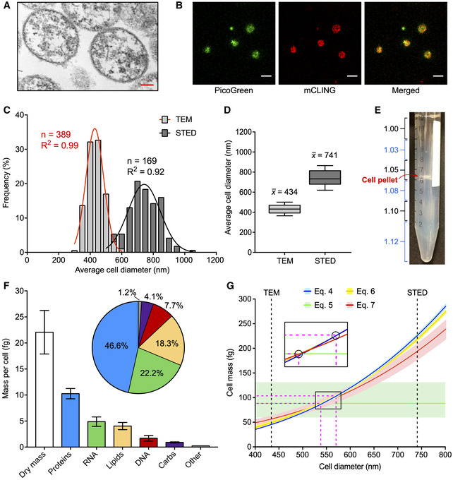
- Representative image of M. florum cells observed by transmission electronic microscopy (TEM). Scale: 100 nm.
- Representative image of PicoGreen (DNA) and mCLING (cellular membrane) double‐stained M. florum cells observed by stimulated emission depletion (STED) microscopy. Scale: 1 µm.
- Frequency distribution of M. florum average cell diameter measured by TEM and STED as shown in A and B, respectively. The average cell diameter was obtained by averaging the minor and major axis values measured for each cell. A Gaussian curve fit is indicated for each method, and the calculated correlation coefficients are shown. Bins: 50 nm.
- Boxplots showing the median and interquartile range of the average cell diameter calculated from 389 and 169 individual cells analyzed by TEM and STED, respectively. Whiskers indicate the 10–90 percentile range.
- Picture of M. florum cells analyzed by discontinuous density gradient centrifugation in Percoll. The approximative density of each Percoll layer is indicated (g/ml) and colored in blue if trypan blue was added to the layer. The position of the cell pellet is marked.
- M. florum biomass quantification. The mass of each macromolecular constituent is shown as well as its relative fraction in the quantified cellular dry mass. Bars represent the mean and standard deviation values obtained from three independent biological replicates (dry mass) or four technical replicates (proteins, RNA, lipids, DNA, carbs). The “Other” category bar represents the residual mass obtained by the subtraction of all quantified macromolecule masses from the total dry mass value.
- Graph showing the relation between the M. florum cell diameter (d) and its cell mass (CM) according to cell mass Equations (4), (5), (6), (7), (4.1), (4.2), (4.3) (see Materials and Methods). For each equation, the mean cell mass (CMmean) is indicated by a colored line, and the range of probable values (CMmin − CMmax) is shown by a light‐colored shading. The mean values of the average cell diameter measured by TEM and STED (see panel D) are indicated by black dashed lines. The portion of the graph where all the CMmean curves converge is enlarged and devoid of colored shadings for representation purposes. CMmean interception points encompassing all other interception points are encircled, and their corresponding x and y coordinates are indicated by fuchsia dashed lines (most probable cell diameter and most probable cell mass ranges).
Measuring the total mass of a cell requires specialized equipment and can be very challenging, especially for small cells (Bryan et al, 2014; Zhao et al, 2014; Rahman et al, 2015). The cell mass can however be estimated using different mathematical equations that involve only a limited number of variables more easily amenable to quantification, including the cell diameter, buoyant density, and dry mass. Since we had already measured the cell diameter of M. florum using TEM and STED microscopy, we next evaluated its buoyant density by discontinuous Percoll density gradient centrifugation. After one or two rounds of centrifugation, the M. florum cell pellet was located at the bottom of the 1.05 g/ml Percoll layer, indicating a buoyant cell density lying between 1.05 and 1.08 g/ml (Fig 2E and Table 1). We next determined the M. florum cell dry mass using conventional weighting procedures performed on exponential‐phase batch cultures (see Materials and Methods and Fig EV1), and observed a total dry mass of 22.1 ± 4.2 fg per cell (Fig 2F and Table 1). The measured buoyant cell density and cell dry mass were then used to infer the most probable M. florum cell mass using four different equations (see Equations (4), (5), (6), (7), (4.1), (4.2), (4.3) in Materials and Methods section). Three of those equations also require the total dry mass fraction and the dry mass density to estimate the total mass of the cell, which were assumed to be within typical ranges found in bacteria, i.e., 20–30% and 1.3–1.5 g/ml, respectively (Bakken & Olsen, 1983; Bratbak & Dundas, 1984; Bratbak, 1985; Fischer et al, 2009; Bionumbers, 2015). Interestingly, all four equations converged to a relatively tight range of cellular mass (88.2–103.3 fg), which corresponded to a cell diameter (538–570 nm) positioned in‐between average values obtained by TEM and STED microscopy and within the overlapping portion of their relative distribution (Fig 2C and G, and Table 1). Refining the cell diameter also allowed the estimation of the most probable cell volume (0.082–0.097 µm3), cell surface area (0.911–1.021 µm2), and surface area to volume ratio (SA:V; 10.5–11.1 µm−1) using Equations (1), (2), (3) (see Materials and Methods), respectively (Table 1).
Table 1.
Summary of Mesoplasma florum biomass composition and physical characteristics measured or estimated in this study.
| Cellular biomass | Mean ± SD (fg) | Physical parameters | Most probable values |
|---|---|---|---|
| Dry mass | 22.1 ± 4.2 | Density | 1.05–1.08 g/ml a |
| Proteins | 10.3 ± 1.0 | Cell diameter | 538–570 nm b |
| RNA | 4.9 ± 0.9 | Cell mass | 88.2–103.3 fg b |
| Lipids | 4.0 ± 0.7 | Cell volume | 0.082–0.097 µm3 c |
| DNA | 1.7 ± 0.5 | Cell surface area | 0.911–1.021 µm2 c |
| Carbohydrates | 0.9 ± 0.1 | SA:V | 10.5–11.1 µm−1 c |
Figure EV1. Overview of the experimental procedures used to determine the total dry mass of M. florum as well as the mass of its principal cellular macromolecules.
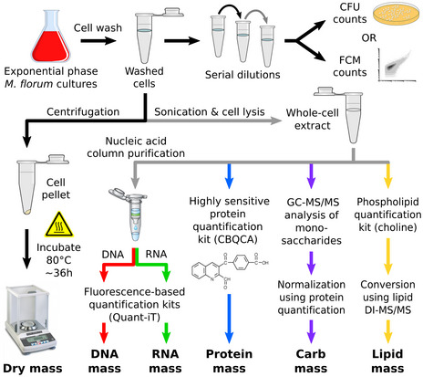
Each constituent is quantified using high sensitivity assays or mass spectrometry methods. Quantification results are normalized according to the number of cells used for each experiment. See Appendix Supplementary Methods for further details.
The vast majority of the cell dry mass can be divided into four classes of macromolecules: proteins, lipids, nucleic acids, and carbohydrates (Cooper & Hausman,). To better define the M. florum dry mass, we quantified each of these macromolecules using different high sensitivity quantification assays and gas chromatography‐mass spectrometry (GC‐MS) methods (see Materials and Methods and Fig EV1). According to our analysis, nearly two‐thirds of the total dry mass was composed of proteins and RNA, with a relative abundance of approximately 46.6 and 22.2%, respectively (Fig 2F and Table 1). The remaining fraction of the dry mass was divided as follows: 18.3% for lipids (Dataset EV8), 7.7% for DNA, and 4.1% for carbohydrates. Overall, these results are comparable to fractions observed in other Mollicutes species (Razin et al, 1963). The majority of the M. florum carbohydrate fraction most probably accounts for the polysaccharidic layer observed by TEM. Carbohydrates detected by mass spectrometry were mainly composed of galactose (0.50 ± 0.07 fg), glucose (0.19 ± 0.03 fg), rhamnose (0.18 ± 0.01 fg), and mannose (0.04 ± 0.01 fg), representing approximately 54.9, 20.6, 20.0, and 4.5% of the total carbohydrate mass, respectively. Interestingly, the residual dry mass, i.e., the difference between the quantified dry mass and the sum of all quantified macromolecules, represented only 1.2% (0.26 fg) of the total dry mass, most likely accounting for small molecules, metabolites, cofactors, and ions (Fig 2F).
Genome‐wide identification of promoters
Transposon mutagenesis and gene conservation datasets have recently been published for M. florum and allowed the proposition of different genome reduction scenarios for this bacterium (Baby et al, 2018). However, these predictions did not account for promoter organization and therefore retained all intergenic regions in the reduced genome designs. The identification of all M. florum promoters and corresponding transcription units (TUs) would certainly improve the quality and accuracy of these predictions, in addition to providing highly valuable information about the transcriptome of this near‐minimal cell. We therefore proceeded to the cartography of all M. florum transcription start sites (TSSs) at single nucleotide resolution using a previously described genome‐wide 5′‐rapid amplification of cDNA ends (5′‐RACE) method (Carraro et al, 2014; Matteau & Rodrigue, 2015). Following Illumina sequencing (see Appendix Table S1 for a summary of library statistics), the number of read starts per million of mapped reads (RSPM) was calculated for each genomic position in a strand‐specific manner, resulting in a frequency distribution reminiscent of a Poisson distribution (Appendix Fig S4A). Out of 1,586,448 possible sites (genome size multiplied by two to account for both strands), a total of 68,650 sites had a non‐null TSS signal, of which 1,514 (< 0.1% of all sites) displayed a significant intensity (see Appendix Fig S4B and Materials and Methods for further details). This resulted in the identification of 605 candidate TSSs distributed throughout the M. florum chromosome (Fig 3A). DNA sequence analysis using the MEME software (Bailey & Elkan, 1994) revealed a conserved promoter motif present in 422 candidate TSSs highly reminiscent of promoter sequences identified in other Mollicutes species (Fig 3B and C), including M. pneumoniae, Mycoplasma hyopneumoniae, Acholeplasma laidlawii, and Mycoplasma gallisepticum (Weiner III, 2000; Güell et al, 2009; Weber et al, 2012; Yus et al, 2012; Mazin et al, 2014; Lloréns‐Rico et al, 2015; Fisunov et al, 2016). More precisely, this promoter motif contained a −10 box typical of the sequences recognized by the principal σ factor in most bacteria (TAWAAT) (Helmann, 1995; Shultzaberger et al, 2007), as well as a partially degenerated TGN extension of the −10 box (EXT element) (Fig 3B). No clear evidence of a conserved −35 box emerged from the analysis. The occurrence of this promoter sequence was validated in ~ 85% (357) of cases using the MAST software (Bailey & Gribskov, 1998), which also provided evidences for an additional 10 sites not initially included in the MEME constructed motif, for a grand total of 432 motif‐associated TSSs (Fig 3C and Dataset EV1). No promoter motif could be identified for the remaining TSS candidates, suggesting a higher sequence variability at these sites or experimental artifacts.
Figure 3. Identification and analysis of M. florum promoters.
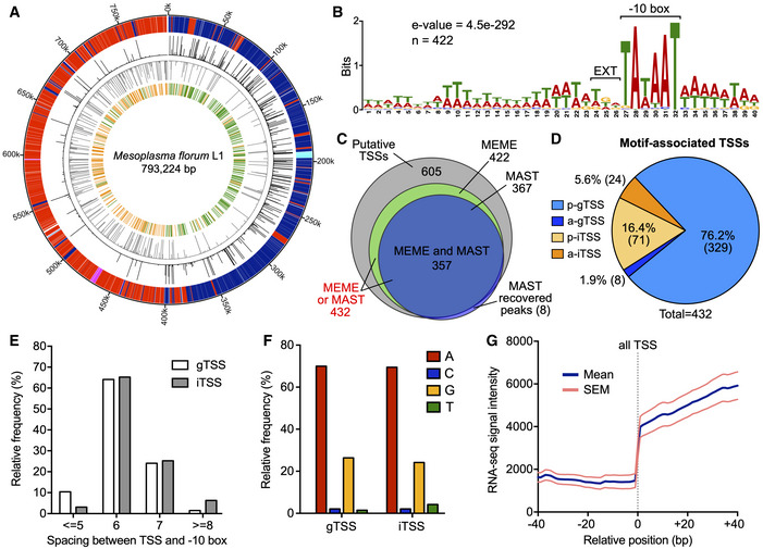
- Circular representation of the M. florum L1 chromosome enhanced with 5′‐RACE data generated in this study. Outer to inner circle: genomic coordinates (kbp); genes encoded on the positive (blue for coding sequences, turquoise for RNAs) and negative (red for coding sequences, fuchsia for RNAs) DNA strands; raw 5′‐RACE signal (0–1,000 read starts scale) observed at each genomic position for the positive (black) and negative (gray) DNA strands; putative transcription start sites (TSSs) identified on the positive (green) and negative (orange) DNA strands from significant 5′‐RACE peaks.
- M. florum promoter motif determined using the MEME software (Bailey & Elkan, 1994) from the DNA sequence located upstream the 605 putative TSSs identified by 5′‐RACE. A total of 422 sites across the genome were included in the motif. The position of the −10 box (TAWAAT) and the extended element (EXT) is indicated.
- Venn diagram illustrating the number of TSSs associated with a conserved promoter motif (see panel B) found by MEME, MAST, or both software compared with the total number of putative TSSs passing filters (605). Eight additional putative TSSs were added to the initial set according to the MAST search.
- Localization and orientation of TSSs associated with a MEME or MAST promoter motif. p‐gTSS, parallel intergenic TSS; a‐gTSS, antiparallel intergenic TSS; p‐iTSS, parallel internal TSS; a‐iTSS, antiparallel internal TSS. For gTSSs, the orientation was defined according to the closest downstream gene, while the overlapping gene was used in the case of iTSSs.
- Relative frequency distribution of the spacing between TSSs and their associated promoter −10 box.
- Nucleotide identity at the transcription initiation site (+1) for gTSSs and iTSSs associated with a promoter motif.
- Aggregate profile showing the mean RNA‐seq read coverage observed at and around all motif‐associated TSSs identified in this study. The calculated SEM is also shown. The aggregate profile was centered on the TSSs coordinates (relative position 0 bp), indicated by a gray dashed line.
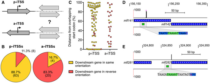
As expected, the vast majority (78.0%) of motif‐associated TSSs were located within intergenic regions of the chromosome (gTSSs), even though these regions occupy only ~ 6.1% of the genome (Fig 3D) (Baby et al, 2018). Interestingly, putative TSSs devoid of a promoter motif were located within coding sequences (CDS) in more than 90% of all instances, clearly contrasting with motif‐associated TSSs (Fig EV2A). In most cases (76.2%), motif‐associated gTSSs were in the same orientation (parallel) as their closest downstream gene (p‐gTSS), with only a few cases (1.9%) of antiparallel downstream associated gene (a‐gTSS) (Fig 3D). The remaining TSSs (22.0%) were found to be internal to coding regions of the genome (iTSS), most of the time in the same orientation as the overlapping gene in which they occur (p‐iTSS). In total, nearly 12% of M. florum genes contained at least one motif‐associated iTSS (Fig EV3D). p‐iTSSs were found to be remarkably enriched near the end of their overlapping gene (Fig EV4C), with several instances separated by less than 100 bp from the next correctly oriented downstream gene (see Fig EV4D for a visual example). A few cases of p‐iTSS were also precisely located on the first base of translation start codons, suggesting the transcription of leaderless mRNA (Weiner III, 2000; Moll et al, 2002; Zheng et al, 2011; Nakagawa et al, 2017) (Fig EV4C and E). gTSSs and iTSSs shared approximately the same distribution regarding their relative spacing with the conserved promoter motif, predominantly separated by 6 or 7 bases from the −10 box most proximal extremity (Fig 3E). Both TSS types were also located preferentially on coordinates corresponding to purine nucleotides (A or G), yet with an important bias for adenine (~ 70% of cases), reflecting the low G‐C nature of the M. florum genome (Fig 3F). Despite these similarities, motif‐associated gTSSs displayed a significantly higher signal intensity compared with motif‐associated iTSSs, the latter group being not significantly different from TSSs without promoter motif (Fig EV2B). TSSs lacking the M. florum promoter motif were however not enriched for purine nucleotides like motif‐associated gTSSs and iTSSs (Fig EV2C). Further information about the genetic context of gTSSs and iTSSs can be found in Appendix Supplementary Text and in Figs EV3, EV4.
Figure EV2. Principal characteristics of transcription start sites (TSSs) not associated with the M. florum promoter motif.

- Localization and orientation of TSSs without a MEME or MAST promoter motif. p‐gTSS, parallel intergenic TSS; a‐gTSS, antiparallel intergenic TSS; p‐iTSS, parallel internal TSS; a‐iTSS, antiparallel internal TSS. For gTSSs, the orientation was defined according to the closest downstream gene, while the overlapping gene was used in the case of iTSSs.
- Comparison of the read start per million of mapped reads (RSPM) signal intensity of gTSSs, iTSSs, and TSSs without any promoter motif. The median and interquartile range are shown for each group. Distributions were compared using a Kruskal–Wallis test with Dunn’s multiple comparison post‐test (****P‐value < 0.0001).
- Nucleotide identity at the transcription initiation site (+1) for TSSs without a promoter motif.
Figure EV3. Additional information concerning the genetic context of motif‐associated TSSs.
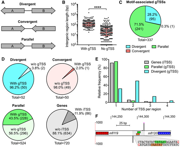
- Types of intergenic regions based on surrounding genes orientation.
- Length of intergenic regions with or without gTSS. The median and interquartile range are shown for each group. Distributions were compared using a Mann–Whitney test (two‐sided, ****P‐value < 0.0001).
- Total number of gTSSs for each of the three intergenic region groups depicted in A.
- Proportion of divergent, convergent, and parallel intergenic regions hit by at least one gTSS relative to their respective total number across the genome. The proportion of genes hit by iTSSs is also shown.
- Relative frequency distribution of the number of motif‐associated TSSs detected per gene, parallel intergenic region or divergent intergenic region.
- Genomic locus showing a representative case of two divergent genes expressed from two back‐to‐back overlapping promoters identified by 5′‐RACE. Genomic coordinates are indicated at the top of the panel. Strand‐specific 5′‐RACE signals are shown by black bars (0–1,000 read starts scale). Peaks above 1,000 read starts are cut and marked by fuchsia dots. The position of −10 boxes attributed to 5′‐RACE peaks are indicated by green and orange rectangles for positive and negative DNA strands, respectively. The genomic coordinates containing the identified TSSs and −10 boxes is enlarged and its corresponding DNA sequence is illustrated. Bases corresponding to +1 sites are colored in red. Bases corresponding to the −10 boxes are highlighted in green and orange for positive and negative DNA strands.
Figure EV4. Additional information about the genetic context of motif‐associated iTSSs.
-
AClassification of iTSSs according to the orientation of the overlapping gene in which they are located. p‐iTSS, parallel internal TSS; a‐iTSS, antiparallel internal TSS. Depending on the orientation of downstream genes, both TSS types could contribute to their expression.
-
Bp‐iTSSs and a‐iTSSs orientation relative to the nearest downstream gene.
-
CDistance from overlapping gene start codon for p‐iTSSs and a‐iTSSs. Distance was normalized according to the overlapping gene length. Yellow and red dots indicate iTSSs located upstream genes of the same and reverse orientation, respectively.
-
D, EGenomic loci showing representative cases of p‐iTSS located at less than 100 bp from the most immediate downstream gene (D) and p‐iTSS located directly on a translation start codon (E). Details are as in Fig EV3F.
To validate promoters identified by 5′‐RACE, we performed directional RNA sequencing (RNA‐seq) on three exponential‐phase M. florum steady‐state cultures and evaluated read coverage across the genome. RNA‐seq libraries were prepared in duplicate for each biological replicate, resulting in a total of six replicates. A statistical summary of RNA‐seq libraries is presented in Appendix Table S1. We observed excellent correlations between the read coverage of the different replicates calculated on non‐overlapping 1 kb windows (average Pearson correlation of 0.92), indicating a very good reproducibility of the method (Appendix Fig S5A). More importantly, coordinates of motif‐associated TSSs coincided with a sharp increase in RNA‐seq signal intensity calculated over the merged replicates, corroborating 5′‐RACE identification results (Fig 3G). This feature was also observed for gTSSs and iTSSs analyzed independently, but to a much lesser extent in the case of iTSSs because of their intragenic context (Appendix Fig S6). Taken together, these results showed that motif‐associated iTSSs and gTSSs share similar features and could both be responsible for the transcription of downstream genes.
Reconstruction of transcription units
Having identified the key features of the M. florum promoters as well as the genomic coordinates of TSSs, we leveraged this information to reconstruct TUs of this quasi‐minimal bacterium. A TU consists of a DNA segment transcribed into a single mRNA molecule from one promoter to a transcription termination site (TTS) and encoding for zero, one or many open reading frames (ORFs). In Mollicutes, termination of transcription is believed to occur through a Rho‐independent mechanism since no Rho protein homologue is detected in their genomes (de Hoon et al, 2005; D’Heygère et al, 2013). This mechanism involves structured terminators that can be reliably predicted from the DNA sequence and genes annotation, reaching excellent sensitivity for many species such as M. florum (de Hoon et al, 2005). We therefore used an updated version of an algorithm developed by de Hoon and colleagues to predict the position of terminators in M. florum according to our previously published genome annotation (de Hoon et al, 2005; Baby et al, 2018). In total, 298 different Rho‐independent terminators were predicted for the entire genome (Dataset EV2). As expected, the positions of the predicted terminators concurred with an important decrease in the RNA‐seq signal intensity, supporting the predictions made by the algorithm (Appendix Fig S7). We then used the 432 motif‐associated TSSs (gTSSs and iTSSs) identified by 5′‐RACE along with the predicted transcription terminators to reconstruct all possible TUs (see Appendix Fig S8 and Materials and Methods for a detailed description of the procedure). After manual curation, a total of 387 TUs, each responsible for the expression of at least one gene, were reconstructed (Dataset EV3). These TUs encompassed more than 90% of all annotated M. florum genes (652), including all rRNA and tRNA genes, leaving only 68 genes out of 720 without an associated promoter (orphan gene). TUs start and stop coordinates coincided with a steep increase and decrease in the average RNA‐seq read coverage (Fig 4A). Almost half of reconstructed TUs contained only a single gene, with up to 21 genes transcribed within a single RNA molecule, for an average of approximately 2.2 genes per TU (Fig 4B). The size of gene‐associated TUs ranged from 112 bp to 12.5 kb and showed an average length of ~ 2.4 kb (Fig 4C) with 5′ and 3′ untranslated regions (UTR) of 58 and 51 bp, respectively (Fig 4D). Representative M. florum TUs are depicted in Fig 4E along with the associated 5′‐RACE, terminators, and RNA‐seq data.
Figure 4. Analysis of reconstructed M. florum transcription units (TUs).
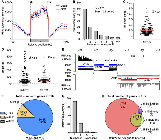
- Aggregate profile showing the mean RNA‐seq read coverage observed for all reconstructed TUs and their surrounding DNA regions. The calculated SEM is also shown. The aggregate profile was centered on the TUs start and stop coordinates, corresponding to transcription start site (TSS) and termination site (TTS), respectively.
- Relative frequency distribution of the number of genes per TU. The average and the maximal number of genes per TU are indicated.
- Scatter plot showing the length of all reconstructed TUs. The mean and associated SD are shown.
- Scatter plot showing the 5′ and 3′ untranslated regions (UTR) length of reconstructed TUs. The mean and associated SD are shown for each UTR type.
- Genomic locus showing a representative example of reconstructed TUs. Genomic coordinates are indicated at the top of the panel (kb). From innermost to outermost tracks: terminators predicted on the positive (purple) and negative (orange) DNA strands; coordinates of TUs on the positive (black) and negative (gray) DNA strands; M. florum genes encoded on the positive (blue) and negative (red) DNA strands; position of motif‐associated TSSs identified on the positive (green) and negative (red) DNA strands; RNA‐seq and 5′‐RACE signals observed on the positive and negative DNA strands, colored‐coded identically to TUs and identified TSSs, respectively. Illustrated RNA‐seq and 5′‐RACE signals represent the number of read and read starts observed for a given position, respectively. RNA‐seq signal was smoothed using a 5 pixels window (UCSC Genome Browser integrated function). RNA‐seq and 5′‐RACE peaks above 20,000 reads and 1,000 read starts are cut and marked by fuchsia dots, respectively.
- Proportion of TUs per TSS type. a‐gTSS are by definition excluded from the analysis since they are facing the nearest downstream gene.
- Relative frequency distribution of the number of TUs per M. florum gene.
- Venn diagram showing the total number of genes included in TUs generated from the different TSS types.
As expected, most gene‐encoding TUs were transcribed from gTSSs (86.6%) since they constitute the majority of TSSs identified in M. florum (Figs 3D and 4F). The remaining TUs were associated with p‐iTSS (12.9%) and a‐iTSS (0.5%). Both gTSS and iTSS‐driven TUs showed enrichment of RNA‐seq coverage, yet with a less defined 5′ border for iTSS TUs (Appendix Fig S9). A small number of mapped TSSs (56), mostly iTSSs (45 out of 56), could not be attributed to any downstream gene according to their genetic context. These TSSs were either (i) located within an intergenic region immediately upstream a predicted terminator; (ii) located within a gene positioned at the end of a TU; or (iii) facing a gene in the opposite direction. The two first cases were categorized as non‐coding TUs, whereas TSSs facing a gene in the opposite direction were classified as orphan TSSs (Dataset EV4). Nonetheless, orphan TSSs and gTSSs located immediately before a terminator coincided with a small (~ 50–75 bp) RNA‐seq signal enrichment (Appendix Fig S10). Some of these TSSs could be responsible for the expression of small non‐coding RNAs (sRNAs) or antisense RNAs (asRNAs). Of the 652 genes covered by TUs, nearly two‐thirds were individually included in only one TU, i.e., being transcribed from a single promoter (Fig 4G). The remaining genes were found to be comprised in up to four different TUs each. Interestingly, the vast majority of genes associated with an iTSS were also found to be transcribed from a gTSS, revealing only 15 genes exclusively transcribed from iTSSs (Figs 4H and EV4E). In fact, every gene associated with more than one TUs was part of a gTSS TU, and only about half of them (45.4%) were also transcribed from an iTSS TU. Overall, this suggests that iTSSs might have only a secondary role in the transcription of downstream genes. Nevertheless, iTSSs could still be involved in the transcription of other elements such as sRNAs.
Estimation of intracellular levels of protein and nucleic acid species
We then estimated the intracellular levels of M. florum nucleic acid and protein species using our macromolecular biomass quantification data, starting with the DNA fraction. In M. florum L1, the genome is organized as a single and circular chromosome of 793,224 bp (Baby et al, 2013, 2018). Based on its sequence, this chromosome has a predicted molecular weight of 489,954 kDa. The number of chromosome copies can then be directly estimated from the DNA mass per cell in respect with its molecular weight. Given that M. florum contains 1.70 ± 0.54 fg of DNA per cell during the exponential phase (Table 1), we estimated that the average M. florum cell should contain the equivalent of 2.1 chromosome copies under these growth conditions, which is practically identical to the amount estimated in E. coli but twice as in JCVI‐syn3A (Table EV1).
In cells, RNA can be subdivided into three major classes, i.e., rRNA, tRNA, and mRNA. In both bacteria and eukaryotes, rRNA constitutes the predominant form of cellular RNA, representing approximately 80% of the total RNA mass (Westermann et al, 2012; Bionumbers, 2015). Prokaryotes rRNA is composed of the 5S, 16S, and the 23S rRNA, which are typically organized as a co‐transcribed operon and produced by the cleavage of a long precursor transcript. In M. florum, two copies of the rRNA locus are present in the genome. Our 5′‐RACE results confirmed that M. florum rRNA genes are indeed transcribed as single polycistronic transcripts corresponding to TU_090 and TU_229 (Datasets EV3 and EV5). The remaining proportion of cellular RNA is composed of tRNA (~ 15%), mRNA (~ 5%), and other less abundant species such as sRNA and asRNA (< 1%) (Westermann et al, 2012). According to our macromolecular quantification results (see Table 1) and supposing that the proportions of RNA classes are conserved in M. florum, rRNA, tRNA, and mRNA have a total mass of 3.91, 0.73, and 0.24 fg, respectively (Dataset EV5). If we assume that the 5S, 16S, and 23S rRNAs are found at equimolar ratios, the calculated rRNA mass and estimated molecular weight suggest that roughly 4,900 rRNA molecules are present in a single M. florum cell (see Dataset EV5). Using the same assumption for tRNA species, approximately 18,000 tRNA molecules would also be present. Given the most probable M. florum cell volume (Table 1), this means that rRNAs and tRNAs would be found at a concentration of ~ 5.4 × 10⁴ rRNAs/µm3 and ~ 2.0 × 10⁵ tRNAs/µm3, respectively (Table EV1). tRNAs would thus be almost four times more abundant than rRNA molecules even though they occupy only ~ 15% of the total RNA mass.
We next used our RNA‐seq data to estimate the intracellular abundance of each M. florum mRNA species (Dataset EV5). We observed excellent correlations between replicates (average Pearson correlation of 0.91) when considering the number of fragments per kilobase per million of mapped reads (FPKM) calculated for all M. florum CDS (Appendix Fig S5B). The FPKM values averaged over all replicates followed a typical Poisson distribution, with two‐thirds of CDS (453/685) siting between 0 and 1,000 FPKM (Fig 5A and Appendix Fig S5C). A total of 660 CDS showed a detectable expression level (FPKM > 0), and 314 of these were expressed at a higher level than if all the reads were equally distributed across the M. florum genome (FPKM > 630) (Fig 5A and Appendix Fig S5D). Many metabolic genes involved in glycolysis showed particularly high expression levels, notably peg.600 (mfl596; L‐lactate dehydrogenase), peg.583 (mfl578; glyceraldehyde‐3‐phosphate dehydrogenase), and peg.582 (mfl577; phosphoglycerate kinase) (Fig 5A and Dataset EV5). Interestingly, three of the ten most expressed genes were annotated as hypothetical proteins, suggesting that important cellular functions are still unidentified in the current genome annotation. We also observed a striking difference in the transcription levels of CDS included in TUs compared with orphan CDS, which displayed significatively lower expression values (Fig 5B). However, we did not observe any clear correlation between the TSS signal intensity of a TU and the expression of its associated genes. According to the measured RNA mass (Table 1) and calculated FPKM values, we estimated that a total of approximately 420 mRNA molecules are expected to be present at any moment within an exponential‐phase M. florum cell growing in rich medium (Dataset EV5). If we normalize this value according to the most probable M. florum volume (Table 1), this represents ~ 4.7 × 10³ mRNAs per µm3 of cell volume, which is similar to numbers found in M. pneumoniae and E. coli (Table EV1). The expression value of most CDS (553/685) corresponded to less than one mRNA copy per cell, suggesting heterogenous expression levels between cells of the population and dynamic control of gene expression. Considering that M. florum has a doubling time of approximately 32 min (see Fig 1C) and that most bacterial mRNA have a very short half‐life (less than 7 min for Bacillus subtilis (Hambraeus et al, 2003) or between 3 and 8 min for E. coli (Bernstein et al, 2002)), it is fair to assume that the entire M. florum mRNA pool of is almost completely renewed after one generation. In fact, more than 1,000 mRNA molecules are expected to be synthetized during a single‐cell cycle. mRNA transcribed at less than one copy per cell could thus be expressed at substantial levels at some points during the cell cycle. For mRNAs that may not be expressed at each cycle, the corresponding proteins could still exert their functions over many generations since the half‐life of bacterial proteins is typically ~ 20 h (Levy & Koch, 1955; Borek et al, 1958; Maier et al, 2011).
Figure 5. Expression levels of M. florum protein‐coding genes and enrichment of functional categories.
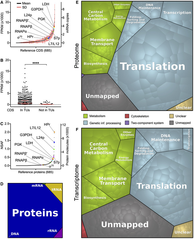
- Transcription levels of all M. florum coding sequences (CDS) quantified by RNA‐seq. Transcription levels were calculated according to the number of fragments per kilobase per million of mapped reads (FPKM) observed over six replicates. The corresponding numbers of mRNA copies per cell, estimated from the measured M. florum RNA mass, are also indicated. CDS were sorted from least to most transcribed. The transcription level of selected genes of importance is presented. LDH, L‐lactate dehydrogenase (peg.600/mfl596); G3PDH, glyceraldehyde‐3‐phosphate dehydrogenase (peg.583/mfl578); PGK, phosphoglycerate kinase (peg.582/mfl577); L24p and L7/L12, large subunit ribosomal proteins L24p (peg.133/mfl134) and L7/L12 (peg.605/mfl601); S7p, small subunit ribosomal protein S7p (peg.626/mfl623); RNAPβ, RNAPβ′, and RNAPα, RNA polymerase subunits β, β′, and α (peg.601/mfl597, peg.602/mfl598, and peg.149/mfl150); HPr, phosphotransferase system phosphocarrier protein HPr (peg.570/mfl565); σ70, RNA polymerase sigma factor RpoD (peg.269/mfl270).
- Transcription level of CDS included in transcription units (TUs) compared with CDS not attributed to any TU (orphan CDS). The median and interquartile range are shown for both groups. The mean rank of each group was compared using a Mann–Whitney test (two‐sided, ****P‐value < 0.0001).
- Expression levels of all M. florum reference proteins quantified by two‐dimensional liquid chromatography‐tandem mass spectrometry (2D LC‐MS/MS). Abundance was estimated according to the normalized spectral abundance factor (NSAF) calculated for each protein. A NSAF value of 0 was assigned to undetected proteins. The corresponding number of protein molecules per cell (derived from the biomass data) is indicated. Proteins were sorted from least to most abundant. The selected genes of importance presented in panel A are also highlighted.
- Overall DNA, tRNA, rRNA, mRNA, and protein proportions in terms of intracellular abundances in M. florum.
- Voronoi diagram illustrating the relative abundance of M. florum reference proteins grouped into different functional categories. Each polygon represents a specific protein weighted by its expression level quantified by 2D LC‐MS/MS. Functions were attributed based on the KEGG Orthology (KO) database (Kanehisa et al, 2016a). The unmapped category regroups proteins for which no KO identifier could be assigned, while the unclear category contains proteins with KO numbers matching to unclear functions.
- As panel E but for mRNA abundances quantified by RNA‐seq.
We previously showed that proteins occupy nearly half (46.6%) of the total M. florum dry mass (Fig 2F and Table 1). However, this macromolecular quantification did not provide information about the identity and specific abundance of the different proteins produced by the cell, which is highly relevant in the context of whole‐cell modeling approaches such as GEMs. We therefore performed two‐dimensional liquid chromatography–tandem mass spectrometry (2D LC‐MS/MS) on an exponential‐phase M. florum culture and analyzed the resulting spectra using three different search engines to maximize the identification of peptides matching the genome annotation (see Materials and Methods). More than 6,400 unique validated peptides were identified, altogether supported by more than 40,000 validated spectra at 1% false‐discovery rate (FDR). Both the validated peptides and matching spectra showed very high average confidence rates (98.9%). More importantly, the detected peptides matched with 481 different M. florum ORFs, with each corresponding protein supported by an average of 84.3 peptides (13.2 validated peptides), for a mean protein coverage of ~ 33.0% (Dataset EV6). For 402 out of the 481 detected proteins (~ 84%), the region immediately upstream the corresponding ORF contained a ribosome binding site motif very similar to the Shine‐Dalgarno consensus sequence (Fig EV5). The detected proteins also showed a very high average confidence rate (99.8%), and similarly to the estimated transcription levels, the normalized spectral abundance factor (NSAF) associated with each protein followed a Poisson distribution (Fig 5C and Dataset EV6). Indeed, a very low numbers of proteins, mainly ribosomal proteins, were detected at strikingly high levels, while most proteins showed medium to relatively low expression levels. Nonetheless, the correlation between transcription (FPKM) and protein expression (NSAF) levels was relatively modest (Spearman r = 0.61), a tendency also observed in other organisms (Greenbaum et al, 2003; Maier et al, 2009; Yang et al, 2014; Mazin et al, 2014; Kuchta et al, 2018). Using the molecular weight calculated for each protein and the total protein mass (Table 1), we converted the associated NSAF into absolute molecular quantities. According to our data, the average M. florum cell should contain approximately 250,000 protein molecules, with the most abundant protein present at almost 10,000 copies (peg.570/Mfl565, HPr PTS phosphocarrier protein) (Fig 5C and Dataset EV6). This represents more than ten times more molecules compared with the RNA fraction of the cell, for roughly twice the mass (Fig 5D). If we normalize the number of protein molecules per unit of cell volume, this represents roughly 2.8 × 10⁶ proteins/µm3, which is comparable to protein concentrations reported for JCVI‐syn3A, M. pneumoniae, and E. coli (Table EV1).
Figure EV5. Mesoplasma florum ribosome binding site motif.
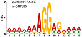
The motif was determined from the DNA region located immediately upstream (≤ 20 bp) the translation initiation codon of every reference open reading frame. A total of 548 upstream regions were included in the motif (out of 685).
Overview of expressed cellular functions
Finally, we used our proteomic quantification data to visualize what cellular functions were predominantly expressed by M. florum. We therefore assigned KEGG Orthology (KO) identifiers (Kanehisa et al, 2016a) to M. florum reference ORFs and retrieved the associated functional categories. A KO number was successfully attributed to a total of 435 M. florum proteins, of which 22 showed unclear function (Dataset EV7). Since the same protein can be assigned to multiple functional categories, we then curated the assigned categories based on the non‐redundant Proteomap functional hierarchy (Liebermeister et al, 2014). This allowed the creation of a curated tree‐like functional hierarchy for 413 different M. florum annotated proteins (Table 2 and Dataset EV7). The predicted functions of these proteins could be regrouped in just 27 different functional categories, illustrating the striking simplicity of this organism. We then used weighted Voronoi diagrams to visualize the relative importance of the assigned functional categories (Liebermeister et al, 2014). Unsurprisingly, the largest portion of the M. florum proteome was occupied by proteins implicated in translation processes, representing almost half (49.0%) of the total protein molecules of the cell and 33.5% of the total protein mass (Fig 5E, Datasets EV6 and EV7). Central carbon metabolism and membrane transport categories also displayed particularly important proteome fractions, accounting for 7.5 and 7.4% of the M. florum protein diversity, respectively (Fig 5E). On the other hand, only very limited proteome allocation (< 1%) was devoted to cytoskeleton and two‐component system functional categories. More importantly, proteins assigned to functional categories (excluding the unclear function category) comprised 86.0% of the total estimated protein molecules per cell, representing 82.1% of the M. florum protein mass (Fig 5E, Datasets EV6 and EV7). Functional categories weighted with the estimated mRNA abundances also showed the same overall picture, with however a slightly larger portion occupied by metabolism and unmapped categories (Fig 5F). Additional experiments would be required to determine the role of proteins with unknown or hypothetical function, and therefore assign the remaining protein fraction to the appropriate functional categories. Interestingly, our protein quantification data and functional category assignments can be used to estimate the abundance of conserved protein complexes, the bacterial ribosome for example. According to our analysis, we estimated that each M. florum cell should contain between 1,600 and 2,100 ribosomes. This corresponds to approximately 18,000 to 24,000 ribosomes per µm3 of cell volume, concentrations in range with values reported for M. mycoides and E. coli (Table EV1). We also estimated that ~ 270 core RNA polymerase (RNAP) should be present in the average M. florum cell (~ 3,000 RNAP/µm3), which nearly matches the number of σ70 factor per cell (~ 230).
Table 2.
Curated functional hierarchy tree of Mesoplasma florum annotated ORFs.
| Functional category | Subcategory | Sub subcategory | Number of ORFs | % of total ORFs |
|---|---|---|---|---|
| Cellular processes | Cytoskeleton | Cytoskeleton proteins | 2 | 0.3 |
| Environmental information processing | Signal transduction | Two‐component system | 1 | 0.1 |
| Genetic information processing | DNA maintenance | DNA repair | 23 | 3.4 |
| DNA replication and partition | 30 | 4.4 | ||
| Subtotal | 53 | 7.7 | ||
| Folding, sorting and degradation | Chaperones and folding catalysts | 7 | 1.0 | |
| Nucleases | 11 | 1.6 | ||
| Peptidases | 9 | 1.3 | ||
| Protein export | 7 | 1.0 | ||
| Sulfur relay system | 2 | 0.3 | ||
| Subtotal | 36 | 5.3 | ||
| Transcription | RNA polymerase | 5 | 0.7 | |
| Transcription factors | 6 | 0.9 | ||
| Subtotal | 11 | 1.6 | ||
| Translation | Ribosome | 51 | 7.4 | |
| Ribosome biogenesis | 29 | 4.2 | ||
| Translation factors | 11 | 1.6 | ||
| tRNA loading and maturation | 30 | 4.4 | ||
| Subtotal | 121 | 17.7 | ||
| Total | 221 | 32.3 | ||
| Metabolism | Biosynthesis | Amino acid metabolism | 5 | 0.7 |
| Cofactor biosynthesis | 16 | 2.3 | ||
| Lipid and steroid metabolism | 8 | 1.2 | ||
| Purine and pyrimidine metabolism | 23 | 3.4 | ||
| Subtotal | 52 | 7.6 | ||
| Central carbon metabolism | Glycolysis and carbohydrate metabolism | 35 | 5.1 | |
| Other central metabolism enzymes | 6 | 0.9 | ||
| Pentose phosphate metabolism | 8 | 1.2 | ||
| Subtotal | 49 | 7.2 | ||
| Energy metabolism | Oxidative phosphorylation | 9 | 1.3 | |
| Membrane transport | PTS system | 13 | 1.9 | |
| Secretion system | 2 | 0.3 | ||
| Transport | 42 | 6.1 | ||
| Subtotal | 57 | 8.3 | ||
| Other enzymes | Other enzymes | 22 | 3.2 | |
| Total | 189 | 27.6 | ||
| Not mapped | – | – | 250 | 36.5 |
| Unclear | – | – | 22 | 3.2 |
| Grand total | 685 | 100.0 |
Discussion
Due to its interesting characteristics, M. florum is an attractive model organism for synthetic genomics and systems biology. This near‐minimal bacterium possesses a genome smaller than those of most current model organisms (e.g. E. coli, M. pneumoniae, M. mycoides), grows rapidly in standard laboratory conditions, and is classified as a BSL‐1 organism. The flip side of being non‐pathogenic is that until recently, only little attention had been given to M. florum, although it was isolated almost 40 years ago (McCoy et al, 1980; Whitcomb et al, 1982; McCoy et al, 1984). Consequently, practically no quantitative data about the physiology of M. florum was available in the literature, and many important aspects of its cellular mechanisms and metabolism remained uncharacterized. Here, we measured several physical, physiological, and molecular characteristics of M. florum and integrated the generated data to estimate parameters difficult to evaluate using conventional laboratory equipment. A summary of the characterization reported in this study is presented in Fig 6. More specifically, we precisely evaluated the M. florum growth kinetics in rich medium (Fig 1) and measured the cell diameter, buoyant density, and dry mass to infer the most probable cell mass, volume, and surface area (Fig 2 and Table 1). We also quantified the macromolecular mass fractions of the cell (Figs 2F and EV1) and proceeded to the first experimental cartography of M. florum TUs based on 5′‐RACE TSSs identification results and Rho‐independent terminator predictions (Figs 3 and 4, and EV2, EV3, EV4, Appendix Figs S4–S10, and Datasets [Link], [Link], [Link], [Link]). Finally, we quantified the transcription and protein expression levels of all M. florum reference CDS, used the macromolecular quantification results to estimate absolute mRNA and protein abundances, and exploited these estimations to evaluate the relative importance of protein functional categories (Fig 5, Table 2, and Datasets [Link], [Link], [Link]).
Figure 6.
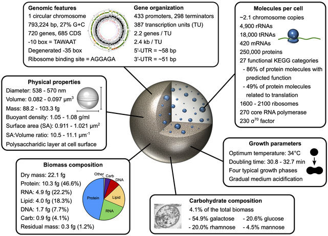
Overview of the M. florum characterization reported in this study.
While M. florum has never been associated with any disease, this does not completely rule out the possibility that this bacterium could be pathogenic in yet unidentified circumstances or for specific organisms. However, since the growth of M. florum L1 is impaired at 36°C and completely abolished at 38°C, the probability that it infects warm‐blooded animals is very low. In addition, no known virulence factor is predicted from its genome sequence. The exact nature of the primary niche of this bacterium remains unclear, but the previous isolation of various M. florum strains from insects suggests that it could be a commensal of the digestive tract of these organisms (Tully et al, 1987; Baby et al, 2018). This is further supported by the fact that many members of the Entomoplasmatales group, including several species of the Mesoplasma, Spiroplasma and Entomoplasma genera, have been isolated from or are associated with arthropods (Tully et al, 1993; Tully et al, 1994; Funaro et al, 2011; Brown & Bradbury, 2014; Sapountzis et al, 2018). This would also explain the presence of M. florum on plants (McCoy et al, 1980, 1984; Whitcomb et al, 1982; Baby et al, 2018). The digestive tract of insects would provide a unique environment in which M. florum would have continuous access to complex nutrients such as lipids and peptides to palliate for its metabolic deficiencies, as well as to various sugar sources depending on the diet of its host. Additional data are however required to confirm this hypothesis.
In our growth experiments, M. florum exhibited the four typical bacterial growth phases (Fig 1B–D). The measured OD560 nm signal, which was shown to correlate with the growth medium pH (Matteau et al, 2015), showed a progressive acidification during exponential phase. Since the main route for energy production in M. florum is predicted to be the glycolysis pathway and that no tricarboxylic acid (TCA) cycle is present, this gradual acidification is most probably caused by the accumulation of fermentation products (lactate and acetate) in the medium (Pollack et al, 1997; Halbedel et al, 2007; Caspi et al, 2014, 2016). The decrease in OD560 nm eventually reached a plateau, corresponding to a medium pH of ~ 6.0, which also coincided with the beginning of the death phase. At that point, the high concentration of protons in the medium is most likely toxic, but the underlying mechanisms resulting in M. florum death remain unknown. Compared with most Mollicutes, M. florum showed a remarkably fast doubling time (~ 32 min) (Fig 1C). For example, M. mycoides subspecies capri exhibits a doubling time of ~ 60 min in rich medium (Gibson et al, 2010; Hutchison et al, 2016), while it is estimated to be around 90 min for M. capricolum subspecies capricolum (Seto & Miyata, 1998) and 8–20 h for M. pneumoniae (Yus et al, 2009; Wodke et al, 2013). Intriguingly, M. genitalium, which possesses the smallest genome among all Mollicutes (~ 580 kb), has an extremely slow growth rate corresponding to a doubling time of ~ 16 h (Jensen et al, 1996; Hutchison et al, 2016). Clearly, the doubling time of Mollicutes is not correlated with their genome size, and the factors contributing to a fast‐growing phenotype are still elusive. This trait is most likely related to the selective pressures and evolutionary strategies adopted by specific species in their natural habitat. The utilization of a GEM that integrates the metabolic fluxes, nutrients availability, growth rate, and ATP production rate of M. florum, and more importantly its comparison with other Mollicutes GEMs, might yield more specific hypotheses on the underlying genetic factors contributing to the fast‐growing phenotype of M. florum.
Using TEM and STED microscopy, we measured an average M. florum cell diameter of 434 and 741 nm, respectively (Fig 2A–D). This range of cell diameter was refined to 538–570 nm using a mathematical approach that integrates other physical parameters such as the buoyant cell density and the cell dry mass (Fig 2E–G). Overall, M. florum cells are slightly bigger than the reported size of M. mycoides subspecies capri, JCVI‐syn1.0 and JCVI‐syn3A (~ 400 nm) (Gibson et al, 2010; Breuer et al, 2019), but within typical ranges observed for most Mollicutes (~ 200–600 nm). More importantly, the determination of the most probable cell diameter allowed us to estimate the cell mass, volume, surface area, and SA:V ratio of M. florum (Table 1). According to our analysis, M. florum is expected to have a volume between 0.082 and 0.097 µm3 during the exponential phase, which is nearly 50 times smaller than E. coli growing in similar conditions (~ 4 µm3) (Volkmer & Heinemann, 2011; Dai & Zhu, 2018). This important difference in cell volume is also apparent in the respective SA:V ratio of the two bacteria, with values approaching 10 µm−1 for M. florum compared with ~ 4 µm−1 for E. coli. Recent publications showed that bacteria exhibit robust SA:V ratio homeostasis in response to different types of perturbations, including nutritional shifts and genetic alterations (Harris & Theriot, 2016, 2018; Ojkic et al, 2019). Since Mollicutes have lost the ability to synthetize many important metabolites, their high SA:V ratios could represent a physical adaptation to increase their capacity of importing complex nutrients from the environment. Interestingly, this difference in SA:V ratios between M. florum and E. coli is also apparent when comparing the macromolecular mass fractions associated with each bacterium (Fig 2F). In M. florum, we showed that approximately 18% of the dry mass comes from lipids and 47% from proteins, whereas these fractions typically represent ~ 9 and ~ 55% of the E. coli dry mass (Dennis & Bremer, 1974; Feist et al, 2007; Bionumbers, 2015).
According to our TEM pictures and biomass quantification results (Fig 2A and F), M. florum produces a surface polysaccharide layer primarily composed of galactose (54.9%) and glucose (20.6%). This suggests the presence of a biosynthesis pathway similar to what is found in M. mycoides and M. capricolum (Razin et al, 1963; Bertin et al, 2013; Gaurivaud et al, 2014; Daubenspeck et al, 2014; Bertin et al, 2015). However, the genetic determinants responsible for the synthesis of this polysaccharidic layer, its biological function, and the precise organization of its sugar monomers remains to be identified in M. florum. Additionally, it is still unclear whether this thin layer constitutes capsular polysaccharides (CPS) covalently bound to the cell surface or exopolysaccharides (EPS) secreted in the culture medium that passively coat M. florum cells. In fact, both forms could exist and be subjected to regulation depending on environmental conditions or specific signals. Since M. florum cells were washed several times prior biomass quantification and TEM examination, the sole presence of EPS would be unlikely. In the environment, this layer could potentially serve as a protection against desiccation outside of its host. This would provide increased survivability on plant surfaces and contribute to its dissemination across insect populations. An important proportion (20.0%) of the M. florum carbohydrate mass also consisted of rhamnose, a monosaccharide commonly found in mycoplasmas and involved in the attachment of proteins on the cell membrane (Jordan et al, 2013; Daubenspeck et al, 2016). This anchoring process is thought to provide cytoplasmic proteins with additional functions, giving them the ability to moonlight on the cell surface. The considerable amount of rhamnose present in M. florum biomass indicates that this mechanism could also play a role in this species.
In this study, we combined 5′‐RACE and RNA‐seq methodologies to draw a first portrait of the M. florum transcriptome. The analysis of 5′‐RACE reads revealed 432 TSSs associated with a promoter motif sharing important similarities with previously characterized Mollicutes promoters, including a highly conserved Pribnow box (TAWAAT), a partially conserved EXT element, and a highly degenerated −35 box (Fig 3B, E and F, and Dataset EV1) (Weiner III, 2000; Güell et al, 2009; Weber et al, 2012; Yus et al, 2012; Mazin et al, 2014; Lloréns‐Rico et al, 2015; Fisunov et al, 2016). Since no other motif could be found and that only one σ factor is predicted in M. florum (σ70), this promoter motif is most likely responsible for the transcription of nearly all M. florum genes. Overall, these observations strengthen once again the idea that the −35 box and the EXT element could be less important for promoter recognition in Mollicutes compared with other bacteria such as B. subtilis or E. coli. In M. gallisepticum, for instance, only 122 mapped TSSs (out of 1,061) were shown to be associated with a −35 box motif (Mazin et al, 2014), while in M. pneumoniae, attempts to determine a clear −35 box motif were apparently unsuccessful (Weiner III, 2000; Güell et al, 2009; Lloréns‐Rico et al, 2015). The EXT element was also shown to be absent from the core promoter of M. gallisepticum and M. pneumoniae, whereas it appears to be fairly conserved in M. hyopneumoniae and in A. laidlawii (Weiner III, 2000; Güell et al, 2009; Weber et al, 2012; Yus et al, 2012; Mazin et al, 2014; Lloréns‐Rico et al, 2015; Fisunov et al, 2016). In some cases, the EXT element could compensate the absence of the −35 box as previously demonstrated with B. subtilis and Streptococcus pneumoniae (Sabelnikov et al, 1995; Voskuil & Chambliss, 1998). Still, many Mollicutes promoters seem to rely only on the −10 box to properly interact with the RNA polymerase and initiate transcription at the +1 site. Other regions such as A‐T rich region located between the −35 position and the EXT element might play a role in promoter recognition and in the formation of the open promoter complex. High‐throughput approaches using randomized promoter libraries could be an efficient strategy to analyze the importance of promoter elements and explore the diversity of sequence enabling transcription initiation in M. florum (Mutalik et al, 2013; Guiziou et al, 2016).
In A‐T rich genomes, the number of spurious Pribnow boxes arising at unexpected genomic positions such as within coding regions is expected to be particularly high (Lloréns‐Rico et al, 2016; Wade & Grainger, 2018). These cryptic elements contribute to a genome‐wide and low level transcriptional noise, a phenomenon referred as pervasive transcription (Wade & Grainger, 2014, 2018). Interestingly, our 5′‐RACE data revealed 181 putative TSSs, mostly located within coding regions of the genome, which could not be associated with the identified M. florum promoter motif (Figs 3A and C, and EV2A). Additional efforts to search for promoter sequence similarities among these TSSs were unsuccessful. These 5′‐RACE peaks are probably the result of low affinity‐binding events of the σ70 subunit to sequences faintly resembling to promoter elements, resulting in the initiation of transcription at spurious sites. However, since the intensity of these TSSs is globally very low (Fig EV2B), the energetic cost related to the synthesis of the associated transcripts as well as their potential impact on the normal transcription of overlapping genes is most likely negligible (Lloréns‐Rico et al, 2016). Even though pervasive transcription seems to be widespread across bacterial species (Dornenburg et al, 2010; Chao et al, 2012; Nicolas et al, 2012; Lybecker et al, 2014; Mazin et al, 2014; Haycocks & Grainger, 2016; Lloréns‐Rico et al, 2016), its putative biological function remains controversial. Spurious promoters might in fact serve as a reservoir on which natural selection can operate to produce functional transcripts such as sRNAs and asRNAs, thus participating to the overall transcriptome plasticity of cells (Jose et al, 2019). We indeed observed that a small proportion (22%) of identified motif‐associated TSSs were located within coding regions of the M. florum chromosome (iTSSs) (Fig 3D and Dataset EV1), and many of them could not be attributed to any downstream gene (non‐coding TUs and orphan TSSs), suggesting the presence of sRNAs or asRNAs (Dataset EV4). Motif‐associated iTSSs were however characterized by weaker associated 5′‐RACE and RNA‐seq signal intensities compared with intergenic TSSs (gTSSs) (Fig EV2B, Appendix Figs S6 and S10). Many of these putative transcripts might be only expressed at substantial levels under specific conditions or stresses, as observed in other bacteria (Dornenburg et al, 2010; Chao et al, 2012; Nicolas et al, 2012; Lybecker et al, 2014; Mazin et al, 2014; Haycocks & Grainger, 2016; Lloréns‐Rico et al, 2016). In some instances, these transcripts could even encode for alternative open reading frames (AltORFs) (Vanderperre et al, 2013; Mouilleron et al, 2016) or small ORFs (≤ 100 amino acids) (Lluch‐Senar et al, 2015; Ravikumar et al, 2018; Miravet‐Verde et al, 2019). The analysis of mass spectrometry data using a six‐frame translated database could provide significant evidences in that context.
Using the identified motif‐associated TSSs and the predicted Rho‐independent terminators, we reconstructed 387 TUs in M. florum, encompassing more than 90% of all annotated genes (Fig 4, Appendix Fig S8, and Dataset EV3). Since many motif‐associated iTSSs were properly disposed to drive the expression of downstream genes (Figs 3D and EV4) and displayed very similar characteristics compared with gTSSs (Fig 3E–G), these TSSs were also included in the reconstruction of M. florum TUs (Fig 4F). Although about half of TUs were shown to contain only a single gene, many TUs were polycistronic, and about 25% of M. florum genes were included in more than one TU (Fig 4B and G). This resulted in a surprisingly complex transcriptome architecture comparable to previous characterizations conducted in M. pneumoniae and M. gallisepticum (Güell et al, 2009; Mazin et al, 2014), with many overlapping TUs and an important fraction of genes apparently transcribed from multiple promoters. Curiously, the majority of genes located downstream of iTSSs were apparently also transcribed from a gTSS (Fig 4H). In fact, of the 15 genes strictly transcribed from iTSSs, nine happened to be expressed from iTSSs located exactly on translation start codons (leaderless mRNA), leaving only six genes controlled by true internal promoters. The actual role of intragenic promoters in M. florum is puzzling. In some cases, they could simply be the results of acquired mutations that were not counter‐selected because of the absence of any deleterious effect on the transcription of neighboring genes. In other situations, they could be important for the optimal expression of certain genes via the production of supplementary mRNA isoforms. Some of these promoters could actually constitute regulatory platforms for the biding of transcriptional factors modulating transcription upon specific signals. While our results demonstrate an impressive transcriptome complexity, our TU reconstructions were also based on the assumption that all predicted terminators were 100% efficient, which almost certainly underestimates the full transcriptome diversity in M. florum. Recent studies showed that transcription terminators are often not entirely efficient, allowing transcriptional readthrough and thus contributing to the transcription of downstream elements (Nicolas et al, 2012; Wade & Grainger, 2014; Lalanne et al, 2018). Nevertheless, our RNA‐seq data correlated very well with the reconstructed TU boundaries as well as terminator predictions (Fig 4A, Appendix Figs S7 and S9), suggesting that transcriptional readthrough is not predominant in M. florum. Termination readthrough could still be responsible for the very low expression of genes not associated with any of the identified promoters, which represent roughly 10% of all M. florum genes (Fig 5B). Of course, as this represents the very first characterization of the M. florum transcriptome, it will be possible to integrate additional datasets to improve its overall precision and breadth. For example, methods that inform about the 3′ end coordinates of transcripts such as the Rend‐seq (Lalanne et al, 2018) could be used to validate and improve the current terminator predictions, in addition to potentially highlight occurrences of leaky terminators.
Achieving a complete and quantitative description of all constituents of a cell represents one of the most important goals of systems biology. To understand global properties of complex biological systems such as cells, one must clearly identify and quantify their components. In this study, we estimated that the average M. florum cell contains approximately 250,000 proteins, 4,900 rRNAs, 18,000 tRNAs, 420 mRNAs, and 2.1 copies of the chromosome (Fig 5D, Table EV1, and Datasets EV5 and EV6). Considering the functional categories assigned by the KO database, we further estimated that about 1,600 to 2,100 ribosomes, 270 core RNAP, and 230 σ70 factor are expected to be present in the average M. florum cell (Table EV1 and Datasets EV6 and EV7). Overall, the abundance of RNA and protein molecules per cell is comparable to estimates in other Mollicutes but roughly ten times lower compared with E. coli, which is not surprising considering the large difference in respective cell volumes (Table EV1). Still, among the two other Mollicutes species selected for comparison, M. florum shows the highest number of proteins and ribosomes per cell but also has the highest cell volume with almost three times more cytoplasmic space than M. mycoides subspecies capri or JCVI‐syn3A (Table EV1). Yet, M. florum and E. coli show very similar RNA and protein concentrations when normalized for cell volume. The total number of proteins and ribosomes per unit of volume is also very consistent between all the species compared, with the exception of M. pneumoniae that has the lowest concentration of proteins and nearly ten times less ribosomes per µm3 (Table EV1). This disparity between M. pneumoniae and M. florum is also apparent when comparing the relative importance of protein functional categories in each species, with M. pneumoniae displaying significatively reduced investments in translation processes at the benefit of other processes such as cell motility and cytoskeleton (Fig 5E) (Kühner et al, 2009; Liebermeister et al, 2014). Consistently, the overall RNA levels of M. pneumoniae are also remarkably lower compared with M. florum and E. coli. This is not surprising since M. pneumoniae has only one rRNA operon per genome vs. two and seven for M. florum and E. coli, respectively. These observations are in agreement with the important difference between the growth rate of M. florum (~ 32 min) and M. pneumoniae (~ 8–20 h), supporting the idea that M. pneumoniae is not optimized for biomass production but rather depends on more complex strategies for fitness and competition in its natural environment (Yus et al, 2009). Furthermore, GEM reconstruction for M. pneumoniae revealed that most of the energy produced by this pathogenic bacterium is used for maintenance tasks instead of growth, strongly contrasting with M. mycoides subspecies capri (JCVI‐syn3A) for which the complete opposite was observed (Wodke et al, 2013; Breuer et al, 2019).
Since M. florum and JCVI‐syn3A share similar numbers of ribosomes per unit of volume but have different doubling times (~ 32 min vs. ~ 60 min), it would be interesting to compare how they allocate their resources between growth and maintenance tasks. Other parameters such as the overall efficiency of the glycolysis pathway or the efficiency of the gene expression machinery could also play an important role in the difference observed between their respective growth rate. By reconstructing whole‐cell models for M. florum, it will be possible to integrate the data generated in this study to investigate these questions and gain additional knowledge about the global cell functioning of this near‐minimal bacterium. Moreover, since we reconstructed M. florum TUs, we now have the data required to use whole‐cell modeling algorithms such as MinGenome (Wang & Maranas, 2018) to improve the initial genome reduction scenarios based on gene essentiality and conservation (Baby et al, 2018). MinGenome identifies all dispensable contiguous sequences in size descending order and preserves promoter regions needed for proper transcription of the retained genes (Wang & Maranas, 2018). The minimal genome designs inferred by this method could then be systematically analyzed using modeling approaches and compared with the synthetic minimal organism JCVI‐syn3A to highlight differences in their genome composition and retained protein functions. While some differences can probably be attributed to culture medium compositions, many cases could constitute examples of non‐orthologous gene displacement or divergent evolutionary strategies to compete in their natural habitat, thereby shedding light on some of the principles behind minimal genome plasticity. Interesting genome architectures emerging from these analyses could next be subjected to total DNA synthesis and assembly in yeast followed by transplantation into a recipient bacterium. If successful, the transplanted synthetic genomes could be analyzed using the methods described in this study to potentially acquire new knowledge about genome design principles, which are currently lacking and restraining the rational design of synthetic organisms.
Materials and Methods
Bacterial strains and growth conditions
All experiments were performed using M. florum strain L1 (ATCC 33453) grown with shaking in ATCC 1161 medium (1.75% (w/v) heart infusion broth, 4% (w/v) sucrose, 20% (v/v) horse serum, 1.35% (w/v) yeast extract, 0.004% (w/v) phenol red, 200 U/ml penicillin G (Matteau et al, 2017) at a temperature of 34°C (unless stated otherwise).
Doubling time measurement using colorimetric assays
Colorimetric assays used to measure M. florum doubling time were based on growth assays previously developed for spiroplasmas (Konai et al, 1996). Briefly, ATCC 1161 medium was inoculated with an exponential‐phase M. florum preculture to obtain an initial concentration of ~ 1 × 10⁵ CFU/ml. The inoculated medium was then diluted using twofold serial dilutions to obtain a total of four dilutions (1:1, 1:2, 1:4, and 1:8). Each dilution was transferred in triplicate into a 96‐well microplate, and the plate was incubated with shaking at the desired temperature (30, 32, 34, 36 or 38°C) in a Multiskan GO microplate reader (Thermo Scientific). Bacterial growth was monitored by measuring the OD560 nm every 10 min for ~ 16 h. The metabolic activity of M. florum was previously shown to result in the acidification of the ATCC 1161 growth medium, causing changes in the absorbance of phenol red at 560 nm that correlate with the number CFUs (Matteau et al, 2015). To calculate doubling times, linear regressions (R 2 > 0.999) were traced on the linear portion of the OD560 nm curves, and the amount of time separating each dilution curve was calculated according to the linear regression equations.
Growth kinetics assays
Growth kinetics assays were performed in triplicate by monitoring the cell concentration of three independent M. florum cultures using CFU and FCM counts. Briefly, ATCC 1161 medium was inoculated with an exponential‐phase M. florum preculture to obtain an initial concentration of ~ 1 × 10⁵ CFU/ml. Inoculated medium was incubated at 34°C with shaking for ~ 24 h in an orbital shaker incubator. Aliquots were harvested every ~ 2 h and the OD560 nm was immediately measured in duplicate using a Multiskan GO microplate reader (Thermo Scientific). CFUs were evaluated in duplicate by spotting serial dilutions of the aliquots (in PBS1×) on ATCC 1161 solid medium and counting colonies after an incubation of 24–48 h at 34°C. 37% (w/v) formaldehyde was then added and mixed to each dilution to obtain a final concentration of 1% (w/v), and the plate was incubated at room temperature (RT) for ~ 25 min. SYBR Green I (Invitrogen) dye was added to a final concentration of 1×, mixed, and samples were incubated again at RT for ~ 25 min. Cell concentration was finally measured in duplicate using a BD Accuri C6 Plus flow cytometer (BD Biosciences) equipped with a 488 nm laser. FSC‐H and FL1‐H (FITC) channel thresholds were set at 100 and 750, respectively. Fluidics were set to high speed, and a maximum of 40 µl or 1 × 10⁶ events were collected for each sample. We validated that cell concentrations were well correlated with culture dilutions diluted in PBS1× (Appendix Fig S2), and appropriate controls were performed (PBS1× without cells, unstained cells, etc.).
Cell viability assay
Cell viability of M. florum was assessed by SYTO 9 and PI double staining (Boulos et al, 1999). M. florum cells were centrifuged at 10°C for 2 min at 21,100 × g, and washed once with cold PBS1×. Cells were centrifuged again and then resuspended in PBS1× containing 5 µM SYTO 9 (Molecular Probes) and 10 µg/ml PI (Biotium). Cells were stained at RT for ~ 20 min. A fixed‐cells control was also performed by incubating a M. florum washed cell aliquot with 1% (w/v) formaldehyde at RT for ~ 25 min. Fixed cells were centrifuged at 10°C for 2 min at 21,100 × g, resuspended in PBS1× containing 0.1% (v/v) Triton X‐100, and incubated at RT for 2 min. Cells were centrifuged again and finally resuspended in PBS1× containing 5 µM SYTO 9 (Thermo Fisher Scientific) and 10 µg/ml PI (Biotium). Samples were immobilized on agarose pad slides and examined by widefield fluorescence microscopy using an Axio Observer Z1 inverted microscope (Zeiss) equipped with an AxioCam 506 mono (Zeiss) camera and a 100×/NA1.4 Plan‐Apochromat oil immersion objective. SYTO 9 and PI were excited and acquisitioned using GFP and Cy3 excitation/emission filters, respectively. Images were captured with Zeiss Zen 2.0 imaging software and analyzed using Fiji (Schindelin et al, 2012).
Stimulated emission depletion microscopy
Stimulated emission depletion (STED) microscopy was performed using double‐stained (membrane and DNA) M. florum cells. Briefly, an exponential‐phase M. florum culture was centrifuged at 10°C for 2 min at 21,100 × g and washed twice with cold electroporation buffer [272 mM sucrose, 1 mM HEPES (pH 7.4)]. Washed cells were then immobilized on a poly‐l‐lysine‐coated glass slide (Poly‐Prep Slide, Sigma‐Aldrich) and incubated at RT for 5 min. Cells were washed on slide twice with PBS1×, and then stained, fixed, permeabilized, and stained again for 5 min each at RT using the following solutions (all reagents diluted in PBS1×, with two PBS1× washes between each step): (i) 0.5 µM mCLING‐ATTO 647N‐labeled dye (Synaptic Systems); (ii) 4% (w/v) formaldehyde and 0.2% (w/v) glutaraldehyde; (iii) 0.1% (v/v) Triton X‐100; and (iv) 1/100 dilution (100×) of PicoGreen concentrate reagent (Molecular Probes). Cells were washed twice again with PBS1× and then finally mounted for STED microscopy using ProLong Diamond Mountant (Molecular Probes). Two‐color STED microscopy was performed using a DMi8 STED microscope (Leica TCS SP8) equipped with a 100×/NA1.4 HC Plan‐Apochromat CS2 oil immersion objective and operated with the LAS X imaging software (version 3.1.1.15751, Leica). mCLING‐ATTO 647N and PicoGreen were excited using a pulsed white light laser set at 646 and 488 nm, respectively, and depleted using 775 and 592 nm depletion lasers. Signals were acquisitioned using HyD SMD hybrid detectors (Leica) set at 658–698 nm for the ATTO 647N channel and 505–565 nm for the PicoGreen channel. Images were acquisitioned using a 4× zoom factor and deconvolved using Huygens Professional with STED optical option (version 18.04, Scientific Volume Imaging). Images and cell diameter were analyzed using Fiji (Schindelin et al, 2012). Since cells displayed a variable morphology from ovoid to spherical, minor and major axes were measured and averaged to obtain a single representative cell diameter for each cell. Only cells exhibiting both signals were considered in the analysis.
Transmission electron microscopy (TEM)
Exponential‐phase M. florum cultures were centrifuged at 10°C for 15 min at 7,900 × g and then washed three times with cold PBS1×. The supernatant was discarded, and cells were fixed at RT for 45 min and then overnight at 4°C by adding 1 ml of 2.5% (w/v) glutaraldehyde on top of the cell pellet. Cells were then washed twice with PBS1×, post‐fixed at RT for 90 min using a 1% (w/v) osmium tetroxide solution, and washed twice with water. Cells were then dehydrated through a series of washes (5 min each) with 30, 50, 70, 85, 95%, and three times 100% (v/v) ethanol. Samples were washed again three times using propylene oxide, with a 5‐min incubation at RT after each wash. Samples were then incubated at RT for 1 h with 1:1 propylene oxide:Epon, incubated two times at RT for 180 min with pure Epon, and then overnight at RT with pure Epon. The Epon and cell mixture was embedded within a polyethylene capsule (BEEM) and polymerized by baking at 70°C for 48 h. The block was cut into thin sections (~ 80 nm) and placed on a copper grid, stained sequentially with uranyl acetate and lead citrate (~ 10 min each), and finally examined under a Hitachi H‐7500 TEM microscope operating at an accelerating voltage of 80 kV. Images and cell diameter were analyzed using Fiji (Schindelin et al, 2012). Only cells with a clearly distinguishable cellular membrane, as shown in Fig 2A, were selected for diameter measurement. Since cells displayed a variable morphology from ovoid to spherical, minor and major axes were measured and averaged to obtain a single representative cell diameter for each cell.
Measurement of buoyant cell density
M. florum buoyant cell density was assessed by discontinuous density gradient centrifugation in Percoll (GE Healthcare). First, a Stock Isotonic Percoll (SIP) solution was prepared by mixing nine parts (v/v) of Percoll (GE Healthcare) to one part (v/v) of 1.5 M NaCl, resulting into a 1.12 g/ml solution. The 100% (v/v) SIP solution was then diluted with 0.15 M NaCl to obtain 80, 60, 40, and 20% (v/v) SIP solutions, with corresponding densities of 1.10, 1.08, 1.05, and 1.03 g/ml, respectively. To easily differentiate density gradients, trypan blue was added to half of the dilutions (100, 60, and 20%) to a final concentration of 0.0008%. 2 ml of each dilution was then slowly layered from most concentrated to less into a 15‐ml conical tube to create a discontinuous density gradient varying from 1.12 (100% SIP) to 1.03 g/ml (20% SIP). 20 ml of an exponential‐phase M. florum culture was centrifuged at 10°C for 15 min at 7,900 × g and then washed twice with cold PBS1×. Cells were resuspended in 2 ml of NaCl 0.15 M (1.00 g/ml) and slowly loaded on the top of the density gradient. Cells were then centrifuged two times at 7,900 × g (10°C) for 30 min each, and the position of the cell pellet was noted after each centrifugation.
Biomass quantification
Detailed biomass quantification methods are available in Appendix Supplementary Methods. A summary of the procedures is shown in Fig EV1. Briefly, dry mass was measured by weighting exponential‐phase culture pellets previously dried at 80°C for ~ 36 h. Quantification was performed in quadruplicate and repeated three times. Protein mass was quantified in quadruplicate by fluorescence‐based protein quantification of whole‐cell lysates using the CBQCA Protein Quantitation Kit (Molecular Probes, C‐6667) according to the manufacturer’s specifications. DNA and RNA mass were quantified in quadruplicate by fluorescence‐based nucleic acid quantification performed on purified genomic DNA and purified RNA using the Quant‐iT PicoGreen dsDNA Assay Kit (Thermo Fisher Scientific, P7589) and the Quant‐iT RiboGreen RNA Assay Kit (Thermo Fisher Scientific, R11490), respectively. Carbohydrate mass was measured in quadruplicate by GC‐MS analysis performed on whole‐cell lysates and normalized by the protein concentration of the samples. Lipid mass was quantified using a combination of two different methods: the identification of lipid species by direct infusion‐tandem mass spectrometry (DI‐MS/MS; see Dataset EV8) and the fluorescence‐based quantification of phospholipids using the Phospholipid Assay Kit (Sigma‐Aldrich, MAK122). Lipid quantifications were performed in quadruplicate. All quantifications were normalized by the cell concentration of analyzed samples using CFU or FCM counts.
Protein mass spectrometry
The protein composition of M. florum was determined by 2D LC‐MS/MS from trypsinized protein extracts. Sample preparation and analysis was executed by PhenoSwitch Bioscience (Sherbrooke, Canada). Briefly, an exponential‐phase M. florum culture was centrifuged at 10°C for 2 min at 21,100 × g and washed twice with cold electroporation buffer [272 mM sucrose, 1 mM HEPES (pH 7.4)]. Cells were then resuspended in 0.4% (w/v) sodium deoxycholate and lysed using a Bioruptor UCD‐200 sonication system (Diagenode) set at high intensity and 4°C for 35 cycles (30 s on, 30 s off). Insoluble material was removed by centrifuging the cell lysate at 16,000 × g for 10 min at 4°C, and the supernatant was recovered. Protein concentration was measured using the Bio‐Rad Protein Assay (Bio‐Rad) according to the manufacturer’s specifications and absorbance at 595 nm was measured using a Synergy HT microplate reader (BioTek). The cell lysate was then reduced at 65°C for 15 min with 10 mM dithiothreitol (DTT) in a final pH of 8.0 and then alkylated at RT in the dark for 30 min with 15 mM iodoacetamide. 10 mM of DTT was then added to quench residual iodoacetamide and proteins (~ 200 µg) were digested at 37°C overnight with shaking using 1 µg of trypsin per 30 µg of proteins. The resulting peptides were first separated using a polymeric reversed phase column (Phenomenex, 8E‐S100‐AGB) and eluted into eight fractions with increasing concentration of acetonitrile. ~ 5 µg of each fraction was then injected into a TripleTOF 5600 mass spectrometer (SCIEX) equipped with a HALO ES‐C18 column (0.5 × 150 mm). Peptides were separated with a 60 min gradient of the following two mobile phases: (i) 0.2% (v/v) formic acid and 3% (v/v) DMSO in water; and (ii) 0.2% (v/v) formic acid and 3% (v/v) DMSO in ethanol. Peptides were analyzed in information dependant acquisition (IDA) mode. Raw MS files were analyzed using PeptideShaker software version 1.13.4 (Vaudel et al, 2015) configured to run three different search engines (MS‐GF+, Comet, and OMSSA) via SearchGUI (version 3.1.0) (Barsnes & Vaudel, 2018). SearchGUI parameters were set as follows: maximum precursor charge, 5; maximum number of post‐translational modification per peptide, 4; precursor ion m/z tolerance, 0.006 Da; fragment ion m/z tolerance, 0.1 Da; maximum missed cleavages, 2; minimal peptide length, 8; and maximal peptide length, 30. Carbamidomethylation of C was set as a fixed modification. Acetylation of K, Acetylation of protein N‐term, FormylMet of protein N‐term, Oxidation of M, Phosphorylation of S, Phosphorylation of T, and Phosphorylation of Y were set as variable modifications. Protein search database was defined according to the published M. florum L1 RAST genome annotation (Baby et al, 2018). Peptide spectrum matches, peptides, and proteins were validated using a 1% FDR cut‐off.
Cell equations
For simplicity, we assumed M. florum cells to be of spherical shape in all cell equations described in this study since the observed morphology varied from ovoid to spherical (see Fig 2A). Given a spherical M. florum cell with a certain diameter (d), its volume (V), surface area (A), and surface area to volume ratio (SA:V) can be described according to the following equations:
| (1) |
| (2) |
| (3) |
Additionally, its cell mass (CM) can be described as follows:
| (4) |
| (5) |
| (6) |
| (7) |
where D, DM, DF, and DDM are the cell buoyant density, dry mass, dry mass fraction, and dry mass‐specific density. Detailed description of cell mass equations is given in Appendix Supplementary Methods. For each equation, the mean cell mass (CMmean) was calculated using the mean value associated with each measured or estimated parameter. The minimal (CMmin) and maximal (CMmax) cell mass were calculated using the mean ± SD or the range associated with each parameter. For example, using Equation 4 and considering a cell buoyant density between 1.05 and 1.08 g/ml, the minimal and maximal cell mass values are given by the following expressions:
| (4.1) |
| (4.2) |
And the mean cell mass value is defined as follows:
| (4.3) |
According to typical ranges found in bacteria, the dry mass fraction (DF) and the dry mass specific density (DDM) were estimated to be between 20–30% and 1.3–1.5 g/ml, respectively (Bakken & Olsen, 1983; Bratbak & Dundas, 1984; Bratbak, 1985; Fischer et al, 2009; Bionumbers, 2015). The most probable M. florum cell mass and cell diameter ranges were determined graphically according to the interception points of CMmean curves generated using a variable cell diameter in each equation (see Fig 2G). The most probable cell diameter range was finally used to infer the most probable cell volume (V) using Equation 1, as well as the most probable surface area (A) and surface area to volume ratio (SA:V) ranges using Equations 2 and 3.
5′‐RACE library preparation and analysis
The 5′‐RACE sequencing library was prepared from a M. florum exponential‐phase culture as described previously (Carraro et al, 2014; Matteau & Rodrigue, 2015; Poulin‐Laprade et al, 2015). Library quality and concentration were evaluated using a 2100 Bioanalyzer instrument (Agilent Technologies). Single‐end Illumina sequencing (40 bp) was performed on an Illumina Genome Analyzer IIx instrument at the BioMicroCenter of the Massachusetts Institute of Technology (Cambridge, USA). Reads were trimmed for quality using Trimmomatic version 0.32 (Bolger et al, 2014) and aligned on M. florum L1 genome (NC_006055.1) with Bowtie 2 version 2.3.3.1 (Langmead & Salzberg, 2012). Alignments were processed and filtered to identify all putative TSSs. Analysis details are provided in Appendix Supplementary Methods. A summary of the 5′‐RACE library statistics is shown in Appendix Table S1. Promoter motifs were searched using MEME and MAST version 5.0.3 (Bailey & Elkan, 1994). Strand‐specific 1 bp resolution genome coverage tracks were generated using Bedtools genomecov version 2.27.1 (Quinlan & Hall, 2010).
RNA‐seq libraries preparation and analysis
Total RNA‐seq libraries were prepared in biological triplicate from M. florum steady‐state cultures grown using the Versatile Continuous Culture Device (Matteau et al, 2015). Total RNA was extracted in technical duplicate from each culture replicate using the Direct‐zol RNA MiniPrep Kit (Zymo Research, R2052) as described previously (Carraro et al, 2014), for a total of six RNA‐seq libraries. RNA‐seq libraries were prepared and depleted from ribosomal RNA as described previously (Carraro et al, 2014), with the exception that 200 µg/ml of actinomycin D was added to the reverse transcription reaction to prevent second strand synthesis by the reverse transcriptase (Perocchi et al, 2007). Library quality and concentration were evaluated using a 2100 Bioanalyzer instrument (Agilent Technologies). Paired‐end Illumina sequencing (2 × 50 bp) was performed on a HiSeq 2000 Illumina instrument at the Plateau de biologie moléculaire et génomique fonctionnelle of the Institut de recherches cliniques de Montréal (Montréal, Québec, Canada). Reads were quality trimmed using Trimmomatic version 0.32 (Bolger et al, 2014) and aligned in a strand‐specific manner on the M. florum L1 genome (NC_006055.1) with Bowtie 2 version 2.3.3.1 (Langmead & Salzberg, 2012). Reads with a MAPQ below 10 were discarded using samtools version 1.5 (Li et al, 2009). A summary of the RNA‐seq library statistics is shown in Appendix Table S1. FPKM values were calculated for each M. florum L1 protein‐coding gene (RAST annotation, see Baby et al, 2018) using the GenomicAlignments R package version 1.10.1 (Lawrence et al, 2013). Strand‐specific 1 bp resolution genome coverage tracks were generated using Bedtools genomecov version 2.27.1 (Quinlan & Hall, 2010). Bedtools makewindows and multicov (version 2.27.1) were used to calculate the RNA‐seq coverage on non‐overlapping 1 kb windows for each replicate. Pearson’s correlation coefficients between replicates (1 kb windows coverage as well as gene FPKM) were calculated using GraphPad Prism‐integrated function (version 7.0a).
Reconstruction of transcription units
Rho‐independent terminators were predicted from M. florum L1 DNA sequence and genes annotation (RAST annotation, see Baby et al, 2018) using an updated version of the in‐house Python script developed by de Hoon et al (2005). The main difference between the updated version and the original one is the replacement of the Mfold package (Mathews et al, 1999; Zuker, 2003) by the ViennaRNA package (Lorenz et al, 2011) (version 2.4.11) to calculate the RNA secondary structure. The Python script is available upon request from the author. Only terminators with a calculated score above 0 were considered significant. For each predicted terminator, the TTS was defined as the last base forming the stem‐loop structure since the termination was shown to occur at or near the T‐stretch following the stem‐loop (Gusarov & Nudler, 1999; de Hoon et al, 2005). Strand‐specific term‐to‐term scaffolds were then created according to the genomic position of the TTSs, and the coordinates of the motif‐associated TSSs were used to generate all possible TUs for each scaffold. Genes were attributed to a given TU only if the calculated (5′‐UTR) length was ≤ 500 bp and their coordinates were completely included within the TU, meaning that genes intersected with iTSSs were excluded from the iTSSs‐derived TUs. Generated TUs were manually inspected using the UCSC genome browser (Kent et al, 2002) to correct for different scenarios such as the presence of predicted riboswitches (Kim et al, 2007; Kalvari et al, 2018), the circular topology of the chromosome or the occasional overlap between TSSs and terminator sequences. In the rare cases where no motif‐associated TSSs could be attributed to a gene (orphan gene), the identified TSSs without promoter motif were considered for the expression of a TU encompassing that gene, as long as they initiated transcription on a purine nucleotide and fulfilled the other criteria described previously (signal intensity threshold and 5′‐UTR length). If still no TSS without promoter motif could be found, then TSSs located at the end of the previous term‐to‐term scaffold (thus separated from the orphan gene by a predicted terminator) were considered as putative candidates for the expression of the gene, provided that its expression was non‐null and the 5′‐UTR length was ≤ 500 bp. See the manual curation notes column in Dataset EV3 for further details.
Aggregate profiles
RNA‐seq aggregate profiles were generated using the Versatile Aggregate Profiler (VAP) version 1.0.0 (Coulombe et al, 2014). Aggregate profiles were calculated for each DNA strand independently using the RNA‐seq genome coverage calculated at single bp resolution on all the RNA‐seq replicates merged together. The relative analysis method was used for all cases, along with two reference points and a 1 bp window size. The number of windows for the reference feature was set to 1 in the case of TSSs, whereas this parameter was set to 100 and 40 for TUs and terminators, respectively.
Analysis of Mesoplasma florum ribosome‐binding site
Mesoplasma florum ribosome binding site motif was determined by extracting the DNA sequence (20 bp) immediately upstream the translation initiation codon of each M. florum reference ORF and submitting it to MEME version 5.0.3 (Bailey & Elkan, 1994). The zero or one motif per sequence option was used, with a minimum motif length of 6 bp.
Estimation of molecular abundances
The number of M. florum chromosome copies per cell was estimated from the measured DNA mass and the estimated molecular weight of the chromosome (see Table 1 for the measured DNA, RNA and protein). The molecular weight of the M. florum L1 chromosome (NC_006055.1) was estimated using the Sequence Manipulation Suite server (https://www.bioinformatics.org/sms2/dna_mw.html) (Stothard, 2000). The intracellular abundance of RNA species was calculated from the estimated molecular weight and measured RNA mass by assuming that rRNA, tRNA, and mRNA totalize 80, 15, and 5% of the total RNA mass of the cell (Westermann et al, 2012; Bionumbers, 2015). The molecular weight of RNA species was estimated using in‐house Python scripts. The intracellular levels of protein species were calculated from the estimated molecular weight and the measured protein mass. The molecular weight of proteins was either estimated by PeptideShaker software version 1.13.4 (Vaudel et al, 2015) for proteins detected by mass spectrometry or using the Sequence Manipulation Suite server (https://www.bioinformatics.org/sms2/protein_mw.html) for proteins not detected by mass spectrometry (Stothard, 2000). For rRNAs and tRNAs, the total number of copies per cell was calculated by assuming that each species is found at equimolar ratios. For mRNAs and proteins, molar ratios were normalized according to the expression value of each species, i.e., using the associated FPKM and NSAF values, respectively. Briefly, the FPKM and NSAF values associated to each gene were divided by the sum obtained for all genes, resulting in a relative expression value. This value was then multiplied by the corresponding mRNA or protein molecular weight, producing a normalized molecular weight for each species, which was further divided by the sum of all normalized molecular weights to obtain a fraction of the total mRNA or protein mass for each gene. The mass of each mRNA and protein species was then calculated by multiplying mass fractions by the total mRNA and protein mass in M. florum, which was converted to an absolute number of molecules using their respective molecular weight and the Avogadro number. Calculation details can be found in Datasets EV5 and EV6. The number of ribosomes per cell was estimated using two different approaches: (i) from the average number of protein per cell calculated for all predicted (KO) ribosomal proteins and (ii) by assuming that all rRNA molecules are incorporated into ribosomes, meaning that the estimated number of ribosomes per cell is equivalent to one third of the total number of rRNA molecules per cell (three rRNA molecules per ribosome). The number of RNAP complexes per cell was estimated according to the average number of protein per cell calculated for the α, β, and β′ subunits (see Dataset EV6). The protein stoichiometry of the RNAP complex was taken into account in the calculations (two α, one β, and one β′ subunits per RNAP).
Analysis of functional categories expression
The KO Database was used to assign functional categories to M. florum reference proteins because of its clearly layered structure, and because major efforts were made to associate each KO entry with experimental evidences (Kanehisa et al, 2016a). Moreover, since proteins are assigned to functions via KO identifiers, the comparison between organisms is relatively straightforward. Briefly, the BlastKOALA server (https://www.kegg.jp/blastkoala/) (Kanehisa et al, 2016b) was used to assign KO identifiers to M. florum reference ORFs and retrieve associated functional categories. Since the same protein can be assigned to multiple functional categories, we then curated the assigned categories based on the non‐redundant Proteomap functional hierarchy (Liebermeister et al, 2014). ORFs not matching to any KO identifiers were assigned to the unmapped category. KO entries matching to unclear functions were regrouped into the unclear category. Assigned KO identifiers and functional categories can be found in Dataset EV7. The Proteomap server (https://www.proteomaps.net/index.html) was used to visualize the relative expression of functional categories using either protein or mRNA expression datasets (Liebermeister et al, 2014).
Data visualization
Raw 5′RACE and RNA‐seq profiles, terminator and riboswitch predictions, identified TSSs, reconstructed TUs as well as identified peptide spectrum matches (PSMs) and validated peptides can be visualized using the UCSC genome browser at http://bioinfo.ccs.usherbrooke.ca/M_florum_hub.html.
Author contributions
Manuscript writing and figure preparation: DM; Revision and editing of the manuscript and figures: SR, P‐ÉJ, and J‐CL; Experiments: DM, DG, SG, JMD and KD; Analyses: DM, FG, JMD, and J‐CL; Project design: DM, J‐CL, and SR; Preliminary data and insights on M. florum: TFK.
Conflict of interest
The authors declare that they have no conflict of interest.
Supporting information
Review Process File
Appendix
Expanded View Figures PDF
Table EV1
Dataset EV1
Dataset EV2
Dataset EV3
Dataset EV4
Dataset EV5
Dataset EV6
Dataset EV7
Dataset EV8
Acknowledgements
We thank members of S. Rodrigue and P.‐É. Jacques laboratories for helpful discussions, Joëlle Brodeur, Jean‐Philippe Côté, Alain Lavigueur, Catherine Chamberland, and Pascal Sirand‐Pugnet for their insightful comments on the manuscript, and Jean‐François Lucier and the Centre de Calcul Scientifique at the Université de Sherbrooke for technical assistance. We are also grateful to Michiel J. L. de Hoon for sharing his Rho‐independent terminators prediction script, as well as Charles Bertrand and the Plateforme de microscopie photonique at the Université de Sherbrooke for technical assistance on TEM and STED microscopy, respectively. Access to computational resources was provided in part by Calcul Québec (http://www.calculquebec.ca) and Compute Canada (http://www.computecanada.ca). This research project was funded by the Fonds de recherche du Québec—Nature et technologies: PR‐173580, and by the Natural Sciences and Engineering Research Council of Canada: 386393.
Mol Syst Biol. (2020) 00: e9844
Data availability
The datasets produced in this study are available in the following databases:
RNA‐seq and 5′‐RACE data: Gene Expression Omnibus GSE152985 (https://www.ncbi.nlm.nih.gov/geo/query/acc.cgi?acc=GSE152985)
Proteomics data: PRIDE PXD019922 (https://www.ebi.ac.uk/pride/archive/projects/PXD019922)
References
- Agarwal KL, Büchi H, Caruthers MH, Gupta NK, Khorana HG, Klbppe K, Kumar A, Ohtsuka E, RajBhandary UL, van de Sande JH et al (1970) Total synthesis of the structural gene for an alanine transfer ribonucleic acid from yeast. Nature 227: 27–34 [DOI] [PubMed] [Google Scholar]
- Alper H, Cirino P, Nevoigt E, Sriram G (2010) Applications of synthetic biology in microbial biotechnology. J Biomed Biotechnol 2010: 1–2 [DOI] [PMC free article] [PubMed] [Google Scholar]
- Baby V, Matteau D, Knight TF, Rodrigue S (2013) Complete genome sequence of the Mesoplasma florum W37 strain. Genome Annoucements 1: e00879–13 [DOI] [PMC free article] [PubMed] [Google Scholar]
- Baby V, Labroussaa F, Brodeur J, Matteau D, Gourgues G, Lartigue C, Rodrigue S (2017) Cloning and transplantation of the Mesoplasma florum genome. ACS Synth Biol 7: 209–217 [DOI] [PubMed] [Google Scholar]
- Baby V, Lachance J‐C, Gagnon J, Lucier J‐F, Matteau D, Knight TF, Rodrigue S (2018) Inferring the minimal genome of Mesoplasma florum by comparative genomics and transposon mutagenesis. mSystems 3: e00198–17 [DOI] [PMC free article] [PubMed] [Google Scholar]
- Bailey TL, Elkan C (1994) Fitting a mixture model by expectation maximization to discover motifs in biopolymers. Proc Int Conf Intell Syst Mol Biol 2: 28–36 [PubMed] [Google Scholar]
- Bailey TL, Gribskov M (1998) Combining evidence using p‐values: application to sequence homology searches. Bioinformatics 14: 48–54 [DOI] [PubMed] [Google Scholar]
- Bakken LR, Olsen RA (1983) Buoyant densities and dry‐matter contents of microorganisms: conversion of a measured biovolume into biomass. Appl Environ Microbiol 45: 1188–1195 [DOI] [PMC free article] [PubMed] [Google Scholar]
- Barsnes H, Vaudel M (2018) SearchGUI: a highly adaptable common interface for proteomics search and de novo engines. J Proteome Res 17: 2552–2555 [DOI] [PubMed] [Google Scholar]
- Benders GA, Noskov VN, Denisova EA, Lartigue C, Gibson DG, Assad‐Garcia N, Chuang R‐Y, Carrera W, Moodie M, Algire MA et al (2010) Cloning whole bacterial genomes in yeast. Nucleic Acids Res 38: 2558–2569 [DOI] [PMC free article] [PubMed] [Google Scholar]
- Bernstein JA, Khodursky AB, Lin PH, Lin‐Chao S, Cohen SN (2002) Global analysis of mRNA decay and abundance in Escherichia coli at single‐gene resolution using two‐color fluorescent DNA microarrays. Proc Natl Acad Sci USA 99: 9697–9702 [DOI] [PMC free article] [PubMed] [Google Scholar]
- Bertin C, Pau‐Roblot C, Courtois J, Manso‐Silván L, Thiaucourt F, Tardy F, Le Grand D, Poumarat F, Gaurivaud P (2013) Characterization of free exopolysaccharides secreted by Mycoplasma mycoides Subsp. mycoides . PLoS One 8: e68373 [DOI] [PMC free article] [PubMed] [Google Scholar]
- Bertin C, Pau‐Roblot C, Courtois J, Manso‐Silván L, Tardy F, Poumarat F, Citti C, Sirand‐Pugnet P, Gaurivaud P, Thiaucourt F (2015) Highly dynamic genomic loci drive the synthesis of two types of capsular or secreted polysaccharides within the Mycoplasma mycoides cluster. Appl Environ Microbiol 81: 676–687 [DOI] [PMC free article] [PubMed] [Google Scholar]
- Bionumbers (2015) What is the macromolecular composition of the cell. http://book.bionumbers.org/what‐is‐the‐macromolecular‐composition‐of‐the‐cell/. Accessed 19 March 2019
- Bolger AM, Lohse M, Usadel B (2014) Trimmomatic: a flexible trimmer for Illumina sequence data. Bioinformatics 30: 2114–2120 [DOI] [PMC free article] [PubMed] [Google Scholar]
- Bordbar A, Monk JM, King ZA, Palsson BO (2014) Constraint‐based models predict metabolic and associated cellular functions. Nat Rev Genet 15: 107–120 [DOI] [PubMed] [Google Scholar]
- Borek E, Ponticorvo L, Rittenberg D (1958) Protein turnover in micro‐organisms. Proc Natl Acad Sci USA 44: 369–374 [DOI] [PMC free article] [PubMed] [Google Scholar]
- Boulos L, Prévost M, Barbeau B, Coallier J, Desjardins R (1999) LIVE/DEAD(®) BacLight(TM): application of a new rapid staining method for direct enumeration of viable and total bacteria in drinking water. J Microbiol Methods 37: 77–86 [DOI] [PubMed] [Google Scholar]
- Bratbak G, Dundas I (1984) Bacterial dry matter content and biomass estimations. Appl Environ Microbiol 48: 755–757 [DOI] [PMC free article] [PubMed] [Google Scholar]
- Bratbak G (1985) Biovolume and biomass estimations. Microbiology 49: 1488–1493 [DOI] [PMC free article] [PubMed] [Google Scholar]
- Breuer M, Earnest TM, Merryman C, Wise KS, Sun L, Lynott MR, Hutchison CA, Smith HO, Lapek JD, Gonzalez DJ et al (2019) Essential metabolism for a minimal cell. Elife 8: 1–77 [DOI] [PMC free article] [PubMed] [Google Scholar]
- Brown DR, Whitcomb RF, Bradbury JM (2007) Revised minimal standards for description of new species of the class Mollicutes (division Tenericutes). Int J Syst Evol Microbiol 57: 2703–2719 [DOI] [PMC free article] [PubMed] [Google Scholar]
- Brown DR, Bradbury JM (2014) Contentious taxonomy of mollicutes In Mollicutes: molecular biology and pathogenesis, Browning GF, Citti C. (eds), pp 1–14. Toulouse Cedex: Caister Academic Press; [Google Scholar]
- Bryan AK, Hecht VC, Shen W, Payer K, Grover WH, Manalis SR (2014) Measuring single cell mass, volume, and density with dual suspended microchannel resonators. Lab Chip 14: 569–576 [DOI] [PMC free article] [PubMed] [Google Scholar]
- Cambray G, Mutalik VK, Arkin AP (2011) Toward rational design of bacterial genomes. Curr Opin Microbiol 14: 624–630 [DOI] [PubMed] [Google Scholar]
- Carraro N, Matteau D, Luo P, Burrus V, Rodrigue S (2014) The master activator of IncA/C conjugative plasmids stimulates genomic islands and multidrug resistance dissemination. PLoS Genet 10: e1004714 [DOI] [PMC free article] [PubMed] [Google Scholar]
- Caspi R, Altman T, Billington R, Dreher K, Foerster H, Fulcher CA, Holland TA, Keseler IM, Kothari A, Kubo A et al (2014) The MetaCyc database of metabolic pathways and enzymes and the BioCyc collection of Pathway/Genome Databases. Nucleic Acids Res 42: D459–D471 [DOI] [PMC free article] [PubMed] [Google Scholar]
- Caspi R, Billington R, Ferrer L, Foerster H, Fulcher CA, Keseler IM, Kothari A, Krummenacker M, Latendresse M, Mueller LA et al (2016) The MetaCyc database of metabolic pathways and enzymes and the BioCyc collection of pathway/genome databases. Nucleic Acids Res 44: D471–D480 [DOI] [PMC free article] [PubMed] [Google Scholar]
- Chalkley O, Purcell O, Grierson C, Marucci L (2019) The genome design suite: enabling massive in‐silico experiments to design genomes. bioRxiv 10.1101/681270 [PREPRINT] [DOI] [Google Scholar]
- Chao Y, Papenfort K, Reinhardt R, Sharma CM, Vogel J (2012) An atlas of Hfq‐bound transcripts reveals 3′ UTRs as a genomic reservoir of regulatory small RNAs. EMBO J 31: 4005–4019 [DOI] [PMC free article] [PubMed] [Google Scholar]
- Cooper GM, Hausman RE (2013) The composition of cells In The cell: a molecular approach, 6th edn, pp 43–72. Sunderland MA: Sinauer Associates; [Google Scholar]
- Coulombe C, Poitras C, Nordell‐Markovits A, Brunelle M, Lavoie M‐A, Robert F, Jacques P‐É (2014) VAP: a versatile aggregate profiler for efficient genome‐wide data representation and discovery. Nucleic Acids Res 42: W485–W493 [DOI] [PMC free article] [PubMed] [Google Scholar]
- D’Heygère F, Rabhi M, Boudvillain M (2013) Phyletic distribution and conservation of the bacterial transcription termination factor rho. Microbiology 159: 1423–1436 [DOI] [PubMed] [Google Scholar]
- Dai X, Zhu M (2018) High osmolarity modulates bacterial cell size through reducing initiation volume in Escherichia coli . mSphere 3: e00430–18 [DOI] [PMC free article] [PubMed] [Google Scholar]
- Daubenspeck JM, Jordan DS, Dybvig K (2014) The glycocalyx of mollicutes, In Mollicutes: molecular biology and pathogenesis, Browning G, Citti C. (eds), pp 131–147. Norfolk: Caister Academic Press; [Google Scholar]
- Daubenspeck JM, Liu R, Dybvig K (2016) Rhamnose links moonlighting proteins to membrane phospholipid in mycoplasmas. PLoS One 11: 1–11 [DOI] [PMC free article] [PubMed] [Google Scholar]
- Dennis PP, Bremer H (1974) Macromolecular composition during steady‐state growth of Escherichia coli B‐r. J Bacteriol 119: 270–281 [DOI] [PMC free article] [PubMed] [Google Scholar]
- Dornenburg JJE, DeVita AMA, Palumbo MMJ, Wade JJT (2010) Widespread antisense transcription in Escherichia coli . MBio 1: 1–4 [DOI] [PMC free article] [PubMed] [Google Scholar]
- Durot M, Bourguignon PY, Schachter V (2009) Genome‐scale models of bacterial metabolism: Reconstruction and applications. FEMS Microbiol Rev 33: 164–190 [DOI] [PMC free article] [PubMed] [Google Scholar]
- Dybvig K, Voelker LL (1996) Molecular biology of mycoplasmas. Annu Rev Microbiol 50: 25–57 [DOI] [PubMed] [Google Scholar]
- Ebrahim A, Brunk E, Tan J, O’Brien EJ, Kim D, Szubin R, Lerman JA, Lechner A, Sastry A, Bordbar A et al (2016) Multi‐omic data integration enables discovery of hidden biological regularities. Nat Commun 7: 13091 [DOI] [PMC free article] [PubMed] [Google Scholar]
- Feist AM, Henry CS, Reed JL, Krummenacker M, Joyce AR, Karp PD, Broadbelt LJ, Hatzimanikatis V, Palsson B (2007) A genome‐scale metabolic reconstruction for Escherichia coli K‐12 MG1655 that accounts for 1260 ORFs and thermodynamic information. Mol Syst Biol 3: 1–18 [DOI] [PMC free article] [PubMed] [Google Scholar]
- Feist AM, Palsson BO (2010) The biomass objective function. Curr Opin Microbiol 13: 344–349 [DOI] [PMC free article] [PubMed] [Google Scholar]
- Fischer H, Polikarpov I, Craievich AF (2009) Average protein density is a molecular‐weight‐dependent function. Protein Sci 13: 2825–2828 [DOI] [PMC free article] [PubMed] [Google Scholar]
- Fisunov GY, Garanina IA, Evsyutina DV, Semashko TA, Nikitina AS, Govorun VM (2016) Reconstruction of transcription control networks in mollicutes by high‐throughput identification of promoters. Front Microbiol 7: 1–15 [DOI] [PMC free article] [PubMed] [Google Scholar]
- Fouquet C, Gilles JF, Heck N, Dos SM, Schwartzmann R, Cannaya V, Morel MP, Davidson RS, Trembleau A, Bolte S (2015) Improving axial resolution in confocal microscopy with new high refractive index mounting media. PLoS One 10: 1–17 [DOI] [PMC free article] [PubMed] [Google Scholar]
- Fredens J, Wang K, de la Torre D, Funke LFH, Robertson WE, Christova Y, Chia T, Schmied WH, Dunkelmann DL, Beránek V et al (2019) Total synthesis of Escherichia coli with a recoded genome. Nature 569: 514–518 [DOI] [PMC free article] [PubMed] [Google Scholar]
- Funaro CF, Kronauer DJC, Moreau CS, Goldman‐Huertas B, Pierce NE, Russell JA (2011) Army ants harbor a host‐specific clade of Entomoplasmatales bacteria. Appl Environ Microbiol 77: 346–350 [DOI] [PMC free article] [PubMed] [Google Scholar]
- Gaurivaud P, Lakhdar L, Le Grand D, Poumarat F, Tardy F (2014) Comparison of in vivo and in vitro properties of capsulated and noncapsulated variants of Mycoplasma mycoides subsp. Mycoides strain Afadé: a potential new insight into the biology of contagious bovine pleuropneumonia. FEMS Microbiol Lett 359: 42–49 [DOI] [PubMed] [Google Scholar]
- GE (2018) Application note, KA2388300718AN. DeltaVision microscopes – Sample preparation. 1–10.
- Gibson D, Benders G (2008) Complete chemical synthesis, assembly, and cloning of a Mycoplasma genitalium genome. Science 319: 1215–1220 [DOI] [PubMed] [Google Scholar]
- Gibson DG, Benders GA, Axelrod KC, Zaveri J, Algire MA, Moodie M, Montague MG, Venter JC, Smith HO, Hutchison CA (2008) One‐step assembly in yeast of 25 overlapping DNA fragments to form a complete synthetic Mycoplasma genitalium genome. Proc Natl Acad Sci USA 105: 20404–20409 [DOI] [PMC free article] [PubMed] [Google Scholar]
- Gibson DG, Glass JI, Lartigue C, Noskov VN, Chuang R‐Y, Algire MA, Benders GA, Montague MG, Ma L, Moodie MM et al (2010) Creation of a bacterial cell controlled by a chemically synthesized genome. Science 329: 52–56 [DOI] [PubMed] [Google Scholar]
- Glass JI, Merryman C, Wise KS, Iii CAH, Smith HO (2017) Minimal cells – real and imagined. Cold Spring Harb Perspect Biol 1: 1–12 [DOI] [PMC free article] [PubMed] [Google Scholar]
- Greenbaum D, Colangelo C, Williams K, Gerstein M (2003) Comparing protein abundance and mRNA expression levels on a genomic scale. Genome Biol 4: 117 [DOI] [PMC free article] [PubMed] [Google Scholar]
- Gu C, Kim GB, Kim WJ, Kim HU, Lee SY (2019) Current status and applications of genome‐scale metabolic models. Genome Biol 20: 1–18 [DOI] [PMC free article] [PubMed] [Google Scholar]
- Güell M, van Noort V, Yus E, Chen W‐H, Leigh‐Bell J, Michalodimitrakis K, Yamada T, Arumugam M, Doerks T, Kühner S et al (2009) Transcriptome complexity in a genome‐reduced bacterium. Science 326: 1268–1271 [DOI] [PubMed] [Google Scholar]
- Guiziou S, Sauveplane V, Chang HJ, Clerté C, Declerck N, Jules M, Bonnet J (2016) A part toolbox to tune genetic expression in Bacillus subtilis . Nucleic Acids Res 44: 7495–7508 [DOI] [PMC free article] [PubMed] [Google Scholar]
- Gusarov I, Nudler E (1999) The mechanism of intrinsic transcription termination. Mol Cell 3: 495–504 [DOI] [PubMed] [Google Scholar]
- Halbedel S, Hames C, Stülke J (2007) Regulation of carbon metabolism in the mollicutes and its relation to virulence. J Mol Microbiol Biotechnol 12: 147–154 [DOI] [PubMed] [Google Scholar]
- Hambraeus G, Von Wachenfeldt C, Hederstedt L (2003) Genome‐wide survey of mRNA half‐lives in Bacillus subtilis identifies extremely stable mRNAs. Mol Genet Genomics 269: 706–714 [DOI] [PubMed] [Google Scholar]
- Harris LK, Theriot JA (2016) Relative rates of surface and volume synthesis set bacterial cell size. Cell 165: 1479–1492 [DOI] [PMC free article] [PubMed] [Google Scholar]
- Harris LK, Theriot JA (2018) Surface area to volume ratio: a natural variable for bacterial morphogenesis. Trends Microbiol 26: 815–832 [DOI] [PMC free article] [PubMed] [Google Scholar]
- Haycocks JRJ, Grainger DC (2016) Unusually situated binding sites for bacterial transcription factors can have hidden functionality. PLoS One 11: 1–11 [DOI] [PMC free article] [PubMed] [Google Scholar]
- Helmann JD (1995) Compilation and analysis of Bacillus Subtilis σA‐dependent promoter sequences: evidence for extended contact between RNA polymerse and upstream promoter DNA. Nucleic Acids Res 23: 2351–2360 [DOI] [PMC free article] [PubMed] [Google Scholar]
- de Hoon MJL, Makita Y, Nakai K, Miyano S (2005) Prediction of transcriptional terminators in Bacillus subtilis and related species. PLoS Comput Biol 1: e25 [DOI] [PMC free article] [PubMed] [Google Scholar]
- Hughes RA, Ellington AD (2017) Synthetic DNA synthesis and assembly: putting the synthetic in synthetic biology. Cold Spring Harb Perspect Biol 9: a023812 [DOI] [PMC free article] [PubMed] [Google Scholar]
- Hutchison CA, Chuang RY, Noskov VN, Assad‐Garcia N, Deerinck TJ, Ellisman MH, Gill J, Kannan K, Karas BJ, Ma L et al (2016) Design and synthesis of a minimal bacterial genome. Science 351: aad6253 [DOI] [PubMed] [Google Scholar]
- Jensen JS, Hansen HT, Lind K (1996) Isolation of Mycoplasma genitalium strains from the Male Urethra. J Clin Microbiol 34: 286–291 [DOI] [PMC free article] [PubMed] [Google Scholar]
- Jordan DS, Daubenspeck JM, Dybvig K (2013) Rhamnose biosynthesis in mycoplasmas requires precursor glycans larger than monosaccharide. Mol Microbiol 89: 918–928 [DOI] [PMC free article] [PubMed] [Google Scholar]
- Jose BR, Gardner PP, Barquist L (2019) Transcriptional noise and exaptation as sources for bacterial sRNAs. Biochem Soc Trans 47: 527–539 [DOI] [PubMed] [Google Scholar]
- Kalvari I, Argasinska J, Quinones‐Olvera N, Nawrocki EP, Rivas E, Eddy SR, Bateman A, Finn RD, Petrov AI (2018) Rfam 13.0: shifting to a genome‐centric resource for non‐coding RNA families. Nucleic Acids Res 46: D335–D342 [DOI] [PMC free article] [PubMed] [Google Scholar]
- Kanehisa M, Sato Y, Kawashima M, Furumichi M, Tanabe M (2016a) KEGG as a reference resource for gene and protein annotation. Nucleic Acids Res 44: D457–D462 [DOI] [PMC free article] [PubMed] [Google Scholar]
- Kanehisa M, Sato Y, Morishima K (2016b) BlastKOALA and GhostKOALA: KEGG tools for functional characterization of genome and metagenome sequences. J Mol Biol 428: 726–731 [DOI] [PubMed] [Google Scholar]
- Kent WJ, Sugnet CW, Furey TS, Roskin KM, Pringle TH, Zahler AM, Haussler D (2002) The human genome browser at UCSC. Genome Res 12: 996–1006 [DOI] [PMC free article] [PubMed] [Google Scholar]
- Khalil AS, Collins JJ (2010) Synthetic biology: applications come of age. Nat Rev Genet 11: 367–379 [DOI] [PMC free article] [PubMed] [Google Scholar]
- Kim JN, Roth A, Breaker RR (2007) Guanine riboswitch variants from Mesoplasma florum selectively recognize 2’‐deoxyguanosine. Proc Natl Acad Sci USA 104: 16092–16097 [DOI] [PMC free article] [PubMed] [Google Scholar]
- Kim M, Rai N, Zorraquino V, Tagkopoulos I (2016) Multi‐omics integration accurately predicts cellular state in unexplored conditions for Escherichia coli . Nat Commun 7: 1–12 [DOI] [PMC free article] [PubMed] [Google Scholar]
- King ZA, Lloyd CJ, Feist AM, Palsson BO (2015) Next‐generation genome‐scale models for metabolic engineering. Curr Opin Biotechnol 35: 23–29 [DOI] [PubMed] [Google Scholar]
- Konai M, Clark EA, Camp M, Koeh AL, Whitcomb RF (1996) Temperature ranges, growth optima, and growth rates of Spiroplasma (Spiroplasmataceae, class Mollicutes) species. Curr Microbiol 32: 314–319 [DOI] [PubMed] [Google Scholar]
- Kuchta K, Towpik J, Biernacka A, Kutner J, Kudlicki A, Ginalski K, Rowicka M (2018) Predicting proteome dynamics using gene expression data. Sci Rep 8: 1–13 [DOI] [PMC free article] [PubMed] [Google Scholar]
- Kühner S, Van Noort V, Betts MJ, Leo‐Madas A, Batisse C, Rode M, Yamada T, Maier T, Bader S, Beltran‐Alvarez P et al (2009) Proteome organization in a genome‐reduced bacterium. Science 326: 1235–1240 [DOI] [PubMed] [Google Scholar]
- Lachance J‐C, Rodrigue S, Palsson BO (2019a) Synthetic biology: minimal cells, maximal knowledge. Elife 8: 1–4 [DOI] [PMC free article] [PubMed] [Google Scholar]
- Lachance JC, Lloyd CJ, Monk JM, Yang L, Sastry AV, Seif Y, Palsson BO, Rodrigue S, Feist AM, King ZA et al (2019b) BOFdat: generating biomass objective functions for genome‐scale metabolic models from experimental data. PLoS Comput Biol 15: e1006971 [DOI] [PMC free article] [PubMed] [Google Scholar]
- Lalanne JB, Taggart JC, Guo MS, Herzel L, Schieler A, Li GW (2018) Evolutionary convergence of pathway‐specific enzyme expression stoichiometry. Cell 173: 749–761.e38 [DOI] [PMC free article] [PubMed] [Google Scholar]
- Langmead B, Salzberg SL (2012) Fast gapped‐read alignment with Bowtie 2. Nat Methods 9: 357–359 [DOI] [PMC free article] [PubMed] [Google Scholar]
- Lawrence M, Huber W, Pagès H, Aboyoun P, Carlson M, Gentleman R, Morgan MT, Carey VJ (2013) Software for computing and annotating genomic ranges. PLoS Comput Biol 9: 1–10 [DOI] [PMC free article] [PubMed] [Google Scholar]
- Levy RH, Koch AL (1955) Protein turnover in growing cultures of Escherichia coli . J Biol Chem 217: 947–958 [PubMed] [Google Scholar]
- Li H, Handsaker B, Wysoker A, Fennell T, Ruan J, Homer N, Marth G, Abecasis G, Durbin R (2009) The Sequence Alignment/Map format and SAMtools. Bioinformatics 25: 2078–2079 [DOI] [PMC free article] [PubMed] [Google Scholar]
- Liebermeister W, Noor E, Flamholz A, Davidi D, Bernhardt J, Milo R (2014) Visual account of protein investment in cellular functions. Proc Natl Acad Sci USA 111: 8488–8493 [DOI] [PMC free article] [PubMed] [Google Scholar]
- Lloréns‐Rico V, Lluch‐Senar M, Serrano L (2015) Distinguishing between productive and abortive promoters using a random forest classifier in Mycoplasma pneumoniae . Nucleic Acids Res 43: 3442–3453 [DOI] [PMC free article] [PubMed] [Google Scholar]
- Lloréns‐Rico V, Cano J, Kamminga T, Gil R, Latorre A, Chen W‐HH, Bork P, Glass JI, Serrano L, Lluch‐Senar M (2016) Bacterial antisense RNAs are mainly the product of transcriptional noise. Sci Adv 2: 1–10 [DOI] [PMC free article] [PubMed] [Google Scholar]
- Lluch‐Senar M, Delgado J, Chen W, Lloréns‐Rico V, O’Reilly FJ, Wodke JA, Unal EB, Yus E, Martínez S, Nichols RJ et al (2015) Defining a minimal cell: essentiality of small ORF s and nc RNA s in a genome‐reduced bacterium. Mol Syst Biol 11: 780 [DOI] [PMC free article] [PubMed] [Google Scholar]
- Lorenz R, Bernhart SH, Höner zu Siederdissen C, Tafer H, Flamm C, Stadler PF, Hofacker IL (2011) ViennaRNA package 2.0. Algorithms Mol Biol 6: 122–128 [DOI] [PMC free article] [PubMed] [Google Scholar]
- Lybecker M, Zimmermann B, Bilusic I, Tukhtubaeva N, Schroeder R (2014) The double‐stranded transcriptome of Escherichia coli . Proc Natl Acad Sci USA 111: 3134–3139 [DOI] [PMC free article] [PubMed] [Google Scholar]
- Maier T, Güell M, Serrano L (2009) Correlation of mRNA and protein in complex biological samples. FEBS Lett 583: 3966–3973 [DOI] [PubMed] [Google Scholar]
- Maier T, Schmidt A, Güell M, Kühner S, Gavin AC, Aebersold R, Serrano L (2011) Quantification of mRNA and protein and integration with protein turnover in a bacterium. Mol Syst Biol 7: 1–12 [DOI] [PMC free article] [PubMed] [Google Scholar]
- Maniloff J (2002) Phylogeny and evolution In Molecular Biology and pathogenicity of Mycoplasmas, Razin S, Herrmann R. (Eds.), pp 31–43. New York, NY: Kluver Academic/Plenum Publishers; [Google Scholar]
- Mathews DH, Sabina J, Zuker M, Turner DH (1999) Expanded sequence dependence of thermodynamic parameters improves prediction of RNA secondary structure.pdf. J Mol Biol 288: 911–940 [DOI] [PubMed] [Google Scholar]
- Matteau D, Baby V, Pelletier S, Rodrigue S (2015) A small‐volume, low‐cost, and versatile continuous culture device. PLoS One 10: e0133384 [DOI] [PMC free article] [PubMed] [Google Scholar]
- Matteau D, Rodrigue S (2015) Precise identification of genome‐wide transcription start sites in bacteria by 5′‐Rapid Amplification of cDNA Ends (5′‐RACE), In DNA‐Protein Interactions SE – 9, Leblanc BP, Rodrigue S. (eds.), pp 143–159. New York, NY: Springer; [DOI] [PubMed] [Google Scholar]
- Matteau D, Pepin M, Baby V, Gauthier S, Arango Giraldo M, Knight TF, Rodrigue S (2017) Development of oriC‐based plasmids for Mesoplasma florum . Appl Environ Microbiol 83: 1–16 [DOI] [PMC free article] [PubMed] [Google Scholar]
- Mazin PV, Fisunov GY, Gorbachev AY, Kapitskaya KY, Altukhov IA, Semashko TA, Alexeev DG, Govorun VM (2014) Transcriptome analysis reveals novel regulatory mechanisms in a genome‐reduced bacterium. Nucleic Acids Res 42: 13254–13268 [DOI] [PMC free article] [PubMed] [Google Scholar]
- McCoy RE, Basham HG, Tully JG, Rose DL (1980) Isolation of a new acholeplasma from flowers in Florida In Third Conference of the International Organization for Mycoplasmology [Google Scholar]
- McCoy RE, Basham HG, Tully JG, Rose DL, Carle P, Bové JM (1984) Acholeplasma florum, a new species isolated from plants. Int J Syst Bacteriol 34: 11–15 [Google Scholar]
- Miravet‐Verde S, Ferrar T, Espadas‐García G, Mazzolini R, Gharrab A, Sabido E, Serrano L, Lluch‐Senar M (2019) Unraveling the hidden universe of small proteins in bacterial genomes. Mol Syst Biol 15: 1–17 [DOI] [PMC free article] [PubMed] [Google Scholar]
- Mitchell LA, Ellis T (2017) Synthetic genome engineering gets infectious. Proc Natl Acad Sci USA 114: 11006–11008 [DOI] [PMC free article] [PubMed] [Google Scholar]
- Moll I, Grill S, Gualerzi CO, Bläsi U (2002) Leaderless mRNAs in bacteria: surprises in ribosomal recruitment and translational control. Mol Microbiol 43: 239–246 [DOI] [PubMed] [Google Scholar]
- Montague MG, Lartigue C, Vashee S (2012) Synthetic genomics: potential and limitations. Curr Opin Biotechnol 23: 659–665 [DOI] [PubMed] [Google Scholar]
- Morowitz HJ (1984) The completeness of molecular biology. Isr J Med Sci 20: 750–753 [PubMed] [Google Scholar]
- Mouilleron H, Delcourt V, Roucou X (2016) Death of a dogma: eukaryotic mRNAs can code for more than one protein. Nucleic Acids Res 44: 14–23 [DOI] [PMC free article] [PubMed] [Google Scholar]
- Mushegian A, Koonin E (1996) A minimal gene set for cellular life derived by comparison of complete bacterial genomes. Proc Natl Acad Sci USA 93: 10268–10273 [DOI] [PMC free article] [PubMed] [Google Scholar]
- Mutalik VK, Guimaraes JC, Cambray G, Lam C, Christoffersen MJ, Mai Q‐A, Tran AB, Paull M, Keasling JD, Arkin AP et al (2013) Precise and reliable gene expression via standard transcription and translation initiation elements. Nat Methods 10: 354–360 [DOI] [PubMed] [Google Scholar]
- Nakagawa S, Niimura Y, Gojobori T (2017) Comparative genomic analysis of translation initiation mechanisms for genes lacking the Shine‐Dalgarno sequence in prokaryotes. Nucleic Acids Res 45: 3922–3931 [DOI] [PMC free article] [PubMed] [Google Scholar]
- Navas‐Castillo J, Laigret F, Tully J, Bové JM (1992) Mollicute Acholeplasma florum possesses a gene of phosphoenolpyruvate sugar phosphotransferase system and it uses UGA as tryptophan codon. C R Acad Sci III 315: 43–48 [PubMed] [Google Scholar]
- Nicolas P, Mäder U, Dervyn E, Rochat T, Leduc A, Pigeonneau N, Bidnenko E, Marchadier E, Hoebeke M, Aymerich S et al (2012) Condition‐dependent transcriptome reveals high‐level regulatory architecture in Bacillus subtilis . Science 335: 1103–1106 [DOI] [PubMed] [Google Scholar]
- Norsigian CJ, Pusarla N, McConn JL, Yurkovich JT, Dräger A, Palsson BO, King Z (2020) BiGG Models 2020: multi‐strain genome‐scale models and expansion across the phylogenetic tree. Nucleic Acids Res 48: D402–D406 [DOI] [PMC free article] [PubMed] [Google Scholar]
- O’Brien EJ, Monk JM, Palsson BO (2015) Using genome‐scale models to predict biological capabilities. Cell 161: 971–987 [DOI] [PMC free article] [PubMed] [Google Scholar]
- O’Brien EJ, Palsson BO (2015) Computing the functional proteome: recent progress and future prospects for genome‐scale models. Curr Opin Biotechnol 34: 125–134 [DOI] [PMC free article] [PubMed] [Google Scholar]
- Oberhardt MA, Palsson B, Papin JA (2009) Applications of genome‐scale metabolic reconstructions. Mol Syst Biol 5: 1–15 [DOI] [PMC free article] [PubMed] [Google Scholar]
- Ojkic N, Serbanescu D, Banerjee S (2019) Surface‐to‐volume scaling and aspect ratio preservation in rod‐shaped bacteria. Elife 8: 1–11 [DOI] [PMC free article] [PubMed] [Google Scholar]
- Perocchi F, Xu Z, Clauder‐Münster S, Steinmetz LM (2007) Antisense artifacts in transcriptome microarray experiments are resolved by actinomycin D. Nucleic Acids Res 35: e128 [DOI] [PMC free article] [PubMed] [Google Scholar]
- Peterson BM, Mermelstein PG, Meisel RL (2015) Impact of immersion oils and mounting media on the confocal imaging of dendritic spines. J Neurosci Methods 242: 106–111 [DOI] [PMC free article] [PubMed] [Google Scholar]
- Pettersson B, Johansson K‐E (2002) Taxonomy of mollicutes, In Molecular biology and pathogenicity of mycoplasmas, Razin S, Herrmann R. (eds.), pp 1–30. New York, NY: Springer; [Google Scholar]
- Pollack JD, Williams MV, McElhaney RN (1997) The comparative metabolism of the mollicutes (Mycoplasmas): the utility for taxonomic classification and the relationship of putative gene annotation and phylogeny to enzymatic function in the smallest free‐living cells. Crit Rev Microbiol 23: 269–354 [DOI] [PubMed] [Google Scholar]
- Poulin‐Laprade D, Matteau D, Jacques P‐É, Rodrigue S, Burrus V (2015) Transfer activation of SXT/R391 integrative and conjugative elements: unraveling the SetCD regulon. Nucleic Acids Res 43: 2045–2056 [DOI] [PMC free article] [PubMed] [Google Scholar]
- Quinlan AR, Hall IM (2010) BEDTools: a flexible suite of utilities for comparing genomic features. Bioinformatics 26: 841–842 [DOI] [PMC free article] [PubMed] [Google Scholar]
- Rahman MH, Ahmad MR, Takeuchi M, Nakajima M, Hasegawa Y, Fukuda T (2015) Single cell mass measurement using drag force inside lab‐on‐chip microfluidics system. IEEE Trans Nanobiosci 14: 927–934 [DOI] [PubMed] [Google Scholar]
- Ravikumar V, Nalpas NC, Anselm V, Krug K, Lenuzzi M, Šestak MS, Domazet‐Lošo T, Mijakovic I, Macek B (2018) In‐depth analysis of Bacillus subtilis proteome identifies new ORFs and traces the evolutionary history of modified proteins. Sci Rep 8: 17246 [DOI] [PMC free article] [PubMed] [Google Scholar]
- Razin S, Argaman M, Avigan J (1963) Chemical composition of mycoplasma cells and membranes. J Gen Microbiol 33: 477–487 [DOI] [PubMed] [Google Scholar]
- Rees‐Garbutt J, Chalkley O, Landon S, Purcell O, Marucci L, Grierson C (2020) Designing minimal genomes using whole‐cell models. Nat Commun 11: 1–12 [DOI] [PMC free article] [PubMed] [Google Scholar]
- Revelo NH, Kamin D, Truckenbrodt S, Wong AB, Reuter‐Jessen K, Reisinger E, Moser T, Rizzoli SO (2014) A new probe for super‐resolution imaging of membranes elucidates trafficking pathways. J Cell Biol 205: 591–606 [DOI] [PMC free article] [PubMed] [Google Scholar]
- Richardson SM, Mitchell LA, Stracquadanio G, Yang K, Dymond JS, DiCarlo JE, Lee D, Huang CLV, Chandrasegaran S, Cai Y et al (2017) Design of a synthetic yeast genome. Science 355: 1040–1044 [DOI] [PubMed] [Google Scholar]
- Sabelnikov AG, Greenberg B, Lacks SA (1995) An extended‐10 promoter alone directs transcription of the DpnII operon of Streptococcus pneumoniae . J Mol Biol 250: 144–155 [DOI] [PubMed] [Google Scholar]
- Sapountzis P, Zhukova M, Shik JZ, Schiott M, Boomsma JJ (2018) Reconstructing the functions of endosymbiotic mollicutes in fungus‐growing ants. Elife 7: 1–31 [DOI] [PMC free article] [PubMed] [Google Scholar]
- Schindelin J, Arganda‐Carreras I, Frise E, Kaynig V, Longair M, Pietzsch T, Preibisch S, Rueden C, Saalfeld S, Schmid B et al (2012) Fiji: an open‐source platform for biological‐image analysis. Nat Methods 9: 676–682 [DOI] [PMC free article] [PubMed] [Google Scholar]
- Schindler D, Dai J, Cai Y (2018) Synthetic genomics: a new venture to dissect genome fundamentals and engineer new functions. Curr Opin Chem Biol 46: 56–62 [DOI] [PMC free article] [PubMed] [Google Scholar]
- Seto S, Miyata M (1998) Cell reproduction and morphological changes in Mycoplasma capricolum . J Bacteriol 180: 256–264 [DOI] [PMC free article] [PubMed] [Google Scholar]
- Shultzaberger RK, Chen Z, Lewis KA, Schneider TD (2007) Anatomy of Escherichia coli σ70 promoters. Nucleic Acids Res 35: 771–788 [DOI] [PMC free article] [PubMed] [Google Scholar]
- Sirand‐Pugnet P, Citti C, Barré A, Blanchard A (2007) Evolution of mollicutes: down a bumpy road with twists and turns. Res Microbiol 158: 754–766 [DOI] [PubMed] [Google Scholar]
- Sleator RD (2010) The story of Mycoplasma mycoides JCVI‐syn1.0. Bioeng Bugs 1: 231–232 [DOI] [PMC free article] [PubMed] [Google Scholar]
- van der Sloot A, Tyers M (2017) Synthetic genomics: rewriting the genome chromosome by chromosome. Mol Cell 66: 441–443 [DOI] [PubMed] [Google Scholar]
- Stothard P (2000) The sequence manipulation suite: JavaScript programs for analyzing and formatting protein and DNA sequences. Biotechniques 28: 1102–1104 [DOI] [PubMed] [Google Scholar]
- Tully J, Rose D, Whitcomb RF, Hackett KJ, Clark TB, Henegar RB, Clark E, Carle P, Bove JM (1987) Characterization of some new insect‐derived acholeplasmas. Isr J Med Sci 23: 699–703 [PubMed] [Google Scholar]
- Tully JG, Bove JM, Laigret F, Whitcomb RF (1993) Revised taxonomy of the class mollicutes: proposed elevation of a monophyletic cluster of arthropod‐associated mollicutes to ordinal rank (Entomoplasmatales ord. nov.), with provision for familial rank to separate species with nonhelical morphology (Entomoplasmataceae fam. nov.) from Helical species (Spiroplasmataceae), and emended descriptions of the order mycoplasmatales, Family Mycoplasmataceae. Int J Syst Bacteriol 43: 378–385 [Google Scholar]
- Tully JG, Whitcomb RF, Hackett KJ, Rose DL, Henegar RB, Bové JM, Carle P, Williamson DL, Clark TB (1994) Taxonomic descriptions of eight new non‐sterol‐requiring Mollicutes assigned to the genus Mesoplasma . Int J Syst Bacteriol 44: 685–693 [DOI] [PubMed] [Google Scholar]
- Vanderperre B, Lucier J‐F, Bissonnette C, Motard J, Tremblay G, Vanderperre S, Wisztorski M, Salzet M, Boisvert F‐M, Roucou X (2013) Direct detection of alternative open reading frames translation products in human significantly expands the proteome. PLoS One 8: e70698 [DOI] [PMC free article] [PubMed] [Google Scholar]
- Vaudel M, Burkhart JM, Zahedi RP, Oveland E, Berven FS, Sickmann A, Martens L, Barsnes H (2015) PeptideShaker enables reanalysis of MS‐derived proteomics data sets. Nat Biotechnol 33: 22–24 [DOI] [PubMed] [Google Scholar]
- Volkmer B, Heinemann M (2011) Condition‐Dependent cell volume and concentration of Escherichia coli to facilitate data conversion for systems biology modeling. PLoS One 6: 1–6 [DOI] [PMC free article] [PubMed] [Google Scholar]
- Voskuil MI, Chambliss GH (1998) The ‐16 region of Bacillus subtilis and other gram‐positive bacterial promoters. Nucleic Acids Res 26: 3584–3590 [DOI] [PMC free article] [PubMed] [Google Scholar]
- Wade JT, Grainger DC (2014) Pervasive transcription: illuminating the dark matter of bacterial transcriptomes. Nat Rev Microbiol 12: 647–653 [DOI] [PubMed] [Google Scholar]
- Wade JT, Grainger DC (2018) Spurious transcription and its impact on cell function. Transcription 9: 182–189 [DOI] [PMC free article] [PubMed] [Google Scholar]
- Wang L, Maranas CD (2018) MinGenome: an in silico top‐down approach for the synthesis of minimized genomes. ACS Synth Biol 7: 462–473 [DOI] [PubMed] [Google Scholar]
- Weber SDS, Sant'Anna FH, Schrank IS (2012) Unveiling Mycoplasma hyopneumoniae promoters: sequence definition and genomic distribution. DNA Res 19: 103–115 [DOI] [PMC free article] [PubMed] [Google Scholar]
- Weiner J III (2000) Transcription in Mycoplasma pneumoniae . Nucleic Acids Res 28: 4488–4496 [DOI] [PMC free article] [PubMed] [Google Scholar]
- Westermann AJ, Gorski SA, Vogel J (2012) Dual RNA‐seq of pathogen and host. Nat Rev Microbiol 10: 618–630 [DOI] [PubMed] [Google Scholar]
- Whitcomb RF, Tully JG, Rose DL, Stephens EB, Smith A, McCoy RE, Barile MF (1982) Wall‐less prokaryotes from fall flowers in central United States and Maryland. Curr Microbiol 7: 285–290 [Google Scholar]
- Wodke JAH, Puchałka J, Lluch‐Senar M, Marcos J, Yus E, Godinho M, Gutierrez‐Gallego R, dos Santos VAPM, Serrano L, Klipp E et al (2013) Dissecting the energy metabolism in Mycoplasma pneumoniae through genome‐scale metabolic modeling. Mol Syst Biol 9: 653 [DOI] [PMC free article] [PubMed] [Google Scholar]
- Xavier JC, Patil KR, Rocha I (2014) Systems Biology Perspectives on Minimal and Simpler Cells. Microbiol Mol Biol Rev 78: 487–509 [DOI] [PMC free article] [PubMed] [Google Scholar]
- Yang M, Yang Y, Chen Z, Zhang J, Lin Y, Wang Y, Xiong Q, Li T, Ge F, Bryant DA et al (2014) Proteogenomic analysis and global discovery of posttranslational modifications in prokaryotes. Proc Natl Acad Sci USA 111: E5633–E5642 [DOI] [PMC free article] [PubMed] [Google Scholar]
- Yus E, Maier T, Michalodimitrakis K, van Noort V, Yamada T, Chen W‐HW‐H, Wodke JAHJAH, Güell M, Martínez S, Bourgeois R et al (2009) Impact of genome reduction on bacterial metabolism and its regulation. Science 326: 1263–1268 [DOI] [PubMed] [Google Scholar]
- Yus E, Güell M, Vivancos AP, Chen WH, Lluch‐Senar M, Delgado J, Gavin AC, Bork P, Serrano L (2012) Transcription start site associated RNAs in bacteria. Mol Syst Biol 8: 1–7 [DOI] [PMC free article] [PubMed] [Google Scholar]
- Zhang Y, Huang T, Jorgens DM, Nickerson A, Lin LJ, Pelz J, Gray JW, López CS, Nan X (2017) Quantitating morphological changes in biological samples during scanning electron microscopy sample preparation with correlative super‐resolution microscopy. PLoS One 12: 1–15 [DOI] [PMC free article] [PubMed] [Google Scholar]
- Zhao Y, Lai HSS, Zhang G, Bin LG, Li WJ (2014) Rapid determination of cell mass and density using digitally controlled electric field in a microfluidic chip. Lab Chip 14: 4426–4434 [DOI] [PubMed] [Google Scholar]
- Zheng X, Hu GQ, She ZS, Zhu H (2011) Leaderless genes in bacteria: clue to the evolution of translation initiation mechanisms in prokaryotes. BMC Genom 12: 361 [DOI] [PMC free article] [PubMed] [Google Scholar]
- Zuker M (2003) Mfold web server for nucleic acid folding and hybridization prediction. Nucleic Acids Res 31: 3406–3415 [DOI] [PMC free article] [PubMed] [Google Scholar]
Associated Data
This section collects any data citations, data availability statements, or supplementary materials included in this article.
Supplementary Materials
Review Process File
Appendix
Expanded View Figures PDF
Table EV1
Dataset EV1
Dataset EV2
Dataset EV3
Dataset EV4
Dataset EV5
Dataset EV6
Dataset EV7
Dataset EV8
Data Availability Statement
The datasets produced in this study are available in the following databases:
RNA‐seq and 5′‐RACE data: Gene Expression Omnibus GSE152985 (https://www.ncbi.nlm.nih.gov/geo/query/acc.cgi?acc=GSE152985)
Proteomics data: PRIDE PXD019922 (https://www.ebi.ac.uk/pride/archive/projects/PXD019922)


