Abstract
Purpose
The purpose of this study was to characterize the impact of lighting changes on gait in elderly patients with glaucoma and evaluate whether associations are mediated by fear of falling (FOF).
Methods
Gait initiation and parameters measured with the GAITRite Electronic Walkway were captured in normal indoor light, then in dim light, and again in normal light (normal post dim [NPD]). Participants’ right and left eye visual fields (VFs) were merged into integrated VF (IVF) sensitivities. FOF was evaluated using a Rasch-analyzed questionnaire. Multivariable regression models evaluated whether IVF sensitivity was associated with lighting-dependent gait changes and if this relationship was mediated by FOF.
Results
In 213 participants (mean age = 71.4 years), gait initiation in dim light took longer with more VF damage (P = 0.02). Greater VF damage was associated with slower gait in dim (P < 0.001) and NPD (P = 0.003) lighting, as well as shorter strides (P = 0.02), broader stance (P = 0.003), and more variable stride velocity and length in all lighting (all P < 0.03). When moving from normal to dim lighting, those with more VF damage slowed gait and cadence, shortened stride length, and lengthened double support time (all P < 0.001). Velocity, cadence, and double support time did not return to baseline in NPD lighting (all P < 0.05). Fear of falling did not appear to mediate the relationship between IVF sensitivity and lighting-dependent gait changes.
Conclusions
Patients with more VF damage demonstrate gait degradation in extreme or changing lighting, which is not mediated by FOF.
Translational Relevance
Quantitative spatiotemporal gait evaluation reveals lighting-associated impairment, supporting patient-reported difficulty with nonideal lighting and equipping providers to advise patients about limitations.
Keywords: glaucoma, visual field (VF), gait, lighting, fear of falling (FOF)
Introduction
Early visual field (VF) loss from glaucoma has traditionally been portrayed as asymptomatic. However, patients with glaucoma, including those with mild disease, frequently report poor visual performance under extreme (high or low luminance) or changing lighting conditions.1–4 More than 80% of patients completing the original Glaucoma Symptom Scale (GSS) complained of difficulty in low light, and > 40% of patients newly diagnosed with glaucoma in the Collaborative Initial Glaucoma Treatment Study (CIGTS) reported visual impairment in very bright or changing light.1,3 In a recently published survey of patients with open-angle glaucoma and controls, patients with glaucoma subjective difficulty performing several tasks (driving, walking, and reading) in nonideal lighting (extreme luminance or a sudden change in luminance) increased with worsening VF damage.5
Prior studies describe the impact of reduced contrast on driving and reading performance in patients with glaucoma.6,7 However, there has been little to no objective study of mobility in nonideal lighting in this population, and data are needed to better substantiate and understand patient complaints. Even among individuals with normal vision, simulated VF constriction impairs navigation more under scotopic conditions.8 Furthermore, dim lighting impairs gait precision in individuals with visual impairment from other eye diseases, like macular degeneration, reducing media opacity with cataract extraction increases gait velocity independent of other patient factors, and we have found that gait changes associated with falls are seen in persons with glaucoma.9–13 However, no prior study has analyzed the impact of nonideal lighting on gait in persons with glaucomatous VF damage.
Understanding gait features in nonideal lighting is of practical importance, as daily life frequently requires navigating in suboptimal lighting conditions. Persons face a wide range of lighting conditions and changes in lighting that are beyond their control, as when entering or exiting a building or traffic tunnel, driving into glare from headlights, shopping in a supermarket, or reading a restaurant menu.4,14 Even at home, lighting conditions are often not ideal: an assessment of the home environments of persons with suspected or diagnosed glaucoma found that 98.9% of homes had at least one room with hazardously low ambient lighting (< 300 lux).15
Here, to objectively measure patient-reported challenges with nonideal lighting, we characterize lighting-induced gait changes in elderly patients with glaucoma along the spectrum of disease severity. Given the greater degree of fear of falling (FOF) observed in glaucoma,11,12,16–21 and the significant downstream consequences of FOF,22,23 we also investigate whether FOF mediates the association between VF damage and lighting-depending gait changes. Prior research shows that individuals who fall tend to walk more slowly and more variably, have shorter stride length, and spend a higher proportion of their gait cycle in double-leg support.12,24,25 In this study, we measure these gait features (Table 1) across a sequence of lighting conditions: normal indoor lighting, dim lighting after room lights are turned off, and lighting after room lights have been turned back on (normal-post-dim [NPD]). We hypothesize that dimming the lights will bring out changes in gait reflecting cautious ambulation in most participants, and that such changes will be more extreme for persons with greater VF damage. We expect that recovery of normal gait when moving from dim back into bright light will be less complete in persons with greater VF damage.
Table 1.
List of Studied Gait Parameters
| Parameters | Units | Explanation |
|---|---|---|
| Time to gait initiation | S | Time elapsed between verbal instruction/lighting cue to begin walking and initiation of first step (any movement) |
| Velocity | cm/s | Distance traveled divided by ambulation time |
| Cadence | steps/min | Step rate, defined as the average number of steps taken per minute |
| Stride length | cm | Distance between heel centers of two consecutive footfalls of the dominant leg |
| Stride time | S | Time elapsed between first contact of two consecutive footfalls of the dominant leg |
| Stride velocity | cm/s | Stride length divided by stride time |
| Stance time | S | Time elapsed between first contact and last contact for a single footfall of the dominant leg |
| Swing time | S | Time elapsed between last contact of the dominant leg footfall and first contact of the next dominant leg footfall |
| Base of support | cm | Distance between the heel center of the dominant foot and the line of progression created by two subsequent heel strikes of the nondominant leg |
| Double support % cycle time | % | Percentage of stride time during which both feet are contacting the ground |
Methods
Study Design and Study Population
The Falls in Glaucoma Study (FIGS) recruited a prospective longitudinal cohort of patients with glaucoma or suspected glaucoma between September 2013 and March 2015 from the Glaucoma Center of Excellence at the Johns Hopkins Wilmer Eye Institute. Inclusion criteria were as follows: (1) age 60 years by completion of the planned 3-year study; (2) diagnosis of suspected or confirmed glaucoma not secondary to another condition (e.g. neovascular or uveitic glaucoma); (3) residence within a 60-mile radius of the Wilmer Eye Institute; and (4) ability to perform static automated perimetry VF testing. Exclusion criteria included: (1) presence of visually significant concurrent eye disease reducing visual acuity below 20/40 in either eye; (2) ocular or nonocular surgery in the preceding 2 months; (3) hospitalization in the preceding month; (4) confinement to a bed or wheelchair; and (5) history of stroke or other neurological disorders causing VF loss. Study procedures were approved by the Johns Hopkins Institutional Review Board and performed in accordance with the tenets of the Declaration of Helsinki. All participants provided written informed consent.
Gait Evaluation
Temporal and spatial gait features were collected using the GAITRite Electronic Walkway (14 feet, 4.27 meters length; CIR System Inc., Franklin, NJ), while a stopwatch was used to capture gait initiation time.26–29 Gait features and initiation were collected under three different lighting conditions: normal (1000 lux; office lighting), dim (2 lux; equivalent of deep twilight), and NPD (return to 1000 lux, office lighting). For each lighting condition, participants walked on the electronic walkway barefoot at a normal pace wearing their habitual vision correction. To measure gait initiation time, participants were instructed to begin walking whenever they were comfortable doing so after an initial prompt – a verbal instruction for normal lighting, or the lighting change for the dim and NPD conditions. The time between the prompt and when the participant started to move was recorded.
The following gait parameters were captured for analysis using the GAITRite, based on evidence of their importance with regards to falls, or their association with VF damage: velocity, cadence, base of support, stride length, and percent of time spent in double support (Table 1).30–35 In addition, stride-to-stride variability for stride length, stride time, stride velocity, stance time, and swing time were assessed by calculating coefficients of variation (CV; ratio of the standard deviations to the mean, multiplied by 100; see Table 1).31,36,37 All gait metrics were converted to z-score units.
Visual Assessment
The Humphrey Field Analyzer II (Carl Zeiss Meditec, Inc., Dublin, CA) was used for all VF testing, as described by Odden et al.38 VFs were obtained using the Swedish Interactive Threshold Algorithm (SITA) standard 24-2 protocol at either the first FIGS study visit or a recent clinic visit. One glaucoma specialist (P.R.) screened VFs for reliability, absence of artifacts, and consistency with prior VF performance. An integrated VF (IVF) score was calculated as follows: sensitivities of spatially corresponding points in the right and the left eyes were integrated by selecting the greater sensitivity at each spatial coordinate, exponentiating decibel (dB) sensitivities to derive raw sensitivity values at each integrated coordinate, arithmetically averaging across all points, then transforming back to dB values to derive a mean IVF sensitivity.10 Visual acuity was assessed in participants’ habitual distance correction using a back-lit Early Treatment Diabetic Retinopathy Study (ETDRS) chart at a 4 m distance, and converted to the logarithm of the minimum angle of resolution (logMAR) values for analysis. In addition, contrast sensitivity (CS) was assessed in participants’ habitual corrections using a Mars test (Mars Perceptrix, Chappaqua, NY) at a 40 cm distance, and stereoacuity assessed using the Distance Randot Stereotest (Stereo Optical, Chicago, IL).
Evaluation of Covariates
Standardized questionnaires were used to gather participants’ age, gender, and race, and to assess the presence of comorbidities from a list of 15 relevant conditions.39 Those with more than five of these conditions (n = 9) were reclassified as having five comorbidities. Medication information was collected by directly observing medication bottles when possible, or otherwise by patient report; polypharmacy was defined as five or more daily prescription medications, excluding eye drops.40 The Jamar Hand Dynamometer (Sammons Preston Rolyan, Bolingbrook, IL) and MicroFET2 Dynamometer (Hoggan Scientific LLC, West Jordan, UT) were used to measure grip strength and leg strength, respectively (both in kilograms of force).
Evaluation of Fear of Falling
FOF was evaluated using a previously validated questionnaire, administered orally during an in-person interview with each participant.41 Study participants were asked how worried they would be about falling while performing each of 18 different tasks, regardless of whether they had performed the tasks recently. Tasks were assigned item measure scores denoting difficulty. Four possible responses were accepted for each question: “not worried,” “a little worried,” “moderately worried,” or “very worried”; “a little worried” and “moderately worried” were combined into a single category. Rasch analysis, conducted using Winsteps Rasch statistical package version 3.91.2 (Winsteps, Chicago, IL), estimated person measure scores from the participants’ responses; higher scores reflected greater FOF. Person and item measure scores were expressed using log-odds (logits).
Statistical Analysis
All analyses were conducted using STATA version 15.0 (College Station, TX). Outcome measures considered in this analysis included time to gait initiation, gait parameters (velocity, cadence, base of support, stride length, and percent of cycle time spent in double support), and stride-to-stride variability metrics (coefficients of variation for stride length, stride time, stride velocity, stance time, and swing time). Each outcome measure was first evaluated under each individual lighting condition: normal, dim, and NPD lighting. Next, lighting-dependent changes in outcome measures between consecutive lighting conditions (e.g. velocity in dim lighting – velocity in normal lighting and velocity in NPD lighting – velocity in dim lighting), as well as between NPD and normal lighting, were calculated and considered as outcomes in separate regression models. Time to gait initiation measurements under each lighting condition were log transformed for analysis. Linear regression models were used for all gait parameters, with robust regression used when considering CVs under each lighting condition and differences in gait across consecutive lighting conditions as outcomes. IVF sensitivity was the primary independent variable in all models. Additional analyses to examine FOF as a potential mediator between IVF sensitivity and changes in gait across lighting conditions added FOF to models as an additional independent variable. All models controlled for age, gender, race, polypharmacy, and burden of medical comorbidities.40,42,43
Results
Description of Study Population
A total of 213 participants completed the visual and gait testing. Nearly half (47%) of participants were women, 28% were African American, and average age was 71.4 years (Table 2). A majority (63%) had more than one comorbid illness and 37% used 5 or more systemic prescription medications. Median IVF sensitivity was 27.94 (interquartile range [IQR] = 26.15 to 29.67), with 31 dB and above representing normal VFs.
Table 2.
Falls in Glaucoma Study Population Characteristics
| Demographics | Values (n = 213) |
|---|---|
| Age, y, mean (SD) | 71.4 (7.2) |
| African American race, n (%) | 61 (28) |
| Female gender, n (%) | 100 (47) |
| Employed, n (%) | 75 (35) |
| Lives alone, n (%) | 42 (20) |
| Education, n (%) | |
| Less than high school | 7 (3) |
| High school | 24 (11) |
| Some college | 29 (14) |
| Bachelor's degree | 50 (24) |
| More than bachelor's degree | 102 (48) |
| Health | |
| Comorbid illnesses > 1, n (%) | 135 (63) |
| Polypharmacy, n (%) | 79 (37) |
| Body mass index, kg/m^2, mean (SD) | 27.5 (5.4) |
| Height, cm, mean (SD) | 170.5 (10.2) |
| Weight, kg, mean (SD) | 80.1 (17.9) |
| Grip strength, kg, mean (SD) | 32.4 (10.2) |
| Lower body strength, kg, mean (SD) | 17.9 (6.0) |
| Vision | |
| IVF sensitivity, dB, median (IQR) | 27.94 (26.15 to 29.67) |
| MD better-eye, median (IQR) | −2.62 (−5.40 to −0.69) |
| MD worse-eye, median (IQR) | −5.72 (−13.35 to −2.64) |
| Better-eye acuity-logMAR, median (IQR) | 0.06 (0 to 0.14) |
SD = standard deviation; n = number; kg = kilogram; m = meter; IVF = integrated visual field; dB = decibel; IQR = interquartile range; MD = mean deviation; logMAR = logarithm of the minimum angle of resolution.
Associations Between Gait Initiation and VF Damage Under Various Lighting Conditions
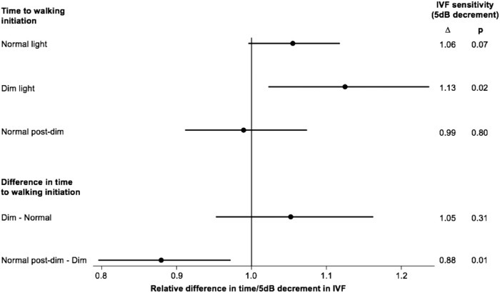
Average (SD) times to walking initiation under normal, dim, and NPD conditions were 0.97 (0.33), 2.93 (2.10), and 1.80 (1.02) seconds. Each 5 dB decrement in total IVF sensitivity was associated with a 13% (95% confidence interval [CI] = 2% to 24%, P = 0.02) longer gait initiation time in dim lighting, although there were no associations between VF loss and gait initiation time in normal and NPD lighting conditions (Fig. 1). When differences in walking initiation times were analyzed between consecutively tested conditions, lower IVF sensitivity was associated with significant shortening of initiation time from dim to NPD conditions (P = 0.01), but no significant difference was observed between normal and dim conditions (P = 0.31; see Fig. 1).
Figure 1.
Association between a 5 db decrement in IVF sensitivity and the time to gait initiation under different lighting conditions, as well as the difference in time to gait initiation between consecutive lighting conditions. Values presented come from separate multivariable models that controlled for age, race, gender, comorbidities, and polypharmacy. IVF = integrated visual field; dB = decibel; ∆ = delta; p = significance level.
Associations Between Gait Parameters and VF Damage Under Various Lighting Conditions
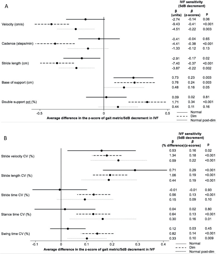
Under all three lighting conditions – normal, dim, and NPD – those with lower IVF sensitivity demonstrated significantly shorter strides (P = 0.02, P < 0.001, and P = 0.002, respectively) and broader base of support (P = 0.003, P = 0.003, and P = 0.05, respectively). For each 5 dB decrease in IVF, walking speed was significantly slower in both dim and NPD light (β = −9.43 cm/s, P < 0.001; β = −4.51 cm/s, P = 0.003), but not in normal lighting (P = 0.06). In dim light, participants with lower IVF sensitivity also walked with lower cadence (β = −4.41 steps/min, P < 0.001) and increased double support time (β = 1.71%, P < 0.001); these parameters were not associated with IVF sensitivity in normal or NPD lighting (Fig. 2a).
Figures 2.
(A) Gait parameters and (B) gait coefficients of variation. Association between IVF sensitivity and gait parameters and coefficients of variation under different lighting conditions. Values presented come from separate multivariable models that controlled for age, race, gender, comorbidities and polypharmacy. A positive z-score represents a higher numeric value for the analyzed metric. IVF = integrated visual field; dB = decibel, β = regression coefficient; p = significance level; pct = percent cycle time; CV = coefficient of variation.
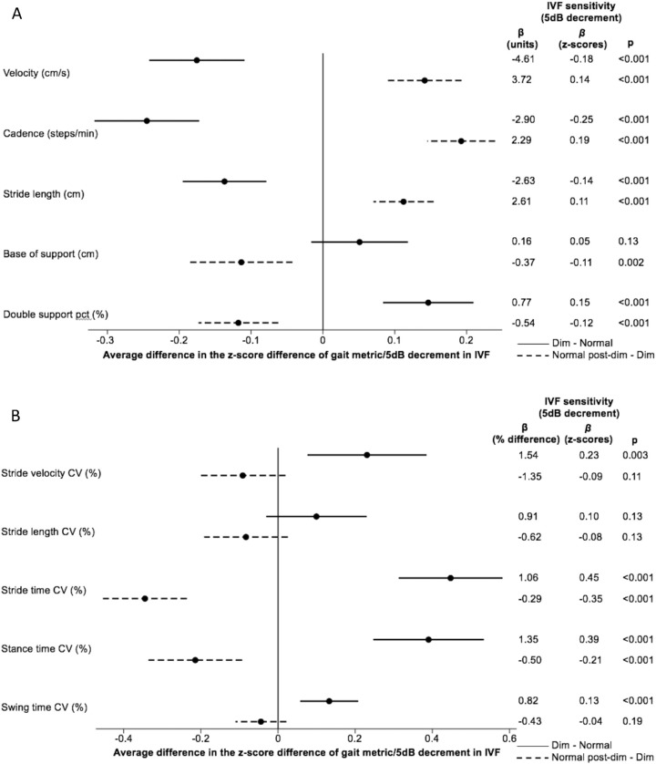
In the transition from normal to dim lighting (“dim – normal”), reduced IVF sensitivity was associated with significant slowing of gait and cadence, shortening of stride length, and lengthening of double support time (P < 0.001 for all); no significant change in base of support was noted with greater VF loss (Fig. 3a). When returning to normal (NPD) lighting after dim lighting (“normal post-dim – dim”), participants appeared to be partially regaining their baseline gait characteristics: among those with lower IVF sensitivity, velocity, cadence, and stride length increased (P < 0.001 for all), whereas base of support and double support time decreased (P < 0.01 for both; see Fig. 3a). However, greater severity of VF damage was still associated with a greater slowing of walking speed and cadence, and a larger increase in double support time, in NPD compared to normal lighting conditions (P < 0.05 for all; data not shown), as well as a greater narrowing of base of support (P = 0.01).
Figure 3.
(A) Difference in gait parameters and (B) difference in gait coefficients of variation. Association between IVF sensitivity and the difference in gait parameters and gait coefficients of variation between different lighting conditions. Values presented come from separate multivariable models that controlled for age, race, gender, comorbidities, and polypharmacy. A positive z-score unit increment represents a higher numeric value for the analyzed metric. IVF = integrated visual field; dB = decibel; β = regression coefficient; p = significance level; pct = percent cycle time; CV = coefficient of variation.
Associations Between Gait Variability and VF Damage Under Various Lighting Conditions
Under all three lighting conditions – normal, dim, and NPD – those with lower IVF sensitivity demonstrated significantly more variability (higher CV%) in their stride velocity (P = 0.02, P < 0.001, and P < 0.001, respectively) and stride length (P < 0.001 for all). Under dim and NPD lighting conditions, but not in normal lighting, those with lower IVF sensitivity showed greater variability in stance time (P < 0.001 and P = 0.01, respectively) and swing time (P < 0.001 and P = 0.009, respectively). In dim light, participants with lower IVF sensitivity also demonstrated greater stride time variability (P < 0.001); this was not seen in normal or NPD light (P > 0.05 for both; see Fig. 2b).
In the transition from normal to dim lighting, lower IVF sensitivity was associated with significant increases in the variability of stride velocity, stride time, stance time, and swing time (P < 0.01 for all), but not stride length (P = 0.13; see Fig. 3b). In the subsequent return to NPD lighting after dim lighting, lower IVF sensitivity was associated with improvement (less variability) in stride time and stance time (P < 0.001 for both), but no change in the variability of stride velocity, stride length, and swing time (P > 0.1 for all; see Fig. 3b). Moreover, those with lower IVF sensitivity showed significantly greater variability in stride velocity, stride time, and swing time under NPD compared to normal lighting conditions (P < 0.03 for all).
Fear of Falling Mediation of the Relationships Between Gait Changes Under Various Lighting Conditions and VF Damage
We investigated whether participants’ FOF mediated the relationship between VF loss and gait response to lighting changes. In this analysis, we did not find any mediation by FOF; observed associations between VF damage and lighting-related gait changes were preserved (i.e. statistically significant) even in models accounting for FOF.
Discussion
Persons with glaucoma demonstrate a more cautious and variable gait than those without VF loss, with irregularities more prominent in dim lighting conditions, and do not promptly recover their baseline gait characteristics when normal lighting is restored. These results corroborate complaints of patients with glaucoma of functional impairment in extreme or changing lighting,5 and demonstrate that such difficulties are associated with the severity of glaucoma damage. Of note, associations between severity of glaucoma damage and gait changes across lighting conditions are not mediated by FOF, suggesting that they reflect a cautiousness in specific conditions not captured by patient-reported mobility concerns (i.e. FOF).
Our findings are consistent with existing literature in that they support the idea that functional impairment in glaucoma is exacerbated by challenging conditions. For instance, prior work has shown that worsening VF loss among patients with glaucoma limits reading in a dose-dependent manner most evident when reading material is low-contrast or content requires sustained reading.44,45 However, the specific impact of VF damage on gait has not been completely characterized. The longitudinal Beaver Dam Eye Study, in which most visual impairments were mild or moderate, reported no association between visual measures and changes in walking speed on an unobstructed walking route. In contrast, the Salisbury Eye Evaluation Project described decreased gait speed in visually impaired individuals only under challenging conditions, such as navigating an obstacle course.46–49 Together, these findings suggested that mild to moderate VF damage (like that seen in our study population, in which average better eye mean deviation was ‒2.62 dB) might not alter gait parameters meaningfully during simple walking, but may impair patients in more challenging situations. One frequently encountered visual challenge is variable lighting, such as moving from outdoors to indoors, or navigating to the restroom in the middle of the night, and patients with glaucoma report increased visual difficulty in these situations.4,11,12 In fact, patients with glaucoma rate “glare” and “adaptation to different levels of lighting” at the top of a list of visual difficulties.2 Thus, to fully appreciate gait impairment in glaucoma, we must examine it under nonideal lighting conditions; examining gait (and perhaps others measures of functionality) under normal lighting may miss glaucoma-related disability.
Our analysis demonstrates that, within the same individual, sudden exposure to nonideal lighting conditions is associated with more hesitant, unsteady gait, and that these gait changes are most pronounced in persons with greater VF loss. Although the full group of participants, regardless of glaucoma status, hesitated before walking in dim light, those with reduced IVF sensitivity took significantly longer to initiate gait under dim conditions. Those with reduced IVF sensitivity took shorter and more wide-based strides under all conditions, even in normal light, perhaps representing an adaptation to perceived unsteadiness. In dim light, however, additional significant gait changes emerged among patients with glaucoma, including reduced velocity and cadence, more time in double support stance, and increased variability. Some of these changes may represent cautious walking, but they also signify less stable gait and poor adaptability, possibly interfering with a person's ability to safety perform daily activities.10 Patients and providers alike could use this information to improve patient safety. For example, providers may want to ensure the lights are on when patients enter examination rooms or VF testing suites and in the final minutes prior to patients exiting rooms.
Our data also demonstrate that gait changes in patients with glaucoma persist immediately after lighting conditions are returned to normal (NPD lighting), as happens when a person first turns on an additional light in a dark room. Although gait parameters trended toward baseline in NPD lighting, velocity and cadence remained significantly slower and double support time longer, compared to normal lighting conditions. Increased variability in stride velocity, stride time, and swing time in NPD, as compared to normal, conditions also remained. Slow, variable gait despite restored normal lighting reflects the subjective difficulty patients with glaucoma report functioning when light levels change in either direction and has important implications, highlighting why improving function for patients with glaucoma is not as simple as recommending brighter light.2,3,5 Patients could improve safety in their home environments by arranging lights so that several contiguous areas are illuminated by a single switch, perhaps one easily reached from a chair or bed.
The associations observed between VF loss and gait changes remained even after accounting for FOF, suggesting they are not attributable to patient-perceived fall risk. We have previously reported that glaucoma severity, represented here by reduced IVF sensitivity, is the most important predictor of falls per step, and that cautious, unsteady walking is associated with a higher risk of falling.50 However, whereas slower, more variable gait under photo-stressed conditions may represent caution in patients with glaucoma, who take shorter wider-based steps at baseline, it appears to be associated with disease severity, but not mediated by FOF. Understanding this position, providers have to address safety effects of VF loss with education about optimal lighting for patients across the spectrum of glaucoma severity. More research is needed to inform fall-prevention interventions and understand how gait impairment impacts patients psychologically or socially.
This study has several limitations. Although we excluded persons with neurological causes of VF loss, we did not account for every potential cause of abnormal gait (vestibular, orthopedic, etc.) in our exclusion criteria. Participants’ gait was assessed barefoot, rather than wearing habitual footwear, and was only measured on a flat surface without any obstruction, which is not representative of all surfaces on which individuals typically walk. A stopwatch is the gold standard for capturing gait initiation, but is known to introduce some intra-operator and inter-operator variability.51 In addition, our unexpected observations that gait initiation improved and base of support narrowed compared to baseline in NPD lighting conditions suggests that walking performance may have improved with practice, as participants repeated the task assigned. As such, our results could underestimate the impact of lighting changes on gait parameters. There may also be recruitment or participation bias in the FIGS population. We previously reported that recruited patients were of similar age, gender, and race, and had similarly severe glaucoma, as study-eligible individuals seen in the Wilmer Eye Institute Glaucoma Center of Excellence who were not recruited.42 However, they were also more likely than other patients to report falling in the 12 months prior to recruitment.42 One can imagine that persons more predisposed to falling might be more likely to participate in this study, or conversely that those with more mobility difficulties might be less likely to enroll due to difficulty attending study visits. It is also possible that the population being followed at the Wilmer Eye Institute is not representative of the entire glaucoma population in the United States. Whether any such bias would alter the relationship between lighting and gait described here is not clear. More importantly, findings in our elderly study population, which reflects the typical age of patients with newly diagnosed glaucoma, may not be generalizable to younger patients.52 Strengths of the study include a large sample size compared with prior studies of gait in visually impaired patients, and rigorous spatiotemporal characterization of numerous gait parameters using the GAITRite Electronic Walkway.10
In summary, among patients with glaucoma, a constellation of gait features was observed to change under extreme or changing lighting conditions, in a dose-dependent manner with respect to VF damage. Further research is needed to understand if specific changes are adaptive (improving safe ambulation) or maladaptive, and additional work is needed to translate these findings into a proactive approach to improve functionality in patients with glaucoma.
Acknowledgments
Supported by National Institutes of Health Grant No. EY022976.
Disclosure: A.K. Bicket, None; A. Mihailovic, None; J.-Y. E, None; A. Nguyen, None; M.R. Mukherjee, None; D.S. Friedman, None; P.Y. Ramulu, None
References
- 1. Lee BL. The Glaucoma Symptom Scale. Arch Ophthalmol. 1998; 116(7): 861. [DOI] [PubMed] [Google Scholar]
- 2. Nelson P, Aspinall P, O'Brien C. Patients’ perception of visual impairment in glaucoma: a pilot study. Br J Ophthalmol. 1999; 83(5): 546–552. [DOI] [PMC free article] [PubMed] [Google Scholar]
- 3. Janz NK, Wren PA, Lichter PR, Musch DC, Gillespie BW, Guire KE. Quality of life in newly diagnosed glaucoma patients: the collaborative initial glaucoma treatment study. Ophthalmology. 2001; 108(5): 887–897. [DOI] [PubMed] [Google Scholar]
- 4. Hu CX, Zangalli C, Hsieh M, et al.. What do patients with glaucoma see? Visual symptoms reported by patients with glaucoma. Am J Med Sci. 2014; 348(5): 403–409. [DOI] [PMC free article] [PubMed] [Google Scholar]
- 5. Bierings RAJM, van Sonderen FLP, Jansonius NM. Visual complaints of patients with glaucoma and controls under optimal and extreme luminance conditions. Acta Ophthalmol. 2018; 96(3): 288–294. [DOI] [PubMed] [Google Scholar]
- 6. Diniz-Filho A, Boer ER, Elhosseiny A, Wu Z, Nakanishi M, Medeiros FA. Glaucoma and driving risk under simulated fog conditions. Transl Vis Sci Technol. 2016; 5(6): 15. [DOI] [PMC free article] [PubMed] [Google Scholar]
- 7. Burton R, Crabb DP, Smith ND, Glen FC, Garway-Heath DF. Glaucoma and reading: exploring the effects of contrast lowering of text. Optom Vis Sci. 2012; 89(9): 1282–1287. [DOI] [PubMed] [Google Scholar]
- 8. Hassan SE, Hicks JC, Lei H, Turano KA. What is the minimum field of view required for efficient navigation? Vision Res. 2007; 47(16): 2115–2123. [DOI] [PubMed] [Google Scholar]
- 9. Alexander MS, Lajoie K, Neima DR, Strath RA, Robinovitch SN, Marigold DS. Effect of ambient light and age-related macular degeneration on precision walking. Optom Vis Sci. 2014; 91(8): 990–999. [DOI] [PubMed] [Google Scholar]
- 10. Mihailovic A, Swenor BK, Friedman DS, West SK, Gitlin LN, Ramulu PY. Gait implications of visual field damage from glaucoma. Transl Vis Sci Technol. 2017; 6(3): 23. [DOI] [PMC free article] [PubMed] [Google Scholar]
- 11. Black AA, Wood JM, Lovie-Kitchin JE, Newman BM. Visual impairment and postural sway among older adults with glaucoma. Optom Vis Sci. 2008; 85(6): 489–497. [DOI] [PubMed] [Google Scholar]
- 12. Nakamura T. Quantitative analysis of gait in the visually impaired. Disabil Rehabil. 1997; 19(5): 194–197. [DOI] [PubMed] [Google Scholar]
- 13. Ayaki M, Nagura T, Toyama Y, Negishi K, Tsubota K. Motor function benefits of visual restoration measured in age-related cataract and simulated patients: case-control and clinical experimental studies. Sci Rep. 2015; 5: 14595. [DOI] [PMC free article] [PubMed] [Google Scholar]
- 14. Sippel K, Kasneci E, Aehling K, et al.. Binocular glaucomatous visual field loss and its impact on visual exploration - a supermarket study. PLoS One. 2014; 9(8): e106089. [DOI] [PMC free article] [PubMed] [Google Scholar]
- 15. Yonge A V., Swenor BK, Miller R, et al. Quantifying fall-related hazards in the homes of persons with glaucoma. Ophthalmology. 2017; 124(4): 562–571. [DOI] [PMC free article] [PubMed] [Google Scholar]
- 16. Ramulu PY, van Landingham SW, Massof RW, Chan ES, Ferrucci L, Friedman DS. Fear of falling and visual field loss from glaucoma. Ophthalmology. 2012; 119(7): 1352–1358. [DOI] [PMC free article] [PubMed] [Google Scholar]
- 17. Van Landingham SW, Massof RW, Chan E, Friedman DS, Ramulu PY. Fear of falling in age-related macular degeneration. BMC Ophthalmol. 2014; 14(1): 1–9. [DOI] [PMC free article] [PubMed] [Google Scholar]
- 18. Haymes SA, LeBlanc RP, Nicolela MT, Chiasson LA, Chauhan BC. Risk of falls and motor vehicle collisions in glaucoma. Investig Opthalmol Vis Sci. 2007; 48(3): 1149. [DOI] [PubMed] [Google Scholar]
- 19. Legood R. Are we blind to injuries in the visually impaired? A review of the literature. Inj Prev. 2002; 8(2): 155–160. [DOI] [PMC free article] [PubMed] [Google Scholar]
- 20. Lord SR, Dayhew J.. Visual risk factors for falls in older people. J Am Geriatr Soc. 2001; 49(5): 508–515. [DOI] [PubMed] [Google Scholar]
- 21. Lamoreux EL, Chong E, Wang JJ, et al.. Visual impairment, causes of vision loss, and falls: the Singapore Malay Eye Study. Investig Opthalmol Vis Sci. 2008; 49(2): 528. [DOI] [PubMed] [Google Scholar]
- 22. Daga FB, Diniz-Filho A, Boer ER, Gracitelli CPB, Abe RY, Medeiros FA. Fear of falling and postural reactivity in patients with glaucoma. PLoS One. 2017; 12(12): e0187220. [DOI] [PMC free article] [PubMed] [Google Scholar]
- 23. Wang MY, Rousseau J, Boisjoly H, et al.. Activity limitation due to a fear of falling in older adults with eye disease. Investig Ophthalmol Vis Sci. 2012; 53(13): 7967–7972. [DOI] [PubMed] [Google Scholar]
- 24. Turano KA, Rubin GS, Quigley HA. Mobility performance in glaucoma. Investig Ophthalmol Vis Sci. 1999; 40(12): 2803–2809. [PubMed] [Google Scholar]
- 25. Hallemans A, Ortibus E, Truijen S, Meire F. Development of independent locomotion in children with a severe visual impairment. Res Dev Disabil. 2011; 32(6): 2069–2074. [DOI] [PubMed] [Google Scholar]
- 26. Bilney B, Morris M, Webster K. Concurrent related validity of the GAITRite® walkway system for quantification of the spatial and temporal parameters of gait. Gait Posture. 2003; 17(1): 68–74. [DOI] [PubMed] [Google Scholar]
- 27. Cutlip RG, Mancinelli C, Huber F, DiPasquale J. Evaluation of an instrumented walkway for measurement of the kinematic parameters of gait. Gait Posture. 2000; 12(2): 134–138. [DOI] [PubMed] [Google Scholar]
- 28. McDonough AL, Batavia M, Chen FC, Kwon S, Ziai J. The validity and reliability of the GAITRite system's measurements: a preliminary evaluation. Arch Phys Med Rehabil. 2001; 82(3): 419–425. [DOI] [PubMed] [Google Scholar]
- 29. Webster KE, Wittwer JE, Feller JA. Validity of the GAITRite® walkway system for the measurement of averaged and individual step parameters of gait. Gait Posture. 2005; 22(4): 317–321. [DOI] [PubMed] [Google Scholar]
- 30. Shimada H, Kim H, Yoshida H, et al.. Relationship between age-associated changes of gait and falls and life-space in elderly people. J Phys Ther Sci. 2010; 22(4): 419–424. [Google Scholar]
- 31. Maki BE. Gait changes in older adults: predictors of falls or indicators of fear? J Am Geriatr Soc. 1997; 45(3): 313–320. [DOI] [PubMed] [Google Scholar]
- 32. Quach L, Galica AM, Jones RN, et al.. The nonlinear relationship between gait speed and falls: the Maintenance of Balance, Independent Living, Intellect, and Zest in the Elderly of Boston Study. J Am Geriatr Soc. 2011; 59(6): 1069–1073. [DOI] [PMC free article] [PubMed] [Google Scholar]
- 33. Toulotte C, Thevenon A, Watelain E, Fabre C. Identification of healthy elderly fallers and non-fallers by gait analysis under dual-task conditions. Clin Rehabil. 2006; 20(3): 269–276. [DOI] [PubMed] [Google Scholar]
- 34. Gehlsen GM, Whaley MH.. Falls in the elderly: part I, gait. Arch Phys Med Rehabil. 1990; 71(10): 735–738. [PubMed] [Google Scholar]
- 35. Wolfson L, Whipple R, Amerman P, Tobin JN. Gait assessment in the elderly: a gait abnormality rating scale and its relation to falls. J Gerontol. 1990; 45(1): M12–M19. [DOI] [PubMed] [Google Scholar]
- 36. Hausdorff JM, Rios DA, Edelberg HK. Gait variability and fall risk in community-living older adults: a 1-year prospective study. Arch Phys Med Rehabil. 2001; 82(8): 1050–1056. [DOI] [PubMed] [Google Scholar]
- 37. Hausdorff JM, Edelberg HK, Mitchell SL, Goldberger AL, Wei JY. Increased gait unsteadiness in community-dwelling elderly fallers. Arch Phys Med Rehabil. 1997; 78(3): 278–283. [DOI] [PubMed] [Google Scholar]
- 38. Odden JL, Mihailovic A, Boland M V, Friedman DS, West SK, Ramulu PY. Evaluation of central and peripheral visual field concordance in glaucoma. Investig Opthalmol Vis Sci. 2016; 57(6): 2797. [DOI] [PMC free article] [PubMed] [Google Scholar]
- 39. Ramulu PY, Maul E, Hochberg C, Chan ES, Ferrucci L, Friedman DS. Real-world assessment of physical activity in glaucoma using an accelerometer. Ophthalmology. 2012; 119(6): 1159–1166. [DOI] [PMC free article] [PubMed] [Google Scholar]
- 40. Gnjidic D, Hilmer SN, Blyth FM, et al.. Polypharmacy cutoff and outcomes: five or more medicines were used to identify community-dwelling older men at risk of different adverse outcomes. J Clin Epidemiol. 2012; 65(9): 989–995. [DOI] [PubMed] [Google Scholar]
- 41. Velozo CA, Peterson EW.. Developing meaningful fear of falling measures for community dwelling elderly. Am J Phys Med Rehabil. 2001; 80(9): 662–673. [DOI] [PubMed] [Google Scholar]
- 42. Ramulu PY, Mihailovic A, West SK, Friedman DS, Gitlin LN. What is a falls risk factor? Factors associated with falls per time or per step in individuals with glaucoma. J Am Geriatr Soc. 2019; 67(1): 87–92. [DOI] [PMC free article] [PubMed] [Google Scholar]
- 43. Campbell AJ, Borrie MJ, Spears GF. Risk Factors for falls in a community-based prospective study of people 70 years and older. J Gerontol. 1989; 44(4): M112–M117. [DOI] [PubMed] [Google Scholar]
- 44. Nguyen AM, van Landingham SW, Massof RW, Rubin GS, Ramulu PY. Reading ability and reading engagement in older adults with glaucoma. Investig Opthalmol Vis Sci. 2014; 55(8): 5284. [DOI] [PMC free article] [PubMed] [Google Scholar]
- 45. Ramulu PY, Swenor BK, Jefferys JL, Friedman DS, Rubin GS. Difficulty with out-loud and silent reading in glaucoma. Investig Ophthalmol Vis Sci. 2013; 54(1): 666–672. [DOI] [PMC free article] [PubMed] [Google Scholar]
- 46. Turano KA, Broman AT, Bandeen-Roche K, Munoz B, Rubin GS, West SK. Association of visual field loss and mobility performance in older adults: Salisbury Eye Evaluation Study. Optom Vis Sci. 2004; 81(5): 298–307. [DOI] [PubMed] [Google Scholar]
- 47. Friedman DS, Freeman E, Munoz B, Jampel HD, West SK. Glaucoma and mobility performance. The Salisbury Eye Evaluation Project. Ophthalmology. 2007; 114(12): 2232–2237. [DOI] [PubMed] [Google Scholar]
- 48. Patel I, Turano KA, Broman AT, Bandeen-Roche K, Muñoz B, West SK. Measures of visual function and percentage of preferred walking speed in older adults: the Salisbury Eye Evaluation Project. Investig Ophthalmol Vis Sci. 2006; 47(1): 65–71. [DOI] [PubMed] [Google Scholar]
- 49. Klein BE., Moss SE, Klein R, Lee KE, Cruickshanks KJ.. Associations of visual function with physical outcomes and limitations 5 years later in an older population. Ophthalmology. 2003; 110(4): 644–650. [DOI] [PubMed] [Google Scholar]
- 50. Mihailovic A, De Luna RM, West SK, Friedman DS, Gitlin LN, Ramulu PY. Gait and balance as predictors and/or mediators of falls in glaucoma. Investig Opthalmology Vis Sci. 2020; 61(3): 30. [DOI] [PMC free article] [PubMed] [Google Scholar]
- 51. Maggio M, Ceda GP, Ticinesi A, et al.. Instrumental and non-instrumental evaluation of 4-meter walking speed in older individuals. PLoS One. 2016; 11(4): 1–10. [DOI] [PMC free article] [PubMed] [Google Scholar]
- 52. Sharma T, Salmon JF.. Ten-year outcomes in newly diagnosed glaucoma patients: mortality and visual function. Br J Ophthalmol. 2007; 91(10): 1282–1284. [DOI] [PMC free article] [PubMed] [Google Scholar]


