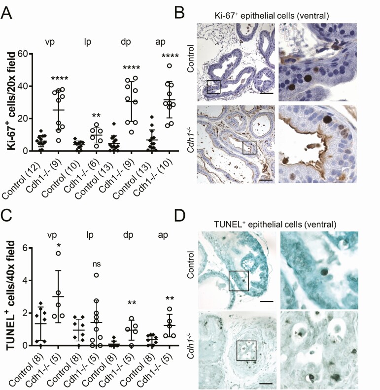
Figure 2.
Effect of Cdh1-deficiency on epithelial proliferation in the murine prostate. A, Quantification of Ki-67+ luminal epithelial cells in the lobes of the prostate from Control and Cdh1-/- mice at 21 to 23 weeks of age. B, Ki-67 immunostaining in prostate ventral lobes. C, Quantification of TUNEL staining in prostate luminal epithelial cells. D, TUNEL staining in prostate ventral lobes, TUNEL-positive apoptotic cells (red arrows). Original magnification, 20×, inset 40×. Scale bars indicate 100 µm 20×, 50 µm in 40×. Data represent mean ± SD, number of mice in each group in parentheses. Lobes that had been washed away during staining process were not quantified. Abbreviations: ap, anterior prostate; dp, dorsal prostate; lp, lateral prostate; ns, nonsignificant; vp, ventral prostate. * P < 0.05; ** P < 0.01; **** P < 0.0001.

