PLOS ONE logoLink to PLOS ONE
. 2020 Dec 17;15(12):e0242215. doi: 10.1371/journal.pone.0242215

Active foot placement control ensures stable gait: Effect of constraints on foot placement and ankle moments

A M van Leeuwen 1,2, J H van Dieën 1, A Daffertshofer 1,2, S M Bruijn 1,2,3,*
Editor: J Lucas McKay4
PMCID: PMC7746185  PMID: 33332421

Abstract

Step-by-step foot placement control, relative to the center of mass (CoM) kinematic state, is generally considered a dominant mechanism for maintenance of gait stability. By adequate (mediolateral) positioning of the center of pressure with respect to the CoM, the ground reaction force generates a moment that prevents falling. In healthy individuals, foot placement is complemented mainly by ankle moment control ensuring stability. To evaluate possible compensatory relationships between step-by-step foot placement and complementary ankle moments, we investigated the degree of (active) foot placement control during steady-state walking, and under either foot placement-, or ankle moment constraints. Thirty healthy participants walked on a treadmill, while full-body kinematics, ground reaction forces and EMG activities were recorded. As a replication of earlier findings, we first showed step-by-step foot placement is associated with preceding CoM state and hip ab-/adductor activity during steady-state walking. Tight control of foot placement appears to be important at normal walking speed because there was a limited change in the degree of foot placement control despite the presence of a foot placement constraint. At slow speed, the degree of foot placement control decreased substantially, suggesting that tight control of foot placement is less essential when walking slowly. Step-by-step foot placement control was not tightened to compensate for constrained ankle moments. Instead compensation was achieved through increases in step width and stride frequency.

Introduction

On every step we take, our center of mass (CoM) accelerates laterally towards the new stance foot. In order not to fall, this motion of the CoM has to be reversed, preventing the CoM to move beyond the lateral border of the base of support [1]. The moment that accelerates the CoM in the opposite direction can be controlled by adjusting the center of pressure (CoP) [2]. During gait, the dominant mechanism to control the CoP is the so-called foot placement strategy [1, 3].

Wang and Srinivasan [4] captured this control strategy in a linear model, indicating that foot placement can be predicted by the CoM kinematic state (i.e. CoM position and velocity) during the preceding swing phase. The association between foot placement and CoM kinematic state was shown to be less pronounced during walking with external lateral stabilization [5], which supports the notion that step-by-step foot placement control promotes mediolateral gait stability.

In healthy individuals, ankle, hip and push-off strategies complement foot placement in maintaining stability [2, 6]. Coordinated recruitment of these control strategies may guarantee stability throughout the gait cycle [7, 8]. For example, the ankle strategy allows for an early response to a perturbation, before the foot placement strategy becomes effective following heel strike [9]. In elderly, pathological, or prosthetic gait, an impairment in one strategy may require individuals to rely more on another one [10, 11]. When aiming for stability improvements, it is hence important to understand the details of these control strategies as well as their interplay. In the current study, we focused on the foot placement strategy in healthy individuals during steady-state treadmill walking. Following Wang and Srinivasan [4], we expected that mediolateral foot placement can be predicted by CoM state, reflective of a step-by-step control strategy (E1). And, in line with Rankin et al. [12], we expected mediolateral foot placement to correlate with hip ab- and adductor muscle activity during the preceding swing phase (E2), supporting the active nature of this control mechanism. We tested these two expectations during normal and slow walking. We chose two speeds because the implementation of the foot placement strategy has been shown to be speed-dependent [13]. We did not make any speed-related predictions, but the speed-dependent nature of the foot placement strategy could potentially affect the effects of our other experimental conditions. These experimental conditions served to challenge the degree of (active) foot placement control; see below.

Degree of control

The degree of foot placement control can be inferred from its predictability based on the CoM state. The mid-swing CoM state is a better predictor of foot placement than the swing foot state itself, although this prediction is not entirely accurate [4]. Eventual inaccuracies could be attributed to motor noise, or task constraints. In certain contexts, a high degree of foot placement control might not be necessary. As an example, we note that foot placement seems to correlate less with CoM state in externally stabilized gait, and at slower speeds. Apparently, its control is less important in these conditions [5, 13]. In addition, the necessary degree of foot placement control may depend on the availability of alternative control strategies. Arguably, foot placement is most effective in shifting the CoP, though adapting ankle moments allows for a complementary shift during stance. As mentioned above, this can enable early stabilizing responses [9] and/or corrections of inaccurate foot placements [11]. According to Fettrow et al. [8], foot placement and ankle moments are interdependent, i.e. one may compensate for the other. In this context, we hypothesized foot placement to compensate for a limited possibility to shift the CoP underneath the stance foot, through constrained ankle moments (H1). If true, this will be in line with the findings of Hof et al. [11], who demonstrated that more lateral foot placement compensated for the impossibility to induce a CoP shift under a prosthetic leg. Conversely, a lesser degree of foot placement control might be compensated for by ankle moments. Such compensation could facilitate adaptation of foot placement to environmental or task constraints without threatening stability. Accordingly, we hypothesized that constraining the foot placement by stepping onto projected lines will yield diminished foot placement control (H2).

Active control

Whether foot placement emerges mainly from a passive rather than an active mechanism is currently under debate [14], though there is increasing evidence for the latter. An illusory perturbation of the CoM state by manipulating muscle spindle afference has been reported to lead to predictable adjustments in foot placement, which indicates it to be affected by sensory input [15]. Similarly, illusory falls induced by visual or vestibular stimulation seem to trigger reactive foot placement adjustments [7, 16]. Both in response to perturbations [3] as well as during steady-state walking [12], gluteus medius activity (hip abductor muscle) has been associated with step width. In addition to replicating the aforementioned findings of Rankin et al. [12], we therefore hypothesized that constraining ankle moments will yield a larger active contribution to step-by-step variability in mediolateral foot placement, indicative of a compensatory foot placement strategy (H3).

With our expectations, E1 & E2, we aimed at replicating findings [4, 12] of actively controlled step-by-step foot placement during steady-state walking. We also sought to investigate how important this foot placement strategy is, by evaluating whether we can constrain foot placement (H2) during steady-state walking and whether foot placement control would tighten as a compensatory mechanism, when ankle moments are constrained (H1 & H3).

This study’s preregistered hypotheses, protocol and sampling plan can be found on OSF: https://osf.io/74pn5.

Methods

Participants

Only participants capable of walking without difficulty for a longer duration (≥ 60 minutes) were recruited. Participants were excluded when they reported sports injuries or other motor impairments that possibly affected their gait pattern. Participants suffering from (self-reported) balance issues were also excluded.

An initial sample size of ten participants was recruited as we used a Bayesian sequential sampling approach. Subsequently, recruitment of participants continued until a threshold of meaningful evidence was reached [17]. We set this threshold to a BF10 or BF01 of 10 (indicative of strong relative evidence) for either the null or the alternative hypothesis, based on our main outcome measures. Since not all outcome measures reached a BF10 or BF01, we continued recruitment until the pre-determined maximum of 30 participants was attained, while accounting for drop-outs.

A total of 35 healthy participants completed the experiment according to instructions. The data of four participants had to be discarded because of technical malfunctioning of equipment, and data of one participant were excluded in view of pronounced toeing out during normal walking. Finally, data of 30 participants (19 female, 11 male; 30 ± 7 yrs, 70 ± 13 kg, 1.73 ± 0.08 m; mean ± sd) were included in the analysis.

Prior to participation, participants signed an informed consent and ethical approval (VCWE-2018-159) was granted by the ethics review board of the faculty of Behavioural and Movement Sciences at ‘Vrije Universiteit Amsterdam’.

Procedure

Participants were invited to walk on a treadmill during four conditions (see Fig 1) at normal (1.25 × √(leg length) m/s) and slow (0.63 × √(leg length) m/s) walking speeds, normalized to leg length in accordance with Hof [18]. In this study, stride frequency control could be a confounding stability control strategy [19]. Therefore, stride frequency was controlled by means of a metronome, to avoid that participants changed stride frequency between conditions. Participants were instructed to time either their right or their left heel strikes to the metronome beat. The imposed frequency was set to the average preferred stride frequency determined during the final 100 steps of a familiarization trial for each speed (see below).

Fig 1. Conditions performed at normal and slow walking speeds.

Fig 1

We randomized the order of the conditions across participants and speeds. Before beginning the experiment, participants performed a five-minutes familiarization phase (two minutes at normal walking speed, three minutes at slow walking speed) without imposing further constraints; cf. below. Additionally, participants were familiarized with ankle moment constraining shoes (Fig 2) prior to data collection. To ensure that all trials contained at least 200 consecutive strides, trials at normal walking speed lasted five minutes and trials at slow walking speed ten minutes. Between trials, sufficient breaks were provided to prevent fatigue as verified by subjective report.

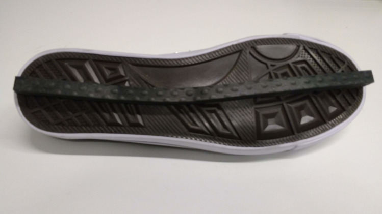
Fig 2. LesSchuh.

Fig 2

Shoe with an ankle moment constraint (width of the ridge is 1 cm).

Constraints

Foot placement constraint

Projections on the treadmill served to constrain the variation in mediolateral foot placement. In brief, first the average step width was derived from the final 100 steps of the familiarization trial, based on the CoP estimated from the force measurement of the instrumented treadmill. Then, this average step width was imposed by projecting beams on the treadmill, and participants were instructed to place their foot in the middle of the beam. For every step, the beam became visible following toe-off in order to prevent modification of the CoM swing phase trajectory by compensatory push-off modulation [20]. Customized labview software allowed us to estimate the toe-off event based on a force threshold, which triggered the projections based on real-time force measurements.

Ankle moment constraint

The mediolateral shift of the CoP underneath the stance foot was minimized using a shoe with a narrow (1 cm) ridge attached to the sole. This so-called LesSchuh limits ankle moments while anteroposterior roll-off and subsequent push-off remain possible, because the material of the ridge bends with the sole in anterior-posterior direction. Participants were asked to walk on the ridge, not touching the ground with the sides of the shoe’s sole. Participants were also instructed to place their feet in a similar orientation as they would without the ankle moment constraint, to avoid a “toeing-out strategy” [21] potentially inducing a mediolateral shift of the center of pressure after foot placement (heel strike) despite the narrow base of support.

Data collection

Force plate

Participants walked on an instrumented dual-belt treadmill (Motek-Forcelink, Amsterdam, Netherlands). Ground reaction forces and moments were recorded from the force plates embedded in the treadmill and sampled at 200 Hz. From these forces, we determined the CoP.

Optotrak

Full body kinematics were measured using two Optotrak cameras (Northern Digital Inc, Waterloo Ontario, Canada) directed at the center of the treadmill (sampling rate was 50 Hz). Cluster markers were attached to the feet, shanks, thighs, pelvis, trunk, upper arms and forearms. Corresponding anatomical landmarks were digitized using a six-marker probe.

Electromyography

Bipolar surface electromyography (EMG) was applied to measure bilateral muscle activity from 16 muscles expected to contribute to gait stability. Here, we concentrated on the m. gluteus medius and m. adductor longus muscle given their hypothesized contribution to foot placement control (for other muscles recorded, we refer to the preregistered protocol: https://osf.io/74pn5). A 16-channel Porti EMG amplifier (TMSi, Enschede, Netherlands) served to record the two muscles bilaterally; signals were sampled at 2 kHz. Surface EMG electrodes with a diameter of 22 mm were positioned on the skin according to the SENIAM guidelines [22].

Data analysis

For all subjects and trials, we analyzed 200 consecutive strides. These were the final 200 strides of every trial, unless data quality urged selection of earlier strides (e.g., better marker visibility, less noise).

Data processing

Gait event detection

Gait events (heel strikes & toe-offs) were detected based on the characteristic “butterfly pattern” of the combined center of pressure as derived from force plate data [23]. We defined a step as the period between toe-off and heel strike. Mid-swing was defined at 50 percent of the step.

Center of mass

For every segment, we estimated the segment’s mass via linear regression including the segment’s length and the segment’s measured circumference as predictors and regression coefficients based on gender [24]. The segment’s CoM was estimated as a percentage of the longitudinal axis of the segment [24, 25]. The full body CoM was derived from a weighted sum of the body segment’s CoMs. Mediolateral CoM displacement was defined along the x-axis of our global coordinate system, of which the x-axis was oriented perpendicular to the direction of the treadmill. We determined the increment of the mediolateral CoM position CoMpos, divided by the time-step, as estimate of the mediolateral CoM velocity CoMvel.

EMG processing

EMG data were high-pass filtered at 20 Hz, rectified, and low-pass filtered at 50 Hz, following Rankin et al. [12]. Strides were time-normalized to 1000 samples. For every stride, we determined EMGgm_swing and EMGal_swing as the median EMG activity during early swing (60–80% of the stride cycle) multiplied by the duration of this episode in seconds (we note here that this is a deviation from our preregistered protocol, see “Deviations from the preregistered plans I”).

Outcome measures

Multiple linear regression models

We used a multiple linear regression with mediolateral foot placement (FP, i.e. the mediolateral foot position at midstance expressed with respect to the mediolateral position of the contralateral foot at midstance (step width), as dependent variable and the CoM state variables (CoMpos, CoMvel), i.e. the CoM position expressed with respect to the position of the stance foot at midstance and CoM velocity, as independent variables (i.e. predictors). The predictors’ time series were time normalized to 51 samples for every step, from toe-off to heel strike. FP, CoMpos and CoMvel were demeaned prior to regression.

In a separate model, mediolateral foot placement (FP2), i.e. the mediolateral foot position at midstance with respect to the CoM at the time of toe-off, was the dependent variable with the median gluteus medius and adductor longus EMG amplitudes from 60–80% of the gait cycle (EMGgm_swing, EMGal_swing) as independent variables. In this model, within subjects and trials, we normalized FP2, EMGgm_swing and EMGal_swing by demeaning and dividing by the standard deviation (we note here that this is a deviation from our preregistered protocol, see “Deviations from the preregistered plans II”).

The first model was created following Wang and Srinivasan [4] and read:

FP=βpos·CoMpos(i)+βvel·CoMvel(i)+ε1(i), (1)

in which ε1 denotes the error in every step cycle, for different phases (i) of the step cycle. Statistical tests were performed for CoM state predictors at mid-swing (mid, i = 25) and terminal swing (ts, i = 51).

The second model had the form:

FP2=βgm_swing·EMGgm_swing+βal_swing·EMGal_swing+ε2. (2)

We quantified the degree of foot placement control via the relative explained variance (R2) of model 1 and the active contribution to step-by-step variability in foot placement via the R2 of model 2.

The data and analysis can be found on Zenodo: 10.5281/zenodo.4229851.

Statistics

All statistical tests were performed in JASP (JASP Team (2019). JASP (Version 0.11.1)).

Step-by-step foot placement control in steady-state walking

The regression coefficients (β) of model (1) were tested against zero in a Bayesian one-sample t-test, to infer whether mediolateral foot placement can be predicted by CoM state during steady-state walking (E1) (we note here that this is a deviation from our preregistered protocol, see “Deviations from the preregistered plans III”). And, to assess whether mediolateral foot placement correlates with hip ab- and adductor muscle activity of the preceding swing phase during steady-state walking (E2), we tested the regression coefficients of model (2) against zero in a Bayesian one-sample t-test.

Step-by-step foot placement control with ankle moment constraints

We performed a Fisher transformation on the R2 values prior to statistical testing. Bayesian equivalents of a 2×2 repeated measures ANOVA with factors Condition (levels: ankle moment constrained/foot placement constrained versus steady-state walking) and Speed (levels: normal versus slow) served to test the effects of the constraints and walking speed, as well as their interaction, on degree of (active) step-by-step foot placement control; relative explained variance of models (1) and (2). Moreover, to test the constrained conditions against the steady-state walking condition, we used Bayesian planned post-hoc assessments.

We tested the R2 values of models (1) and (2) in the ankle moment constrained condition against the steady-state walking condition. By this we could estimate whether constraining the ankle moment led to compensation in the degree of foot placement control. That is, this allowed for testing the hypotheses that compensation will encompass tighter control (i.e. a higher R2 of model (1)) (H1) driven by compensatory muscle activation (i.e. a higher R2 of model (2)) (H3). If both H1 and H3 are true, this would suggest a compensatory tighter coupling between variations in foot placement and CoM state (H1), achieved through compensatory muscle activity (H3). If only H1 is true, this would suggest tighter coupling between variations in foot placement and CoM state (H1), but no evidence that this is actively controlled (in contrast with H3). If only H3 is true, this would suggest that foot placement is more strictly controlled through muscle activity (H3), but not to tighten the coupling between variations in foot placement and CoM state.

Step-by-step foot placement control with foot placement constraints

In order to infer whether the degree of control decreased (i.e. a lower R2 of model (1)) (H2), when constraining foot placement, we tested the R2 of model (1) in the foot placement constrained condition against the steady-state walking condition.

Deviations from the preregistered plans

I) In our preregisterd plans (https://osf.io/74pn5), we intended to take the integral of the EMG signal over the selected time period. To avoid amplification of artefacts by taking the integral, while still retaining the influence of time, we deviated from the preregistered plans and computed the product of the median EMG amplitude and time in seconds.

II) The definition of mediolateral foot placement (FP2) and normalization of FP2, EMGgm_swing and EMGal_swing by both subtracting the mean and dividing by the standard deviation was not included in detail in the preregistered plans, but is in line with Rankin et al. [12].

III) In our preregistration we planned to bootstrap the regression coefficients for each participant. However, although most participants, but not all, demonstrated significant relationships, the final conclusion was based on statistics on group level and included in the results.

Results

Step-by-step foot placement control in steady-state walking

Model 1: Foot placement model

First, we considered the relative explained variance (R2) and tested the regression coefficients of the foot placement model (1) to assess our expectation (E1) that mediolateral foot placement can be predicted by CoM state, as previously shown by Wang and Srinivasan [4]. In the steady-state walking condition (unconstrained), the foot placement model predicted over 60% of the variance at normal walking speed and over 40% at slow walking speed. There was extreme evidence (BF10 > 100), when testing the regression coefficients for CoMpos (at mid-swing & terminal swing) and CoMvel (at mid-swing and terminal swing) against zero (Fig 3), that swing phase CoM state predicts foot placement during steady-state walking (E1).

Fig 3. Mean regression coefficients of the foot placement model (1).

Fig 3

Standard deviations are represented by error bars. Panels A and B represent the results for respectively normal and slow walking speed. The beta coefficients were tested at mid-swing and terminal swing, demonstrating extreme evidence (BF10 > 100) for inclusion of the predictors.

Model 2: Muscle model

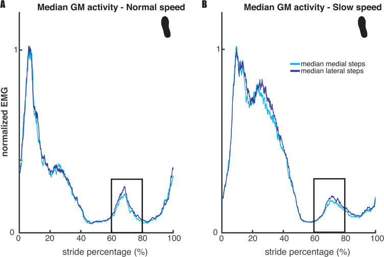
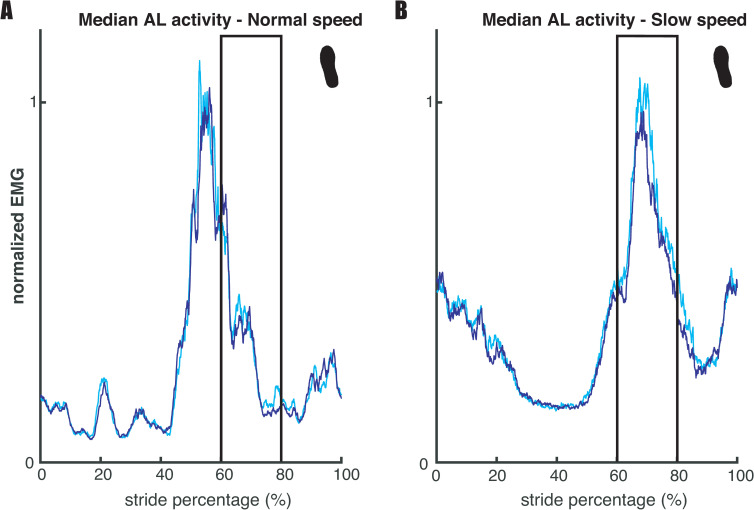
Our second expectation (E2), that mediolateral foot placement correlates with hip ab- and adductor muscle activity during the preceding swing phase, was tested based on the regression coefficients of the muscle model (2), and visualized in Figs 4 and 5 below. Fig 4 illustrates a similar result as in Rankin et al. [12] for the relation between foot placement and gluteus medius (hip abductor) activity. More lateral steps were associated with higher bursts in gluteus medius activity during early swing (60–80% of the stride cycle). Conversely, as shown in Fig 5, higher adductor longus activity was associated with more medial steps.

Fig 4. Median gluteus medius activity across legs and participants.

Fig 4

Panels A and B show the results for respectively normal and slow walking speed. For each participant strides were divided over medial and lateral steps, of which the median was taken respectively. For the median lateral step, there was a higher burst in gluteus medius activity during early swing (60–80%) of the gait cycle. The depicted EMG traces are normalized to average stride peak activity for each speed respectively. The figure serves as a dichotomous illustration of the relationship established through regression and does not show values that were statistically tested.

Fig 5. Median adductor longus activity across legs and participants.

Fig 5

Panels A and B represent the results for respectively normal and slow walking speed. For each participant strides were divided over medial and lateral steps, of which the median was taken respectively. When comparing medial to lateral steps during early swing (60–80% of the gait cycle), higher EMG activity appears to be associated with more medial steps. This is more prominent at slow walking speed. The depicted EMG traces are normalized to average stride peak activity for each speed, respectively. The figure serves as a dichotomous illustration of the relationship established through regression and does not show values that were statistically tested.

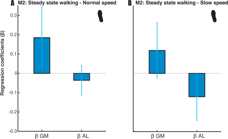
Our results show that, in the steady-state walking condition, m. gluteus medius and m. adductor longus activity predicted foot placement. At normal walking speed, we found extreme (BF10_gm = 50281.309) and moderate evidence (BF10_al = 6.050) when testing the muscle model’s (model 2) regression coefficients against zero (Fig 6A). At slow walking speed, extreme evidence was found for both muscles’ regression coefficients (BF10_gm = 535.867, BF10_al = 4984.586, Fig 6B). The sign of the regression coefficients was as expected, with more gluteus medius activity corresponding to more lateral foot placement (positive sign) and more medial foot placement corresponding to more adductor longus activity (negative sign). Although the mean R2 was low for both speeds (Mnormal_speed = 0.0268; Mslow_speed = 0.0258) see also Fig 8, below), the moderate to extreme evidence for the regression coefficients supports the idea that mediolateral foot placement is determined by hip ab- and adductor muscle activity during the preceding swing phase (E2).

Fig 6. Mean regression coefficients of the muscle model (2).

Fig 6

Standard deviations are represented by error bars. Panels A and B represent the results for respectively normal and slow walking speed. Moderate to extreme evidence (BF10_al > 3 & BF10 _gm> 100) supports the inclusion of the predictors at normal walking speed. Extreme evidence (BF10 > 100) supports the inclusion of the predictors at slow walking speed.

Fig 8. Mean relative explained variance (R2) of the muscle model (2) in the steady-state walking and the ankle moment constrained condition.

Fig 8

Standard deviations are represented by error bars. It remains inconclusive whether there is compensatory muscle activity in the ankle moment constrained condition.

Step-by-step foot placement control with ankle moment constraints

Model 1: Foot placement model

Regarding our first hypothesis (H1), that constrained ankle moments would lead to tighter foot placement control, we tested the relative explained variance (R2) of the foot placement model (1). The Bayesian repeated measures ANOVA of R2 of the foot placement model (model 1, Fig 7) revealed that the best model included only the factor speed. There was extreme evidence for this model as compared to the Null model as tested at mid- and terminal swing (BF10_mid = 4.732·1015, BF10_ts = 2.233·109). Since the inclusion of the factor condition did not improve the model, we could not find evidence for an effect of condition on the R2. Taken together, foot placement did not compensate for the constrained ankle moments by more accurate control (in contrast with H1).

Fig 7. Relative explained variance (R2) of the foot placement model (1) during walking in the steady-state walking and ankle moment constrained condition.

Fig 7

Shaded areas depict the standard deviation. Panels A and B represent the results for respectively normal and slow walking speed. A step was defined from toe-off until subsequent heel strike.

Model 2: Muscle model

Regarding our third hypothesis (H3), that constrained ankle moments would lead to larger active contribution to step-by-step variability in mediolateral foot placement, we tested the relative explained variance (R2) of the muscle model (2). The Bayesian repeated measures ANOVA of R2 of the muscle model (model 2, Fig 8) revealed that the best model included only the factor condition. There was anecdotal evidence for this model as compared to the Null model (BF10 = 1.786). Post-hoc analysis also provided anecdotal evidence (BF10 = 1.374) for compensatory muscle activity (H3) in the ankle moment constrained condition as compared to steady-state walking. As such, the effect of the ankle moment constraint on the relationship between muscle activity and foot placement remains inconclusive.

Step-by-step foot placement control with foot placement constraints

Model 1: Foot placement model

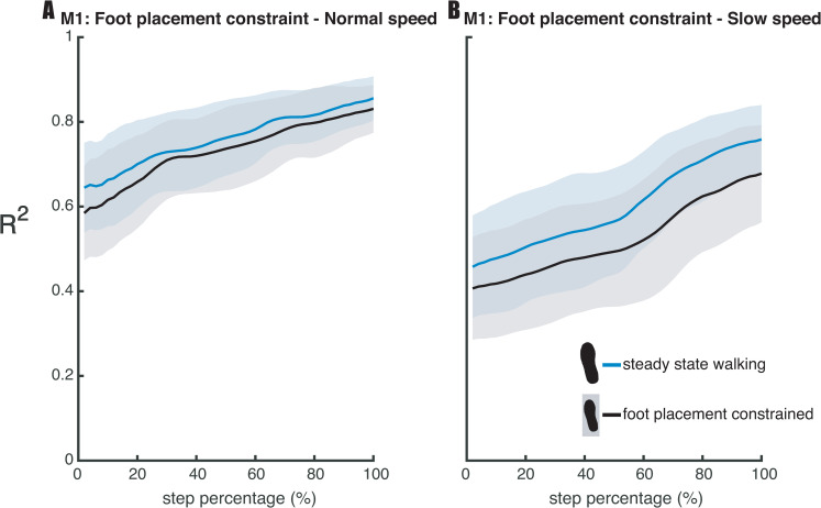
Regarding our second hypothesis (H2), that the degree of foot placement control would decrease, when constraining foot placement, we tested the relative explained variance (R2) of the foot placement model (1). The Bayesian repeated measures ANOVA of the R2 of the foot placement model (model 1, Fig 9) revealed that the best model included the factors condition and speed, when testing for mid-swing. There was extreme evidence for this model as compared to the Null model (BF10_mid = 3.196·1023). Post-hoc analysis provided strong evidence (BF10_mid = 19.125) in favor of a poorer prediction of foot placement by CoM state in the foot placement constrained condition as compared to steady-state walking (H2). When testing the R2 for terminal swing, the best model included the factors condition and speed and their interaction condition × speed. There was extreme evidence for this model as compared to the Null model (BF10_ts = 1.212·1019). In view of the interaction effect, we conducted a one-tailed Bayesian paired samples t-test, for each speed separately. At normal walking speed we found moderate evidence (BF10_ts = 7.352) in favor of a poorer prediction of foot placement by CoM state in the foot placement constrained condition as compared to steady-state walking (H2, Fig 9). At slow walking speed we found extreme (BF10_ts = 2957.043) evidence supporting the R2 to be lower in the foot placement constrained condition as compared to the steady-state walking condition.

Fig 9. Relative explained variance (R2) of the foot placement model (1) in the steady-state walking and foot placement constrained condition.

Fig 9

Shaded areas depict the standard deviation. Panels A and B represent the results for respectively normal and slow walking speed. A step was defined from toe-off until subsequent heel strike.

Discussion

We investigated the degree of (active) foot placement control during steady-state treadmill walking. We successfully replicated the findings of Wang and Srinivasan [4] and Rankin et al. [12] and can support that during steady-state walking foot placement is coordinated to CoM state and is associated with hip ab-/adductor muscle activity. The degree of foot placement control did not tighten when constraining the ankle moments. However, we found that the control strategy can be relaxed, achieving less tight foot placement control when constraining foot placement at a slow walking speed, whereas at a normal walking speed the degree of foot placement control was upheld. Overall, we can underscore the growing body of literature [3, 7, 8, 12, 15, 16, 26] and confirm that the foot placement model as introduced by Wang and Srinivasan [4] reflects an active control strategy, related to mediolateral stability [5].

Ankle moment constrained walking

The ankle moments were constrained by narrowing the surface area underneath the shoe. As a result, the CoP shift was limited to the width of a narrow beam (see S1 Fig for illustration). We hypothesized that foot placement would compensate for the imposed constraint, by tightening control. However, for both normal and slow walking, no difference was found in the relative explained variance of the foot placement model as compared to steady-state walking. The degree of foot placement control was not tightened for compensation. Admittedly, we focused on steady-state gait control, while related studies mostly studied reactive gait control. Yet, this finding was unexpected given previous ones in the literature [7, 8, 11, 27]. For example, Hof et al. [11] demonstrated more lateral steps in prosthetic legs to accommodate for a limited CoP shift. Similarly, Vlutters et al. [27], showed foot placement adjustments in response to anteroposterior perturbations when wearing CoP-shift-limiting ‘pin shoes’. Reimann et al. [7] reported that in response to vestibular stimulation execution of the ankle and foot placement strategy in temporal succession acted as the balance response. This suggests that without complementary ankle moments, a wider step might have been required to accommodate illusory falls, in line with their modelling results. And, Fettrow et al. [8] showed an inverse relationship between the execution of the ankle strategy and foot placement strategy during steady-state walking. Together these studies suggest that in response to natural variations or perturbations, foot placement control can accommodate for limited ankle moments. Here we would like to add that this does not necessarily imply more accurate control. An increased average step width can be used to maintain stability, with less tight foot placement control [28].

An important difference between the aforementioned studies and the current one is that their main outcome measure was based on average adjustments, i.e. on increases in step width or gain, whereas our R2 outcome measure reflects foot placement accuracy in accommodating variations in CoM state. A limitation of our approach is that information provided by the intercept of the model is lost, i.e. the average step width. We hence explored differences in step width between the ankle moment constrained conditions and the steady-state walking conditions. Indeed, extreme evidence (BF > 100) demonstrated increased step width when participants walked with LesSchuh at both speeds (see S2 Fig). While this is likely to reflect a compensatory strategy, a limitation of our study could be that with LesSchuh participants should avoid to step into the middle of the split belt treadmill (as the surface area of the shoes was narrow enough to fit between the two belts). Participants might have increased their step width as a precaution, and these findings should hence be interpreted with care. Nevertheless, increasing step width may be considered an appropriate response to the sustained (and invariable) perturbation of LesSchuh. Given the lower R2 in the ankle moment constrained condition at normal walking speed (Fig 7), this perturbation may not only have perturbed the ankle moments, but foot placement as well. Probably, LesSchuh perturbed stance leg control during swing and consequently led to less accurate foot placement. This suggests that learning how to adapt to this perturbation may improve the degree of foot placement control, similar to earlier findings related to continuous foot placement perturbations [26].

An increased step width has earlier been considered as a general stabilizing strategy, characterizing cautious gait in an unpredictable situation [19, 28, 29]. An increase in step width is a possible temporary compensatory strategy that seems to be used until one is able to develop tighter control of foot placement. The duration of our trials (5 minutes at normal walking speed and 10 minutes at slow walking speed) might have been too short to adapt the degree of control. Recently, it has been shown that repeated exposure to a perturbing force field yielded an adaptation in foot placement control [26]. During later exposures tighter control was manifested as compared to the first 5-minute-perturbed trial. The initial increase in step widths was diminished in these later exposures. We conjecture that longer or multiple trials with LesSchuh can lead to an increased R2 of the foot placement model. An increased degree of control may allow for a reduction of the average step width while maintaining stability. Walking with wider steps has been associated with a higher energy cost, and in normal walking individuals tend to select the step width that minimizes metabolic costs [30]. That is, adaptation over time and reduced step width may lead to a more economic compensatory strategy. Alternatively, increasing step width compared to normal walking might be a more economic strategy when walking with limited ankle moments. An additional energy cost related to actively increasing step-by-step control, might prevent participants to select narrower steps [31].

Further exploratory analysis in slow walking revealed that the imposed ankle moment constraint coincided with increased stride frequency, besides average step width, in spite of the metronome-imposed frequency (see S3 Fig). Increasing stride frequency has already been identified as a strategy to improve gait stability [3234]. The use of an ankle strategy appears less prominent when walking with high as compared to low stride frequency [35]. By modulating stride frequency and increasing average step width, the need for more accurate foot placement control (as reflected by our outcome measure R2) might have been circumvented. In future studies, it is worth looking into the hip and push-off strategies [2, 6, 8, 36, 37] to see whether they worked as compensatory strategies as well.

Whether compensatory hip ab-/adductor muscle activity does play a role in the compensatory response or not still remains inconclusive. We did not find a compensatory increase in step-by-step foot placement accuracy. Thus, it seems likely that hip ab-/adductor activity does not have a higher contribution to the variation in foot placement. However, the average increase in step width could have been driven by increased gluteus medius activity. This may explain why our analysis provided anecdotal (i.e. inconclusive) evidence rather than support for the null hypothesis. The low relative explained variance of the muscle model (2), although partly due to the noisy nature of EMG data, also suggests muscle activity alone cannot accurately predict foot placement. Likely passive dynamics during the swing phase have a large contribution, possibly controlled through push-off mechanisms [20, 36].

Foot placement constrained walking

The foot placement constraint (lines projected on the treadmill) reduced step width variability in both slow and normal walking (see S4 Fig), denoting that the constraint was effective. An earlier study already demonstrated reduced step width modulation by instructing participants to step on continuously projected lines on the treadmill [28]. In the current study the projected line appeared following toe-off. Therefore, we prevented preplanning a CoM trajectory during the preceding step [20, 38] in relation to the projection. Consequently, foot placement adjustments had to be realized during swing, which we expected to undermine the relationship between foot placement and CoM state. Evidence exists that rapid foot placement adjustments are feasible during a stepping task [39], although stability constraints appear to negatively affect responses to large jumps of foot placement targets [40]. In our study, foot placement appeared to be more effectively constrained by the projections during slow walking as compared to normal walking, as reflected by a lower step width variability at the slower speed (see S4 Fig). Regarding the degree of foot placement control, we found a condition × speed interaction effect on the R2 of the foot placement model (model 1). The reduction in step width variability caused a diminished degree of foot placement control according to the foot placement model at a slow walking speed. At a normal walking speed this effect was less pronounced, and R2 remained higher than during slow walking (Fig 9). In other words, at normal walking speed, the relationship between foot placement and CoM state was largely retained. This suggests that at a normal walking speed, stability constraints outweighed the task instruction. We interpret this to indicate that tight foot placement control is more important at normal as compared to slow walking speed. We would like to note that this agrees with previous findings that the R2 of the foot placement model is lower at slower speeds [13]. Yet, our results showed that foot placement remains actively controlled during slow steady-state walking, despite the lower degree of foot placement control. In contrast to an earlier study [28], we did not impose a step width different from average step width. However, from the earlier study we learn that the importance of tight foot placement control does not only differ with speed, but also given the average step width. At wider average step widths, participants demonstrated a lesser degree of step-by-step foot placement control [28]. Combined with the speed interaction effect in our study, these results demonstrate that the degree of foot placement control can be tightened/relaxed to satisfy stability demands under different task constraints.

Conclusion

We found muscle driven step-by-step foot placement control during steady-state walking. This control appears to be more important at normal as compared to slow walking speed, based on the degree of foot placement control and adaptability to a foot placement constraint. When compensating for constrained ankle moments, average step width and stride frequency, rather than the degree of foot placement control were adjusted. Further research is required to unravel what other strategies might have contributed as a compensatory mechanism and whether the compensatory strategy is adapted over time. Perhaps longer exposure to walking with constrained ankle moments will lead to adoption of tighter foot placement control.

Supporting information

S1 Fig. Center of pressure shifts when walking with an unconstrained (normal shoe) left foot and an ankle moment constrained (LesSchuh) right foot.

The most medial (blue) and most lateral (red) shifts are plotted, showing divergence of these shifts when unconstrained (left panel) as compared to overlaying shifts when constrained (right panel). The mediolateral CoP shift is limited by a ± 1-centimeter ridge underneath LesSchuh (Fig 2). The figure presents an example of participant 18. Ankle moment constraint–effect of “LesSchuh”.

(TIF)

S2 Fig. Mean step width during the steady-state walking and ankle moment constrained conditions.

Blue and red bars represent respectively the steady-state walking and ankle moment constrained conditions. The grey lines connect the individual data points. An exploratory Bayesian repeated measures ANOVA, including the steady-state walking and ankle moment constrained condition at both speeds, revealed that the best model included only the factor “Condition” with extreme evidence as compared to the Null model (BF10 = 1.610 * 1015). Post-hoc analysis provided extreme evidence supporting an increase in step width at both speeds to compensate for the ankle moment constraint (BF10 = 1.064*1013). The influence of the ankle moment constrained condition on step width.

(TIF)

S3 Fig. Mean stride frequency during the steady-state walking and ankle moment constrained conditions.

Blue and red bars represent respectively the steady-state walking and ankle moment constrained conditions. The grey lines connect the individual data points. As an exploratory analysis, as well as a protocol check, Bayesian repeated measures ANOVA, including the steady-state walking and ankle moment constrained condition at both speeds, revealed that the best model included the factors “Condition” and “Speed”. Post-hoc analysis provided extreme evidence (BF10 = 7.959*1042) indicating that stride frequency increased in the ankle moment constrained conditions as compared to steady-state walking. The influence of the ankle moment constrained condition on stride frequency.

(TIF)

S4 Fig. Mean step width variability during the steady-state walking and foot placement constrained conditions.

Blue and black bars represent respectively the steady-state walking and foot placement constrained conditions. The grey lines connect the individual data points. Bayesian repeated measures ANOVA, including the steady-state walking and foot placement strategy constrained condition at both speeds, revealed that the best model included the factors “Condition” and “Speed”, with extreme evidence as compared to the Null model (BF10 = 590646.967). Post-hoc analysis provided extreme evidence for a lower step width variability in the foot placement constrained condition as compared to steady-state walking (BF10 = 55714.494). When comparing between speeds, a two-tailed Bayesian paired samples t-test provided extreme evidence demonstrated that in the foot placement constrained condition, the step width variability remained higher at a normal walking speed as compared to the slow walking speed (BF10 = 2091.388). Effectiveness of the foot placement constraint: step width variability.

(TIF)

Acknowledgments

The authors are thankful for all technical support and assistance during the data collection, especially for Leon Schutte, who built LesSchuh.

Glossary

CoM

Center of mass

CoP

Center of pressure

BF10

Bayes factor indicating evidence supporting the alternative hypothesis

BF01

Bayes factor indicating evidence supporting the null hypothesis

FP

The mediolateral (along the global x-axis) foot position (calcaneus position digitized with respect to the foot cluster marker) at midstance expressed with respect to the mediolateral position of the contralateral foot at midstance (i.e. step width). The variable was demeaned prior to the performed regression (model 1)

CoMpos

Mediolateral CoM position (along the global x-axis) expressed with respect to mediolateral position of the stance foot at midstance. This variable was demeaned prior to the performed regression (model 1)

βpos

Regression coefficient defining the relationship between mediolateral CoM position (CoMpos) and step width (FP), as part of the foot placement model (model 1)

CoMvel

Mediolateral CoM velocity (along the global x-axis), calculated as the derivative of the CoM position expressed in the global coordinate system. This variable was demeaned prior to the performed regression (model 1)

βvel

Regression coefficient defining the relationship between mediolateral CoM velocity (CoMvel) and step width (FP), as part of the foot placement model (model 1)

FP2

The mediolateral (along the global x-axis) foot position (calcaneus position digitized with respect to the foot cluster marker) at midstance expressed with respect to the CoM at the time of toe-off in accordance with Rankin et al. [

12

]. The variable was demeaned and divided by the standard deviation prior to the performed regression (model 2)

EMGgm_swing

The median gluteus medius EMG amplitudes over 60–80% of the gait cycle (early swing), multiplied by the duration of this episode in seconds. The variable was demeaned and divided by the standard deviation prior to the performed regression (model 2)

βgm_swing

Regression coefficient defining the relationship between gluteus medius activity during early swing (EMGgm_swing) and step width (FP2), as part of the muscle model (model 2)

EMGal_swing

The median adductor longus EMG amplitudes over 60–80% of the gait cycle (early swing), multiplied by the duration of this episode in seconds. The variable was demeaned and divided by the standard deviation prior to the performed regression (model 2)

βal_swing

Regression coefficient defining the relationship between adductor longus activity during early swing (EMGgm_swing) and step width (FP2), as part of the muscle model (model 2)

R2

The relative explained variance of models (1) and (2). For model 1 this variable is interpreted as a measure of the degree of foot placement control. For model 2 this variable is interpreted as a measure of the active contribution to step-by-step foot placement control

Data Availability

The data and analysis can be found within Zenodo through the following link https://doi.org/10.5281/zenodo.4229851.

Funding Statement

Sjoerd M. Bruijn and Moira van Leeuwen were supported by a grant from the Netherlands Organization for Scientific Research (016.Vidi.178.014), https://www.nwo.nl/en/. The funders had no role in study design, data collection and analysis, decision to publish, or preparation of the manuscript.

References

  • 1.Bruijn SM, van Dieën JH. Control of human gait stability through foot placement. Journal of The Royal Society Interface. 2018;15(143):20170816 10.1098/rsif.2017.0816 [DOI] [PMC free article] [PubMed] [Google Scholar]
  • 2.Hof AL. The equations of motion for a standing human reveal three mechanisms for balance. Journal of biomechanics. 2007;40(2):451–7. 10.1016/j.jbiomech.2005.12.016 [DOI] [PubMed] [Google Scholar]
  • 3.Hof A, Duysens J. Responses of human hip abductor muscles to lateral balance perturbations during walking. Experimental brain research. 2013;230(3):301–10. 10.1007/s00221-013-3655-5 [DOI] [PubMed] [Google Scholar]
  • 4.Wang Y, Srinivasan M. Stepping in the direction of the fall: the next foot placement can be predicted from current upper body state in steady-state walking. Biology letters. 2014;10(9). [DOI] [PMC free article] [PubMed] [Google Scholar]
  • 5.Mahaki M, Bruijn SM, Van Dieën JH. The effect of external lateral stabilization on the use of foot placement to control mediolateral stability in walking and running. PeerJ. 2019;7:e7939 10.7717/peerj.7939 [DOI] [PMC free article] [PubMed] [Google Scholar]
  • 6.Reimann H, Fettrow T, Jeka JJ. Strategies for the Control of Balance During Locomotion. Kinesiology Review. 2018;7(1):18–25. [Google Scholar]
  • 7.Reimann H, Fettrow TD, Thompson ED, Agada P, McFadyen BJ, Jeka JJ. Complementary mechanisms for upright balance during walking. PloS one. 2017;12(2):e0172215 10.1371/journal.pone.0172215 [DOI] [PMC free article] [PubMed] [Google Scholar]
  • 8.Fettrow T, Reimann H, Grenet D, Thompson E, Crenshaw J, Higginson J, et al. Interdependence of balance mechanisms during bipedal locomotion. bioRxiv. 2019:658120 10.1371/journal.pone.0225902 [DOI] [PMC free article] [PubMed] [Google Scholar]
  • 9.Hof A, Duysens J. Responses of human ankle muscles to mediolateral balance perturbations during walking. Human movement science. 2018;57:69–82. 10.1016/j.humov.2017.11.009 [DOI] [PubMed] [Google Scholar]
  • 10.Afschrift M, van Deursen R, De Groote F, Jonkers I. Increased use of stepping strategy in response to medio-lateral perturbations in the elderly relates to altered reactive tibialis anterior activity. Gait & posture. 2019;68:575–82. 10.1016/j.gaitpost.2019.01.010 [DOI] [PubMed] [Google Scholar]
  • 11.Hof AL, van Bockel RM, Schoppen T, Postema K. Control of lateral balance in walking: experimental findings in normal subjects and above-knee amputees. Gait & posture. 2007;25(2):250–8. 10.1016/j.gaitpost.2006.04.013 [DOI] [PubMed] [Google Scholar]
  • 12.Rankin BL, Buffo SK, Dean JC. A neuromechanical strategy for mediolateral foot placement in walking humans. Journal of neurophysiology. 2014;112(2):374–83. 10.1152/jn.00138.2014 [DOI] [PMC free article] [PubMed] [Google Scholar]
  • 13.Stimpson KH, Heitkamp LN, Horne JS, Dean JC. Effects of walking speed on the step-by-step control of step width. Journal of biomechanics. 2018;68:78–83. 10.1016/j.jbiomech.2017.12.026 [DOI] [PubMed] [Google Scholar]
  • 14.Patil NS, Dingwell JB, Cusumano JP. Correlations of pelvis state to foot placement do not imply within-step active control. Journal of biomechanics. 2019;97:109375 10.1016/j.jbiomech.2019.109375 [DOI] [PMC free article] [PubMed] [Google Scholar]
  • 15.Arvin M, Hoozemans M, Pijnappels M, Duysens J, Verschueren SMP, Van Dieen J. Where to step? Contributions of stance leg muscle spindle afference to planning of mediolateral foot placement for balance control in young and older adults. Frontiers in physiology. 2018;9:1134 10.3389/fphys.2018.01134 [DOI] [PMC free article] [PubMed] [Google Scholar]
  • 16.Reimann H, Fettrow T, Thompson ED, Jeka JJ. Neural Control of Balance During Walking. Frontiers in Physiology. 2018;9:1271 10.3389/fphys.2018.01271 [DOI] [PMC free article] [PubMed] [Google Scholar]
  • 17.Wagenmakers E-J. A practical solution to the pervasive problems ofp values. Psychonomic bulletin & review. 2007;14(5):779–804. [DOI] [PubMed] [Google Scholar]
  • 18.Hof AL. Scaling gait data to body size. Gait & posture. 1996;3(4):222–3. [DOI] [PubMed] [Google Scholar]
  • 19.Hak L, Houdijk H, Steenbrink F, Mert A, van der Wurff P, Beek PJ, et al. Stepping strategies for regulating gait adaptability and stability. Journal of biomechanics. 2013;46(5):905–11. 10.1016/j.jbiomech.2012.12.017 [DOI] [PubMed] [Google Scholar]
  • 20.Matthis JS, Barton SL, Fajen BR. The critical phase for visual control of human walking over complex terrain. Proceedings of the National Academy of Sciences. 2017;114(32):E6720–E9. 10.1073/pnas.1611699114 [DOI] [PMC free article] [PubMed] [Google Scholar]
  • 21.Rebula JR, Ojeda LV, Adamczyk PG, Kuo AD. The stabilizing properties of foot yaw in human walking. Journal of biomechanics. 2017;53:1–8. 10.1016/j.jbiomech.2016.11.059 [DOI] [PMC free article] [PubMed] [Google Scholar]
  • 22.Hermens HJ, Freriks B, Merletti R, Stegeman D, Blok J, Rau G, et al. European recommendations for surface electromyography. Roessingh research and development. 1999;8(2):13–54. [Google Scholar]
  • 23.Roerdink M, Lamoth CJ, Beek PJ. Online gait event detection using a large force platform embedded in a treadmill. Journal of biomechanics. 2008;41(12):2628–32. 10.1016/j.jbiomech.2008.06.023 [DOI] [PubMed] [Google Scholar]
  • 24.Zatsiorsky V. Methods of determing mass-inertial characteristics of human body segments. Contemporasy Problems of Biomechnics. 1990. [Google Scholar]
  • 25.De Leva P. Adjustments to Zatsiorsky-Seluyanov's segment inertia parameters. Journal of biomechanics. 1996;29(9):1223–30. 10.1016/0021-9290(95)00178-6 [DOI] [PubMed] [Google Scholar]
  • 26.Reimold NK, Knapp HA, Henderson RE, Wilson L, Chesnutt AN, Dean JC. Altered active control of step width in response to mediolateral leg perturbations while walking. bioRxiv. 2019. [DOI] [PMC free article] [PubMed] [Google Scholar]
  • 27.Vlutters M, van Asseldonk E, van der Kooij H. Reduced center of pressure modulation elicits foot placement adjustments, but no additional trunk motion during anteroposterior-perturbed walking. Journal of biomechanics. 2018;68:93–8. 10.1016/j.jbiomech.2017.12.021 [DOI] [PubMed] [Google Scholar]
  • 28.Perry JA, Srinivasan M. Walking with wider steps changes foot placement control, increases kinematic variability and does not improve linear stability. Royal Society open science. 2017;4(9):160627 10.1098/rsos.160627 [DOI] [PMC free article] [PubMed] [Google Scholar]
  • 29.Wu M, Matsubara JH, Gordon KE. General and specific strategies used to facilitate locomotor maneuvers. PloS one. 2015;10(7). 10.1371/journal.pone.0132707 [DOI] [PMC free article] [PubMed] [Google Scholar]
  • 30.Maxwell Donelan J, Kram R, Arthur D K. Mechanical and metabolic determinants of the preferred step width in human walking. Proceedings of the Royal Society of London Series B: Biological Sciences. 2001;268(1480):1985–92. 10.1098/rspb.2001.1761 [DOI] [PMC free article] [PubMed] [Google Scholar]
  • 31.Donelan JM, Shipman DW, Kram R, Kuo AD. Mechanical and metabolic requirements for active lateral stabilization in human walking. Journal of biomechanics. 2004;37(6):827–35. 10.1016/j.jbiomech.2003.06.002 [DOI] [PubMed] [Google Scholar]
  • 32.Hak L, Houdijk H, Beek PJ, van Dieen JH. Steps to take to enhance gait stability: the effect of stride frequency, stride length, and walking speed on local dynamic stability and margins of stability. PLoS One. 2013;8(12):e82842 10.1371/journal.pone.0082842 [DOI] [PMC free article] [PubMed] [Google Scholar]
  • 33.Young PMM, Dingwell JB. Voluntarily changing step length or step width affects dynamic stability of human walking. Gait & posture. 2012;35(3):472–7. [DOI] [PMC free article] [PubMed] [Google Scholar]
  • 34.Buurke TJ, Lamoth CJ, van der Woude LH, Hof AL, den Otter R. Bilateral temporal control determines mediolateral margins of stability in symmetric and asymmetric human walking. ADAPTIVE CONTROL OF DYNAMIC BALANCE IN HUMAN WALKING. 2019;9:63 10.1038/s41598-019-49033-z [DOI] [PMC free article] [PubMed] [Google Scholar]
  • 35.Fettrow T, Reimann H, Grenet D, Crenshaw J, Higginson J, Jeka J. Walking cadence affects the recruitment of the medial-lateral balance mechanisms. bioRxiv. 2019:658070. [DOI] [PMC free article] [PubMed] [Google Scholar]
  • 36.Kim M, Collins SH. Once-per-step control of ankle-foot prosthesis push-off work reduces effort associated with balance during walking. Journal of neuroengineering and rehabilitation. 2015;12(1):43 10.1186/s12984-015-0027-3 [DOI] [PMC free article] [PubMed] [Google Scholar]
  • 37.Neptune RR, McGowan CP. Muscle contributions to frontal plane angular momentum during walking. Journal of biomechanics. 2016;49(13):2975–81. 10.1016/j.jbiomech.2016.07.016 [DOI] [PMC free article] [PubMed] [Google Scholar]
  • 38.Barton SL, Matthis JS, Fajen BR. Control strategies for rapid, visually guided adjustments of the foot during continuous walking. Experimental brain research. 2019;237(7):1673–90. 10.1007/s00221-019-05538-7 [DOI] [PubMed] [Google Scholar]
  • 39.Zhang Y, Smeets JB, Brenner E, Verschueren S, Duysens J. Fast responses to stepping‐target displacements when walking. The Journal of Physiology. 2020. 10.1113/JP278986 [DOI] [PMC free article] [PubMed] [Google Scholar]
  • 40.Reynolds R, Day B. Rapid visuo-motor processes drive the leg regardless of balance constraints. Current Biology. 2005;15(2):R48–R9. 10.1016/j.cub.2004.12.051 [DOI] [PubMed] [Google Scholar]

Decision Letter 0

J Lucas McKay

24 Aug 2020

PONE-D-20-17797

Step width and frequency to modulate: Active foot placement control ensures stable gait

PLOS ONE

Dear Dr. Bruijn,

Thank you for submitting your manuscript to PLOS ONE. After careful consideration, we feel that it has merit but does not fully meet PLOS ONE’s publication criteria as it currently stands. Therefore, we invite you to submit a revised version of the manuscript that addresses the points raised during the review process.

It has been reviewed carefully and the reviewers have provided some suggestions for improvement. If you could please address these concerns, and provide the journal with a point-by-point rebuttal as described below, I would very much like to see it again. Please note that if you feel that any of the concerns are beyond the scope of the work, you may say this in the rebuttal.

Please submit your revised manuscript by Oct 08 2020 11:59PM. If you will need more time than this to complete your revisions, please reply to this message or contact the journal office at plosone@plos.org. When you're ready to submit your revision, log on to https://www.editorialmanager.com/pone/ and select the 'Submissions Needing Revision' folder to locate your manuscript file.

Please include the following items when submitting your revised manuscript:

  • A rebuttal letter that responds to each point raised by the academic editor and reviewer(s). You should upload this letter as a separate file labeled 'Response to Reviewers'.

  • A marked-up copy of your manuscript that highlights changes made to the original version. You should upload this as a separate file labeled 'Revised Manuscript with Track Changes'.

  • An unmarked version of your revised paper without tracked changes. You should upload this as a separate file labeled 'Manuscript'.

If you would like to make changes to your financial disclosure, please include your updated statement in your cover letter. Guidelines for resubmitting your figure files are available below the reviewer comments at the end of this letter.

If applicable, we recommend that you deposit your laboratory protocols in protocols.io to enhance the reproducibility of your results. Protocols.io assigns your protocol its own identifier (DOI) so that it can be cited independently in the future. For instructions see: http://journals.plos.org/plosone/s/submission-guidelines#loc-laboratory-protocols

We look forward to receiving your revised manuscript.

Kind regards,

J. Lucas McKay, Ph.D., M.S.C.R.

Academic Editor

PLOS ONE

Journal Requirements:

When submitting your revision, we need you to address these additional requirements.

1. Please ensure that your manuscript meets PLOS ONE's style requirements, including those for file naming. The PLOS ONE style templates can be found at

https://journals.plos.org/plosone/s/file?id=wjVg/PLOSOne_formatting_sample_main_body.pdf and

https://journals.plos.org/plosone/s/file?id=ba62/PLOSOne_formatting_sample_title_authors_affiliations.pdf

2. We note that you have stated that you will provide repository information for your data at acceptance. Should your manuscript be accepted for publication, we will hold it until you provide the relevant accession numbers or DOIs necessary to access your data. If you wish to make changes to your Data Availability statement, please describe these changes in your cover letter and we will update your Data Availability statement to reflect the information you provide.

[Note: HTML markup is below. Please do not edit.]

Reviewers' comments:

Reviewer's Responses to Questions

Comments to the Author

1. Is the manuscript technically sound, and do the data support the conclusions?

The manuscript must describe a technically sound piece of scientific research with data that supports the conclusions. Experiments must have been conducted rigorously, with appropriate controls, replication, and sample sizes. The conclusions must be drawn appropriately based on the data presented.

Reviewer #1: Yes

Reviewer #2: Yes

**********

2. Has the statistical analysis been performed appropriately and rigorously?

Reviewer #1: Yes

Reviewer #2: Yes

**********

3. Have the authors made all data underlying the findings in their manuscript fully available?

The PLOS Data policy requires authors to make all data underlying the findings described in their manuscript fully available without restriction, with rare exception (please refer to the Data Availability Statement in the manuscript PDF file). The data should be provided as part of the manuscript or its supporting information, or deposited to a public repository. For example, in addition to summary statistics, the data points behind means, medians and variance measures should be available. If there are restrictions on publicly sharing data—e.g. participant privacy or use of data from a third party—those must be specified.

Reviewer #1: Yes

Reviewer #2: Yes

**********

4. Is the manuscript presented in an intelligible fashion and written in standard English?

PLOS ONE does not copyedit accepted manuscripts, so the language in submitted articles must be clear, correct, and unambiguous. Any typographical or grammatical errors should be corrected at revision, so please note any specific errors here.

Reviewer #1: No

Reviewer #2: Yes

**********

5. Review Comments to the Author

Please use the space provided to explain your answers to the questions above. You may also include additional comments for the author, including concerns about dual publication, research ethics, or publication ethics. (Please upload your review as an attachment if it exceeds 20,000 characters)

Reviewer #1: This manuscript reports the results of one experiment designed to investigate the mechanisms for maintaining gait stability. The study builds upon previous research suggesting that step-by-step variations in the mediolateral position of the foot play a key role in gait stability but that other strategies (e.g., the ankle push-off strategy) play a complementary role. The present study examines the interplay between these strategies by exploring how the degree of foot placement control varies as a function of constraints placed on ankle moments and foot placement. The results support the hypothesis that the degree of foot placement control is dependent on foot placement constraints. However, constraints on ankle moment did not affect the degree of foot placement control.

Overall, this is a solid study that makes a nice empirical contribution that advances our understanding of the role of foot placement control in gait stability. On the positive side, the rationale for the study is solid, the methodological approach is sound, the data are properly analyzed, and the interpretation of findings is reasonable. The authors preregistered their study, identified deviations from the preregistered plan, and will make their data available. There are a few issues that I think should be addressed before a final decision about this manuscript is made.

(1) The logic of the experiment appears to be sound but was not clearly presented. I believe that I was able to figure it out, but it required more effort than should be necessary. The basic aim of the study and the hypotheses are clearly explained in the introduction but the specific predictions are missing. What would be useful is a set of statements that connect the hypotheses (H1, H2, H3) with specific predictions for the analyses that were conducted (e.g., “If H# is true, then the R^2 of Model # should increase/decrease.”).

(2) I understand that the primary manipulations were the ankle moment and foot placement constraints, but it would be helpful if the authors could clarify the purpose of the walking speed manipulation and the predictions associated with this manipulation.

(3) Hypotheses 1 and 3 seem to capture the same idea. What is the difference? Is it possible for one to be true and the other false? Deriving specific predictions (as described in the previous comment) from both hypotheses would help to address this issue. (I wrote this comment after reading the Introduction and Methods. The difference eventually becomes clear to the reader but not until the Results section. The authors should clarify the difference earlier in the manuscript.)

(4) It would help the reader if the authors could begin each section of the results by explaining the question that each analysis is intended to answer.

(5) The manuscript has some grammatical issues, some of which are listed below. These should be addressed in the revision.

SPECIFIC COMMENTS AND MINOR ISSUES

Title: The title is awkwardly worded. I’m not sure what they mean by “Step width and frequency to modulate”.

Line 91: “vis-à-vis” is not properly used in this sentence. I think what the authors mean is “rather than”.

Line 92: Likewise, “accumulates” is not properly used.

Line 141: Replace “conform” with “in accordance with”.

Line 149: Add “phase” after familiarization.

Line 151: “Participants *were* familiarized with…”

Line 153: “…five *minutes*…”

Line 165: Did subjects receive any feedback about the accuracy of their footsteps?

Line 285: Delete “finally”

Reviewer #2: Review of “Step width and step frequency to modulate: active foot placement control ensures stable gate”

My summary of the paper:

In this paper, the author inquires how active foot placement control is compensated when the ability to modulate ankle moment is constrained or when step width is constrained. The author finds that instead of any modulation in foot placement control, the average step width and step frequency are changed. As the author admits, this does not necessarily mean that active foot placement does not adapt when constrained, because the ankle moment constraint imposed here could have inadvertently constrained the ability for active foot placement as well. The paper also replicates two results from past papers.

A summary of my review:

The paper is well-written and the results are discussed and interpreted with care and insightfully, in the context of the literature. Also, it is to be appreciated that the author appropriately preregistered their methods and declared deviations from preregistrations. It is great that the author will make the data available. One main suggestion I have to the author is to explore if there were any systematic trends in the CoM and foot placement states, which needed to be accounted for when demeaning. Secondly, I suggest the author compare the step width constrained control results to Perry and Srinivasan 2017, which doe a similar thing. In the writing, some further details and justifications need to be provided in the methods section of the paper and I provide specific suggestions for these below.

Detailed review (key changes suggested):

When the author says that the hip states and foot placement variables were “demeaned”, do they account for any systematic trends in the states over time? If not, I suggest the author de-trend these variables and perform the regression models again after de-trending to see if this changes the results. This is essential because, if there is a systematic trend over time in these states, it will affect the central results (the control gains) in this paper. I believe this will not take much time to add as it will just need the addition of one line of code.

The constraint condition in which the step width is constrained is similar to the constraint in Perry and Srinivasan 2017. I think it would be appropriate for the author compare and discuss the results here in comparison the results in that paper.

There are some additional details and justifications needed in the methods section. (i) Why did the author choose to constrain the step frequency? Neither of the papers this paper replicates use a metronome (Wang et al. and Rankin et al.). So, the author should justify this experimental choice to constrain step frequency. Also, it is unclear if the metronome was used for all the conditions i.e. steady-state, ankle constraint, and foot placement constraint. (ii) The author use a certain method to calculate the CoM (using regressions), could they provide more details about how the CoM was calculated or refer to a previous paper that calculates the CoM using a similar method? (iii) Further details need to be provided on the setup used for projecting beams onto the treadmill for the foot placement constraint condition. (iv) I suggest the author add a glossary explaining all the variables used in the paper in a single location.

Line-by-line comments (mostly minor):

Title: Can the title be revised for clarity? “step width and step frequency to modulate” sounds a bit unclear. Also, the focus of the paper is on the added constraints so that should feature in the tile in some way.

Abstract:

In line 20, “ankle strategy” is unclear. Maybe say “ankle moment modulation strategy” to show the link to the previous sentence.

In line 31, I think it is misleading to say “foot placement control was not tightened”, because a change in average step width and step frequency (as was found here) is also achieved through foot placement control. Maybe revise to “step-by-step foot placement control was not tightened”.

Introduction:

Very well-written. No changes suggested.

Methods:

Line 141, what is “conform Hof”?

Line 217, I assume COMpos and COMvel are vectors consisting of x, y, and z components? Please elaborate on this.

Line 224, there is a superscript “|” over the word “seconds” and it is not clear what this means. This shows up in other places in the paper as well. Please clarify.

Line 231, contralateral foot is also at midstance?

Line 233, stance foot is also at midstance? In general, please make sure that if states are being measured at a particular gait event, that is mentioned clearly.

Line 233, not sure what the word “predictors” refers to.

Line 238, why is the EMG variable subscripted “swing”? Was the EMG only calculated at swing? The result figures with EMG indicate it was calculated throughout the stride cycle.

Please state If FP2 and the EMG variables in equation 2 are demeaned or not somewhere in the methods section.

In equation 1, if CoM_pos(i) and CoM_vel(i) are written with (i) to indicate step cycle i, should FP also not be FP(i) and not just FP? I have a similar question about equations 2: to my understanding, the EMG are also for one step cycle, so should that also not be (i) ?

Results:

Figure 3 caption states that medial burst is greater than lateral burst, is this difference significant? It looks very small from the figure.

In figures 3 and 4, the y axis does not have any number scale.

Figure 4 caption says “higher emg activity during early swing”, but this appears to be true only for slow speed. Please clarify this in the caption itself.

Line 335, please state the R^2 value there itself. Did Rankin et al. have a similarly low R^2? Please discuss what this could mean.

Discussion:

The discussion is generally very well written and the interpretations are made carefully.

In line 395, maybe use the word “relaxed” instead of “adapted”, to contrast with “tightened"?

Line 414, unclear what “serially coordinated as a balance response” means, please clarify

Line 420, could you elaborate on why you say “this does not necessarily imply more accurate control”

Conclusion:

In line 511, the use of the word “other” seems to indicate that foot placement control compensation was clearly eliminated in this study. However, as the author admits, this is not the case. It is possible that the LeSchuh did not allow for easy foot placement modulation. This can be explored with further experiments to see if the foot placement control compensates with practice with LeSchuh. Please add this caveat in the conclusion.

**********

6. PLOS authors have the option to publish the peer review history of their article (what does this mean?). If published, this will include your full peer review and any attached files.

If you choose “no”, your identity will remain anonymous but your review may still be made public.

Do you want your identity to be public for this peer review? For information about this choice, including consent withdrawal, please see our Privacy Policy.

Reviewer #1: No

Reviewer #2: No

[NOTE: If reviewer comments were submitted as an attachment file, they will be attached to this email and accessible via the submission site. Please log into your account, locate the manuscript record, and check for the action link "View Attachments". If this link does not appear, there are no attachment files.]

While revising your submission, please upload your figure files to the Preflight Analysis and Conversion Engine (PACE) digital diagnostic tool, https://pacev2.apexcovantage.com/. PACE helps ensure that figures meet PLOS requirements. To use PACE, you must first register as a user. Registration is free. Then, login and navigate to the UPLOAD tab, where you will find detailed instructions on how to use the tool. If you encounter any issues or have any questions when using PACE, please email PLOS at figures@plos.org. Please note that Supporting Information files do not need this step.

Attachment

Submitted filename: PLOS_Bruijn_Review.pdf

PLoS One. 2020 Dec 17;15(12):e0242215. doi: 10.1371/journal.pone.0242215.r002

Author response to Decision Letter 0


19 Oct 2020

Reviewer #1: This manuscript reports the results of one experiment designed to investigate the mechanisms for maintaining gait stability. The study builds upon previous research suggesting that step-by-step variations in the mediolateral position of the foot play a key role in gait stability but that other strategies (e.g., the ankle push-off strategy) play a complementary role. The present study examines the interplay between these strategies by exploring how the degree of foot placement control varies as a function of constraints placed on ankle moments and foot placement. The results support the hypothesis that the degree of foot placement control is dependent on foot placement constraints. However, constraints on ankle moment did not affect the degree of foot placement control.

Overall, this is a solid study that makes a nice empirical contribution that advances our understanding of the role of foot placement control in gait stability. On the positive side, the rationale for the study is solid, the methodological approach is sound, the data are properly analyzed, and the interpretation of findings is reasonable. The authors preregistered their study, identified deviations from the preregistered plan, and will make their data available. There are a few issues that I think should be addressed before a final decision about this manuscript is made.

We wish to thank the reviewer for their kind words and helping us to solve remaining issues. We have adapted the manuscript based on the comments raised. Below, we provide a point-by-point reply to each reviewer comment. In addition, we highlighted all changes made in the manuscript.

(1) The logic of the experiment appears to be sound but was not clearly presented. I believe that I was able to figure it out, but it required more effort than should be necessary. The basic aim of the study and the hypotheses are clearly explained in the introduction but the specific predictions are missing. What would be useful is a set of statements that connect the hypotheses (H1, H2, H3) with specific predictions for the analyses that were conducted (e.g., “If H# is true, then the R^2 of Model # should increase/decrease.”).

We understand the need for clarification and added:

‘…(i.e. a higher R2 of model (1))…’ (Page 18, Lines 360-361)

‘…(i.e. a higher R2 of model (2))…’ (Page 18, Lines 361-362)

‘…(i.e. a lower R2 of model (1))…’ (Page 18, Line 371)

to specify our predictions.

(2) I understand that the primary manipulations were the ankle moment and foot placement constraints, but it would be helpful if the authors could clarify the purpose of the walking speed manipulation and the predictions associated with this manipulation.

At a slower walking speed, the degree of foot placement control has been shown to be lower [1]. Apparently, the importance of foot placement control differs at different walking speeds. This could have affected the influence of our other manipulations. We therefore felt that testing at two walking speeds would strengthen our conclusions. We did not however make any specific speed-related predictions regarding foot placement control.

We have now tried to more explicitly state these ideas in the manuscript:

“We did not make any speed-related predictions, but the speed-dependent nature of the foot placement strategy could potentially affect the effects of our other experimental conditions.” (Page 7, Lines 127-129)

Apart from a focus on foot placement control, we also made predictions regarding the mediolateral ankle strategy under the same conditions. This is still work in progress, so for now we refer to the preregistration (https://osf.io/74pn5) for speed-related predictions which were not incorporated in this paper.

(3) Hypotheses 1 and 3 seem to capture the same idea. What is the difference? Is it possible for one to be true and the other false? Deriving specific predictions (as described in the previous comment) from both hypotheses would help to address this issue. (I wrote this comment after reading the Introduction and Methods. The difference eventually becomes clear to the reader but not until the Results section. The authors should clarify the difference earlier in the manuscript.)

We now specify our predictions clearer in the Methods:

“That is, this allowed for testing the hypotheses that compensation will encompass tighter control (i.e. a higher R2 of model (1)) (H1) driven by compensatory muscle activation (i.e. a higher R2 of model (2)) (H3).” (Page 18, Lines 359-362)

Furthermore, we now explain the relation between these two hypotheses as follows:

“If both H1 and H3 are true, this would suggest a compensatory tighter coupling between variations in foot placement and CoM state (H1), achieved through compensatory muscle activity (H3). If only H1 is true, this would suggest tighter coupling between variations in foot placement and CoM state (H1), but no evidence that this is actively controlled (in contrast with H3). If only H3 is true, this would suggest that foot placement is more strictly controlled through muscle activity (H3), but not to tighten the coupling between variations in foot placement and CoM state.” (Page 18, Lines 362-368)

(4) It would help the reader if the authors could begin each section of the results by explaining the question that each analysis is intended to answer.

We have added the following to address this comment:

“First, we considered the relative explained variance (R2) and tested the regression coefficients of the foot placement model (1) to assess our expectation (E1) that mediolateral foot placement can be predicted by CoM state, as previously shown by Wang and Srinivasan [4].” (Page 20, Lines 398-400)

“Our second expectation (E2), that mediolateral foot placement correlates with hip ab- and adductor muscle activity during the preceding swing phase, was tested based on the regression coefficients of the muscle model (2), and visualized in Fig 3 and Fig 4 below.” (Page 21, Lines 409-411)

“Regarding our first hypothesis (H1), that constrained ankle moments would lead to tighter foot placement control, we tested the relative explained variance (R2) of the foot placement model (1).” (Page 24, Lines 437-439)

“Regarding our third hypothesis (H3), that constrained ankle moments would lead to larger active contribution to step-by-step variability in mediolateral foot placement, we tested the relative explained variance (R2) of the muscle model (2).” (Page 25, Lines 450-452)

“Regarding our second hypothesis (H2), that the degree of foot placement control would decrease, when constraining foot placement, we tested the relative explained variance (R2) of the foot placement model (1).“ (Page 26, Lines 465-467)

The manuscript has some grammatical issues, some of which are listed below. These should be addressed in the revision.

SPECIFIC COMMENTS AND MINOR ISSUES

(1) Title: The title is awkwardly worded. I’m not sure what they mean by “Step width and frequency to modulate”.

Also considering the other reviewer’s comments we now rephrased our title to be:

Active foot placement control ensures stable gait: Effect of constraints on foot placement and ankle moments

Line 91: “vis-à-vis” is not properly used in this sentence. I think what the authors mean is “rather than”.

We have replaced “vis-à-vis” with “rather than” (Page 8, Line 156)

(2) Line 92: Likewise, “accumulates” is not properly used.

“though there is increasing” (Page 8, Line 157)

(4) Line 141: Replace “conform” with “in accordance with”.

We have replaced this in the manuscript. (Page 11, Lines 205-206)

(5) Line 149: Add “phase” after familiarization.

We have added this in the manuscript. (Page 11, Line 216)

(6) Line 151: “Participants *were* familiarized with…”

We have added this in the manuscript. (Page 11, Line 218)

(7) Line 153: “…five *minutes*…”

We have added this in the manuscript, (Page 12, Line 220)

(8) Line 165: Did subjects receive any feedback about the accuracy of their footsteps?

They did not receive any feedback other than that they could see themselves where they stepped.

(9) Line 285: Delete “finally”

We have deleted this from the manuscript.

Reviewer #2: Review of “Step width and step frequency to modulate: active foot placement control ensures stable gate”

My summary of the paper:

In this paper, the author inquires how active foot placement control is compensated when the ability to modulate ankle moment is constrained or when step width is constrained. The author finds that instead of any modulation in foot placement control, the average step width and step frequency are changed. As the author admits, this does not necessarily mean that active foot placement does not adapt when constrained, because the ankle moment constraint imposed here could have inadvertently constrained the ability for active foot placement as well. The paper also replicates two results from past papers.

A summary of my review:

The paper is well-written and the results are discussed and interpreted with care and insightfully, in the context of the literature. Also, it is to be appreciated that the author appropriately preregistered their methods and declared deviations from preregistrations. It is great that the author will make the data available. One main suggestion I have to the author is to explore if there were any systematic trends in the CoM and foot placement states, which needed to be accounted for when demeaning. Secondly, I suggest the author compare the step width constrained control results to Perry and Srinivasan 2017, which does a similar thing. In the writing, some further details and justifications need to be provided in the methods section of the paper and I provide specific suggestions for these below.

We wish to thank the reviewer for their kind words and thoughtful suggestions. We have addressed these suggestions in a point-by-point reply below. In addition, we highlighted all changes made in the manuscript.

Detailed review (key changes suggested):

(1) When the author says that the hip states and foot placement variables were “demeaned”, do they account for any systematic trends in the states over time? If not, I suggest the author de-trend these variables and perform the regression models again after de-trending to see if this changes the results. This is essential because, if there is a systematic trend over time in these states, it will affect the central results (the control gains) in this paper. I believe this will not take much time to add as it will just need the addition of one line of code.

We understand detrending could be essential given a drifting position of the participant on the treadmill, and value your concern. However, since we express both the center of mass as well as foot placement with respect to the contralateral stance foot prior to demeaning, we think it is not essential in this case.

To clarify that the demeaning was done after expressing CoMpos and FP with respect to the contralateral stance foot at midstance, we now first describe the variables (i.e. their expression with respect to the contralateral stance foot) in lines 306-312 (pages 15-16), while leaving “demeaning” out of these sentences. Subsequently, at the end of that paragraph we now state:

“FP, CoMpos and CoMvel were demeaned prior to regression.” (Page 16, Line 310)

Moreover, in response to another reviewer comment, we now added a glossary (Pages 2-4, Lines 14-73) to present our variables. In the glossary we mention for the CoM position (CoMpos):

“Mediolateral CoM position (along the global x-axis) expressed with respect to mediolateral position of the stance foot at midstance.” (Page 2, Lines 29-30)

and for foot placement (FP):

“The mediolateral (along the global x-axis) foot position (calcaneus position digitized with respect to the foot cluster marker) at midstance expressed with respect to the mediolateral position of the contralateral foot at midstance (i.e. step width).” (Page 2, Lines 23-286)

We hope these additions clarify that we expressed the variables with respect to the contralateral stance foot, rather than in the global coordinate system. Based on this we believe detrending is not necessary.

(2) The constraint condition in which the step width is constrained is similar to the constraint in Perry and Srinivasan 2017. I think it would be appropriate for the author compare and discuss the results here in comparison the results in that paper.

Thank you for making us aware of this paper. We have added this to our discussion as follows:

“An earlier study already demonstrated reduced step width modulation by instructing participants to step on continuously projected lines on the treadmill [28].” (Page 32, Lines 584-586)

as well as

“In contrast to an earlier study [28], we did not impose a step width different from average step width. However, from the earlier study we learn that the importance of tight foot placement control does not only differ with speed, but also given the average step width. At wider average step widths, participants demonstrated a lesser degree of step-by-step foot placement control [28]. Combined with the speed interaction effect in our study, these results demonstrate the degree of foot placement control can be tightened/relaxed to satisfy stability demands given different task constraints.” (Page 33, Lines 606-613)

Furthermore, we have added this paper as a reference for the following statement we made before:

“An increased step width has earlier been considered as a general stabilizing strategy, characterizing cautious gait in an unpredictable situation [19, 28, 29].” (Page 30, Lines 542-543)

Moreover, we cited this paper to underscore that not only step-by-step foot placement control, but also average step width promotes (mediolateral) gait stability. We did so in the following statement.

“An increased average step width can be used to maintain stability, with less tight foot placement control [28].” (Page 29, Lines 519-520)

(3) There are some additional details and justifications needed in the methods section.

(i) Why did the author choose to constrain the step frequency? Neither of the papers this paper replicates use a metronome (Wang et al. and Rankin et al.). So, the author should justify this experimental choice to constrain step frequency. Also, it is unclear if the metronome was used for all the conditions i.e. steady-state, ankle constraint, and foot placement constraint.

We wanted to constrain stride frequency as we knew from earlier studies [2-4] that varying step frequency is a stability control strategy. More specific, gait can be made more robust by increasing stride frequency. Since we wanted to evaluate the effect of the different constraints on the degree of foot placement control, we attempted to keep potential confounding factors, such as stride frequency constant. We have clarified this in the manuscript as follows:

“In this study, stride frequency control could be a confounding stability control strategy [19]. Therefore, stride frequency was controlled by means of a metronome, to avoid that participants changed stride frequency between conditions.” (Page 11, Lines 206-208)

(ii) The author use a certain method to calculate the CoM (using regressions), could they provide more details about how the CoM was calculated or refer to a previous paper that calculates the CoM using a similar method?

We are happy that you pointed out this mistake in writing. We estimated the segment’s mass using a regression. The CoM however was expressed as a percentage of the longitudinal axis of the segment. We have adjusted the manuscript to include this as follows:

“we estimated the segment’s mass via linear regression including the segment’s length and the segment’s measured circumference as predictors and regression coefficients based on gender [24]. The segment’s CoM was estimated as a percentage of the longitudinal axis of the segment [24, 25].” (Pages 14-15, Lines 284-287)

(iii) Further details need to be provided on the setup used for projecting beams onto the treadmill for the foot placement constraint condition.

We have added more detail to the paragraph below, as highlighted in the manuscript:

“Projections on the treadmill served to constrain the variation in mediolateral foot placement. In brief, first the average step width was derived from the final 100 steps of the familiarization trial, based on the CoP estimated from the force measurement of the instrumented treadmill. Then, this average step width was imposed by projecting beams on the treadmill, and participants were instructed to place their foot in the middle of the beam. For every step, the beam became visible following toe-off in order to prevent modification of the CoM swing phase trajectory by compensatory push-off modulation [20]. Customized labview software allowed us to estimate the toe-off event based on a force threshold, which triggered the projections based on real-time force measurements.” (Page 12, lines 227-235)

(iv) I suggest the author add a glossary explaining all the variables used in the paper in a single location.

Thank you for this suggestion, we feel this overview clarifies our paper a lot. In the manuscript you can find the following glossary following the title page:

“Glossary

CoM Center of mass.

CoP Center of pressure.

BF10 Bayes factor indicating evidence supporting the alternative hypothesis.

BF01 Bayes factor indicating evidence supporting the null hypothesis.

FP The mediolateral (along the global x-axis) foot position (calcaneus position digitized with respect to the foot cluster marker) at midstance expressed with respect to the mediolateral position of the contralateral foot at midstance (i.e. step width). The variable was demeaned prior to the performed regression (model 1).

CoMpos Mediolateral CoM position (along the global x-axis) expressed with respect to mediolateral position of the stance foot at midstance. This variable was demeaned prior to the performed regression (model 1).

βpos Regression coefficient defining the relationship between mediolateral CoM position (CoMpos) and step width (FP), as part of the foot placement model (model 1).

CoMvel Mediolateral CoM velocity (along the global x-axis), calculated as the derivative of the CoM position expressed in the global coordinate system. This variable was demeaned prior to the performed regression (model 1).

βvel Regression coefficient defining the relationship between mediolateral CoM velocity (CoMvel) and step width (FP), as part of the foot placement model (model 1).

FP2 The mediolateral (along the global x-axis) foot position (calcaneus position digitized with respect to the foot cluster marker) at midstance expressed with respect to the CoM at the time of toe-off in accordance with Rankin et al. [12]. The variable was demeaned and divided by the standard deviation prior to the performed regression (model 2).

EMGgm_swing The median gluteus medius EMG amplitudes over 60-80% of the gait cycle (early swing), multiplied by the duration of this episode in seconds. The variable was demeaned and divided by the standard deviation prior to the performed regression (model 2).

βgm_swing Regression coefficient defining the relationship between gluteus medius activity during early swing (EMGgm_swing) and step width (FP2), as part of the muscle model (model 2).

EMGal_swing The median adductor longus EMG amplitudes over 60-80% of the gait cycle (early swing), multiplied by the duration of this episode in seconds. The variable was demeaned and divided by the standard deviation prior to the performed regression (model 2).

βal_swing Regression coefficient defining the relationship between adductor longus activity during early swing (EMGgm_swing) and step width (FP2), as part of the muscle model (model 2).

R2 The relative explained variance of models (1) and (2). For model 1 this variable is interpreted as a measure of the degree of foot placement control. For model 2 this variable is interpreted as a measure of the active contribution to step-by-step foot placement control.“ (Pages 2-4, Lines 14-73)

Line-by-line comments (mostly minor):

(1) Title: Can the title be revised for clarity? “step width and step frequency to modulate” sounds a bit unclear. Also, the focus of the paper is on the added constraints so that should feature in the tile in some way.

We appreciate your comment and tried to clarify the title as follows:

Active foot placement control ensures stable gait: Effect of constraints on foot placement and ankle moments

Abstract:

(2) In line 20, “ankle strategy” is unclear. Maybe say “ankle moment modulation strategy” to show the link to the previous sentence.

Based on your suggestion we have replaced “ankle strategy” by “ankle moment control” in line 82.

(3) In line 31, I think it is misleading to say “foot placement control was not tightened”, because a change in average step width and step frequency (as was found here) is also achieved through foot placement control. Maybe revise to “step-by-step foot placement control was not tightened”.

We agree and have added the suggested ‘step-by-step’ (Page 5, Line 93)

Introduction:

Very well-written. No changes suggested.

Methods:

(4) Line 141, what is “conform Hof”?

Here we meant to refer to the paper by Hof (1996) in which recommendations are given to scale data to body size. Based on the suggestion of the other reviewer, we now rephrased this as

“…walking speeds, normalized to leg length in accordance with Hof [18].” (Page 11, 205-206)

in the manuscript.

(5) Line 217, I assume COMpos and COMvel are vectors consisting of x, y, and z components? Please elaborate on this.

We were interested in the “mediolateral CoM position CoMpos” and the mediolateral CoM velocity CoMvel” (Page 15, Lines 290-291), and hence, the CoMpos and CoMvel are vectors containing only ML elements.

We have now more clearly written this;

“Mediolateral CoM displacement was defined along the x-axis of our global coordinate system, of which the x-axis was oriented perpendicular to the direction of the treadmill.” (Page 15, Lines 288-290)

Lastly, we have emphasized this in the glossary as well (Pages 2-4, Lines 14,-73), specifically in the following lines:

“Mediolateral CoM position (along the global x-axis) …” (Page 2, Line 29)

And

“Mediolateral CoM velocity (along the global x-axis) ...” (Page 3, Lines 37)

(6) Line 224, there is a superscript “|” over the word “seconds” and it is not clear what this means. This shows up in other places in the paper as well. Please clarify.

The superscript refers to the deviations from the preregistered protocol, described later in the manuscript. We now clarified this by using brackets and writing

“(we note here that this is a deviation from our preregistered protocol, see “Deviations from the preregistered plans I”).” (Page 15, Lines 297-299)

and later

“(we note here that this is a deviation from our preregistered protocol, see “Deviations from the preregistered plans II”).” (Page 16, Lines 317-318)

and

“(we note here that this is a deviation from our preregistererd protocol, see “Deviations from the preregistered plans III”).” (Page 17, Lines 342-343)

instead.

(7) Line 231, contralateral foot is also at midstance?

Indeed. We have clarified this in the manuscript by adding “…at midstance” (Page 15, Line 306).

(8) Line 233, stance foot is also at midstance? In general, please make sure that if states are being measured at a particular gait event, that is mentioned clearly.

Again we have added “…at midstance” (Page 15, Line 308) to clarify our procedures.

(9) Line 233, not sure what the word “predictors” refers to.

With “predictors” we mean the independent variables as introduced in the line before. To clarify this we have added “(i.e. predictors)” (Page 15, Line 308).

(10) Line 238, why is the EMG variable subscripted “swing”? Was the EMG only calculated at swing? The result figures with EMG indicate it was calculated throughout the stride cycle.

It is true that the figures show the EMG throughout the stride cycle. However, for the regression we included a measure for the EMG activity from 60-80% of the gait cycle, corresponding to early swing. Hence, we subscripted this variable “swing” for the regression. We have now mentioned this more clearly as “the median gluteus medius and adductor longus EMG amplitudes from 60-80% of the gait cycle” instead of just “gluteus medius and adductor longus EMG amplitudes” (Page 16, Line 314-315).

(11) Please state If FP2 and the EMG variables in equation 2 are demeaned or not somewhere in the methods section.

These variables were also demeaned, as now has been described more explicitly: “we normalized FP2, EMGgm_swing and EMGal_swing by demeaning and dividing by the standard deviation” (Page 16, Lines 316-317).

Moreover, these variables have been described in the glossary (Pages 2-4, Lines 14-73), as follows:

“EMGgm_swing The median gluteus medius EMG amplitudes over 60-80% of the gait cycle (early swing), multiplied by the duration of this episode in seconds. The variable was demeaned and divided by the standard deviation prior to the performed regression (model 2). (Page 3, Lines 52-55)

EMGal_swing The median adductor longus EMG amplitudes over 60-80% of the gait cycle (early swing), multiplied by the duration of this episode in seconds. The variable was demeaned and divided by the standard deviation prior to the performed regression (model 2).” (Page 4, Lines 61-64)

(12) In equation 1, if CoM_pos(i) and CoM_vel(i) are written with (i) to indicate step cycle i, should FP also not be FP(i) and not just FP? I have a similar question about equations 2: to my understanding, the EMG are also for one step cycle, so should that also not be (i) ?

Thank you for noting this mistake in the manuscript. In line 323 we stated “in every step cycle i”, which is wrong. “i” in this case is not a reference to the step cycle, but to an index representing the percentages of the step cycle. We normalized to 51 samples per step, allowing us to implement the model with predictors at different phases of the gait cycle. Therefore, we have rewritten this as:

“…for different phases (i) of the step cycle.” (Page 16, line 321)

Results:

(13) Figure 3 caption states that medial burst is greater than lateral burst, is this difference significant? It looks very small from the figure.

The figures are merely a dichotomous visual representation of the “significant” regression (note that we used Bayesian statistics and strictly cannot use the word significant in this case). In figures 3 and 4 we wanted to illustrate that the relationship between hip ab-/adductor activity and foot placement describes more lateral foot placement with a higher GM burst and a lower AL burst. We statistically tested the beta coefficients of the relationship against zero and found moderate to extreme evidence supporting both muscles predicted foot placement. Note that we did not directly test the differences as presented in the figures.

We agree the burst seem really small, although they are consistent with our expectations, the muscles’ function and earlier results reported by [5]. Despite this small difference we feel the figures illustrate nicely what the relationship represents and therefore we did not make any changes to the manuscript.

(14) In figures 3 and 4, the y axis does not have any number scale.

Since the unit of EMG amplitude is largely arbitrary, as it is affected by individual differences such as muscle mass and skin conductance, we normalized the EMG to the average stride peak activity (i.e. for the GM the large peak during stance) for each speed respectively. We now added a number scale. Since these normalized numbers do not contribute to our message or what we wanted to test, we added a clarification in the caption as follows:

“The figure serves as a dichotomous illustration of the relationship established through regression and does not show values that were statistically tested.” (Pages 21-22, Fig 3&4, captions):

(15) Figure 4 caption says “higher emg activity during early swing”, but this appears to be true only for slow speed. Please clarify this in the caption itself.

By higher we meant the comparison between the medial and lateral steps, and only from 60-80 % of the gait cycle. Although this difference is clearer for the gluteus medius in figure 3, in figure 4 it holds for both speeds that the light blue line is mostly above the dark blue line during early swing. Indeed, based on the figure only, this seems clearer at a slow speed and a bit questionable at a normal speed. We again want to emphasize that the figures are merely a visualization, whereas our conclusions are based on the results of the regression.

However, to make the caption clearer we have added:

“When comparing medial to lateral steps during early swing (60-80% of the gait cycle),” (Page 22, Fig 4, caption) to draw the attention to the period and comparison of interest.

“This is more prominent at slow walking speed.” (Page 22, Fig 4, caption)

to acknowledge it is not that clear at a normal walking speed.

(16) Line 335, please state the R^2 value there itself. Did Rankin et al. have a similarly low R^2? Please discuss what this could mean.

Rankin et al. (2014) did not report the R^2 values in their original paper [5]. However, we corresponded with Dr, Dean (who is the corresponding author of Rankin et al., 2014 [5]). Although he asked me not to cite him on this (he preferred to do a more thorough check of the data to check the exact values they found). He told me their typical R^2 values were ~0.10. This value is within our range of R^2 values, although our mean R^2 is lower.

We have added “(Mnormal_speed = 0.0268; Mslow_speed =0.0258)” (Page 22, lines 429-430) to report the R^2 values.

Furthermore, in the Discussion we have added:

“The low relative explained variance of the muscle model (2), although partly due to the noisy nature of EMG data, also suggests muscle activity alone cannot accurately predict foot placement. Likely passive dynamics during the swing phase have a large contribution, possibly controlled through push-off mechanisms [20, 36].” (Page 31, Lines 577-580)

Discussion:

The discussion is generally very well written and the interpretations are made carefully.

(17) In line 395, maybe use the word “relaxed” instead of “adapted”, to contrast with “tightened"?

Thank you for your suggestion we have replaced “adapted” by “relaxed” (Page 28, Line 493).

(18) Line 414, unclear what “serially coordinated as a balance response” means, please clarify

We have reformulated this in the manuscript as follows:

“…in temporal succession acted as a balance response.” (Page 29, Lines 512-513)

(19) Line 420, could you elaborate on why you say “this does not necessarily imply more accurate control”

We have clarified this statement by adding:

“An increased average step width can be used to maintain stability, with less tight foot placement control [28].” (Page 29, Lines 519-520)

Conclusion:

(20) In line 511, the use of the word “other” seems to indicate that foot placement control compensation was clearly eliminated in this study. However, as the author admits, this is not the case. It is possible that the LeSchuh did not allow for easy foot placement modulation. This can be explored with further experiments to see if the foot placement control compensates with practice with LeSchuh. Please add this caveat in the conclusion.

We have added the following to the conclusion:

“Perhaps longer exposure to walking with constrained ankle moments will lead to adoption of tighter foot placement control”. (Page 33, Lines 622-624)

References

1. Stimpson KH, Heitkamp LN, Horne JS, Dean JC. Effects of walking speed on the step-by-step control of step width. Journal of biomechanics. 2018;68:78-83.

2. Hak L, Houdijk H, Steenbrink F, Mert A, van der Wurff P, Beek PJ, et al. Stepping strategies for regulating gait adaptability and stability. Journal of biomechanics. 2013;46(5):905-11.

3. Buurke TJ, Lamoth CJ, van der Woude LH, Hof AL, den Otter R. Bilateral temporal control determines mediolateral margins of stability in symmetric and asymmetric human walking. ADAPTIVE CONTROL OF DYNAMIC BALANCE IN HUMAN WALKING. 2019;9:63.

4. Young PMM, Dingwell JB. Voluntarily changing step length or step width affects dynamic stability of human walking. Gait & posture. 2012;35(3):472-7.

5. Rankin BL, Buffo SK, Dean JC. A neuromechanical strategy for mediolateral foot placement in walking humans. Journal of neurophysiology. 2014;112(2):374-83.

Attachment

Submitted filename: Response to Reviewers.pdf

Decision Letter 1

J Lucas McKay

29 Oct 2020

Active foot placement control ensures stable gait: Effect of constraints on foot placement and ankle moments

PONE-D-20-17797R1

Dear Dr. Bruijn,

I am pleased to inform you that your manuscript has been judged scientifically suitable for publication and will be formally accepted for publication once it meets all outstanding technical requirements (formatting, etc.). These requirements (if any) are determined by the production office and are separate from the content review provided by the academic editorial process.

Personally, I am happy to congratulate you on a very methodical and thorough study, and commend your group for pre-registration. Very nice work.

Within one week, you’ll receive an e-mail detailing the required amendments. When these have been addressed, you’ll receive a formal acceptance letter and your manuscript will be scheduled for publication.

An invoice for payment will follow shortly after the formal acceptance. To ensure an efficient process, please log into Editorial Manager at http://www.editorialmanager.com/pone/, click the 'Update My Information' link at the top of the page, and double check that your user information is up-to-date. If you have any billing related questions, please contact our Author Billing department directly at authorbilling@plos.org.

If your institution or institutions have a press office, please notify them about your upcoming paper to help maximize its impact. If they’ll be preparing press materials, please inform our press team as soon as possible -- no later than 48 hours after receiving the formal acceptance. Your manuscript will remain under strict press embargo until 2 pm Eastern Time on the date of publication. For more information, please contact onepress@plos.org.

Kind regards,

J. Lucas McKay, Ph.D., M.S.C.R.

Academic Editor

PLOS ONE

Acceptance letter

J Lucas McKay

24 Nov 2020

PONE-D-20-17797R1

Active foot placement control ensures stable gait: Effect of constraints on foot placement and ankle moments

Dear Dr. Bruijn:

I'm pleased to inform you that your manuscript has been deemed suitable for publication in PLOS ONE. Congratulations! Your manuscript is now with our production department.

If your institution or institutions have a press office, please let them know about your upcoming paper now to help maximize its impact. If they'll be preparing press materials, please inform our press team within the next 48 hours. Your manuscript will remain under strict press embargo until 2 pm Eastern Time on the date of publication. For more information please contact onepress@plos.org.

If we can help with anything else, please email us at plosone@plos.org.

Thank you for submitting your work to PLOS ONE and supporting open access.

Kind regards,

PLOS ONE Editorial Office Staff

on behalf of

Dr. J. Lucas McKay

Academic Editor

PLOS ONE

Associated Data

    This section collects any data citations, data availability statements, or supplementary materials included in this article.

    Supplementary Materials

    S1 Fig. Center of pressure shifts when walking with an unconstrained (normal shoe) left foot and an ankle moment constrained (LesSchuh) right foot.

    The most medial (blue) and most lateral (red) shifts are plotted, showing divergence of these shifts when unconstrained (left panel) as compared to overlaying shifts when constrained (right panel). The mediolateral CoP shift is limited by a ± 1-centimeter ridge underneath LesSchuh (Fig 2). The figure presents an example of participant 18. Ankle moment constraint–effect of “LesSchuh”.

    (TIF)

    S2 Fig. Mean step width during the steady-state walking and ankle moment constrained conditions.

    Blue and red bars represent respectively the steady-state walking and ankle moment constrained conditions. The grey lines connect the individual data points. An exploratory Bayesian repeated measures ANOVA, including the steady-state walking and ankle moment constrained condition at both speeds, revealed that the best model included only the factor “Condition” with extreme evidence as compared to the Null model (BF10 = 1.610 * 1015). Post-hoc analysis provided extreme evidence supporting an increase in step width at both speeds to compensate for the ankle moment constraint (BF10 = 1.064*1013). The influence of the ankle moment constrained condition on step width.

    (TIF)

    S3 Fig. Mean stride frequency during the steady-state walking and ankle moment constrained conditions.

    Blue and red bars represent respectively the steady-state walking and ankle moment constrained conditions. The grey lines connect the individual data points. As an exploratory analysis, as well as a protocol check, Bayesian repeated measures ANOVA, including the steady-state walking and ankle moment constrained condition at both speeds, revealed that the best model included the factors “Condition” and “Speed”. Post-hoc analysis provided extreme evidence (BF10 = 7.959*1042) indicating that stride frequency increased in the ankle moment constrained conditions as compared to steady-state walking. The influence of the ankle moment constrained condition on stride frequency.

    (TIF)

    S4 Fig. Mean step width variability during the steady-state walking and foot placement constrained conditions.

    Blue and black bars represent respectively the steady-state walking and foot placement constrained conditions. The grey lines connect the individual data points. Bayesian repeated measures ANOVA, including the steady-state walking and foot placement strategy constrained condition at both speeds, revealed that the best model included the factors “Condition” and “Speed”, with extreme evidence as compared to the Null model (BF10 = 590646.967). Post-hoc analysis provided extreme evidence for a lower step width variability in the foot placement constrained condition as compared to steady-state walking (BF10 = 55714.494). When comparing between speeds, a two-tailed Bayesian paired samples t-test provided extreme evidence demonstrated that in the foot placement constrained condition, the step width variability remained higher at a normal walking speed as compared to the slow walking speed (BF10 = 2091.388). Effectiveness of the foot placement constraint: step width variability.

    (TIF)

    Attachment

    Submitted filename: PLOS_Bruijn_Review.pdf

    Attachment

    Submitted filename: Response to Reviewers.pdf

    Data Availability Statement

    The data and analysis can be found within Zenodo through the following link https://doi.org/10.5281/zenodo.4229851.


    Articles from PLoS ONE are provided here courtesy of PLOS

    RESOURCES