Abstract
Background
Circular RNAs (circRNAs) play a crucial role in a variety of cancers, including colorectal cancer (CRC). This study aimed to explore the role of hsa_circ_0136666 (circ-PRKDC) in CRC and its potential mechanism.
Methods
The levels of circ-PRKDC, miR-198 and discoidin domain receptor 1 (DDR1) were measured using quantitative real-time polymerase chain reaction or Western blot. Cell viability was detected using cell counting kit-8 (CCK-8) assay. Cell apoptosis and cycle were evaluated via flow cytometry. Cell migration and invasion were examined using transwell assay. CyclinD1 protein level was determined via Western blot. The interaction among circ-PRKDC, miR-198 and DDR1 was confirmed by dual-luciferase reporter assay and RNA immunoprecipitation assay. Xenograft assay was performed to analyze tumor growth in vivo.
Results
Circ-PRKDC and DDR1 levels were increased, and miR-198 level was decreased in CRC tissues and cells. Circ-PRKDC depletion inhibited proliferation, migration and invasion, and expedited apoptosis and cell cycle arrest in SW480 and HCT116 cells. Silence of circ-PRKDC impeded CRC progression by sponging miR-198. Overexpression of miR-198 hindered CRC development via targeting DDR1. Moreover, circ-PRKDC silencing suppressed tumor growth in vivo.
Conclusion
Knockdown of circ-PRKDC inhibited CRC progression via modulating miR-198/DDR1 pathway.
Keywords: colorectal cancer, circ-PRKDC, miR-198, DDR1, cell cycle
Introduction
Colorectal cancer (CRC) is one of the most common malignant tumors in the world.1 CRC has caused 1.8 million new cases and 881,000 deaths in 2018, resulting in CRC ranking third in cancer incidence and second in mortality.2 Surgery accompanied by adjuvant chemotherapy and radiotherapy is the first choice for CRC patients.3,4 Nevertheless, the prognosis of CRC remains unsatisfactory, especially in metastatic patients.5 Therefore, discovering novel biomarkers for CRC diagnosis and treatment is urgent.
Circular RNA (circRNA) has a covalent closed-loop structure and no 5ʹ to 3ʹ polarity, which is different from linear RNA.6 CircRNAs exert a critical effect on tumorigenesis and metastasis by acting as microRNA (miRNA) sponges.7 For example, circRBM33 expedited the malignant behaviors of gastric cancer via absorbing miR-149 to elevate IL-6 level.8 In breast cancer, circRNA_002178 contributed to energy metabolism and angiogenesis by sponging miR-328-3p.9 In addition, plentiful studies have demonstrated that circRNAs participate in the occurrence and development of CRC and are promising biomarkers for CRC diagnosis and prognosis.10 In 2019, Jin et al revealed that hsa_circ_0136666 derived from DNA-Dependent Protein Kinase Catalytic Subunit (PRKDC) was overtly up-regulated in CRC through circRNA high-throughput sequencing.11 Chen et al revealed that circ-PRKDC enhanced 5-Fluorouracil resistance in CRC via absorbing miR-375 to up-regulate FOXM1 through Wnt/β-catenin pathway.12 Moreover, Liu et al suggested that circ-PRKDC expedited the malignant behaviors of CRC via competitively binding to miR-1299 and activating CDK6.13 Nevertheless, the precise mechanism of circ-PRKDC in CRC needs further investigation.
Increasing evidence has illuminated that miRNAs were aberrantly expressed in various cancers and participate in tumor occurrence and development via mediating a range of genes and signaling pathways.14,15 Moreover, a large number of miRNAs are related to CRC process.16 For instance, miR-365a-3p overexpression blocked CRC cell progression by targeting ADAM10 and regulating JAK/STAT pathway.17 Additionally, miR-1224-5p restrained CRC cell metastasis via modulating SP1-triggered NF-κB signaling.18 Also, miR-144-3p functioned as a tumor-suppressing factor in CRC through regulation of BCL6/Wnt/β-catenin pathway.19 In previous research, miR-198 level was prominently decreased in CRC.20 Furthermore, Li et al suggested that miR-198 hindered CRC cell progression via inhibiting ADAM28 and blocking JAK/STAT pathway.21 Nevertheless, the association between circ-PRKDC and miR-198 has not been studied.
Discoidin domain receptor 1 (DDR1) is classified as receptor tyrosine kinases family and is activated by binding to collagen.22 Mounting evidence has demonstrated that DDR1 possesses the characteristics of maintaining cell differentiation and promoting cell metastasis.23 Recently, DDR1 has been verified to be dysregulated in various tumors, including oral cancer,24 triple-negative breast cancer25 and thyroid cancer.26 Also, Sirvent et al revealed that depletion of DDR1 contributed to the treatment of metastatic CRC.27 In addition, we discovered that miR-198 might target DDR1 based on bioinformatics analysis. However, whether the targeting relationship exists or plays a role in CRC is unknown.
Herein, we studied the function of circ-PRKDC in CRC cell progression. Furthermore, we unveiled that circ-PRKDC/miR-198/DDR1 axis may provide a novel therapeutic approach for CRC therapy.
Materials and Methods
Specimen Collection
All clinical samples, including CRC tissues (n=30) and matched adjacent non-tumor tissues (n=30), were obtained from 30 patients with CRC who underwent resection surgery at Yan’an People’s Hospital. Non-tumor colorectal tissues were sampled at least 5 cm distal to the tumor, and all tissues were subjected to histological examination. After snap-freezing with liquid nitrogen, all specimens were stored at −80°C. This procedure was ratified by the Ethics Committee of Yan’an People’s Hospital. Written informed consent was collected from all participants. The inclusion criteria were as follows: the subject was diagnosed with CRC and had no history of other tumors; no chemotherapy or radiotherapy prior to surgery; clinical data were complete. The pathological features of CRC patients are presented in Table 1.
Table 1.
Correlation Between Circ-PRKDC Expression and Pathological Features of CRC Patients
| Parameters | No. of Cases | circ-PRKDC | P-value | |
|---|---|---|---|---|
| Low (15) | High (15) | |||
| Ages (years) | ||||
| <60 | 10 | 6 | 4 | 0.4386 |
| ≥60 | 20 | 9 | 11 | |
| Gender | ||||
| Male | 13 | 8 | 5 | 0.269 |
| Female | 17 | 7 | 10 | |
| TNM stage | ||||
| I+II | 13 | 10 | 3 | 0.0099 |
| III+IV | 17 | 5 | 12 | |
| MSI status | ||||
| MSS | 13 | 7 | 6 | 0.7125 |
| MSI | 17 | 8 | 9 | |
Cell Culture
CRC cell lines (SW480 and HCT116) and normal colonic epithelial cell line (FHC) were commercially acquired from American Type Culture Collection (ATCC, Manassas, VA, USA). All cells were cultured in RPMI-1640 medium (Hyclone, Logan, UT, USA) supplemented with 10% fetal bovine serum (FBS; Hyclone) at 37°C with 5% CO2.
Cell Transfection
Circ-PRKDC small interfering RNA (si-circ-PRKDC) and negative control (si-NC), miR-198 mimics (miR-198) and negative control (miR-NC), circ-PRKDC or DDR1 overexpression vector (circ-PRKDC or DDR1) and negative control (pcDNA), miR-198 inhibitor (anti-miR-198) and negative control (anti-miR-NC) were synthesized from Ribobio (Guangzhou, China). Cell transfection was carried out using Lipofectamine 3000 (Invitrogen, Carlsbad, CA, USA) when cell confluence reached ~80%. After 48 h of transfection, cells were harvested for subsequent experiments.
Quantitative Real-Time Polymerase Chain Reaction (qRT-PCR)
RNA was extracted using Trizol reagent (Solarbio, Beijing, China). Then, complementary DNA was synthesized via the specific Reverse Transcription kit (Takara, Dalian, China). Subsequently, qRT-PCR was conducted using SYBR Green PCR Master Mix (LMAI Bio, Shanghai, China). The relative expression was calculated via the 2-ΔΔCt method. The primers included: circ-PRKDC-F: 5ʹ-CAGAGACGATTGGCTGGTGA-3ʹ, circ-PRKDC-R: 5ʹ-TGATAAATTGCCCAACAAAGAGACT-3ʹ; miR-198-F: 5ʹ-GGTCCAGAGGGGAGAT-3ʹ, miR-198-R: 5ʹ-GAATACCTCGGACCCTGC-3ʹ; DDR1-F: 5ʹ-CTTCAGCGAAATCTCCTTCATC-3ʹ, DDR1-R: 5ʹ-CCAACACCCTCCGTTCAGCCT-3ʹ; GAPDH-F: 5ʹ-GGGAAACTGTGGCGTGAT-3ʹ, GAPDH-R: 5ʹ-GAGTGGGTGTCGCTGTTGA-3ʹ; U6-F: 5ʹ-CTCGCTTCGGCAGCACA-3ʹ, U6-R: 5ʹ-AACGCTTCACGAATTTGCGT-3ʹ. GAPDH or U6 was regarded as an internal reference.
Cell Viability Assay
Cells were seeded into 96-well plates at a density of 5×10³ cells per well and incubated at 37°C for 48 h. Briefly, the medium was replaced with fresh medium containing 10 μL cell counting kit-8 (CCK-8) solution (Solarbio). After 4 h of culture, the absorbance was measured using a Microplate Reader (BioTek, Burlington, VT, USA).
Cell Apoptosis Assay
Cell apoptosis was examined using Annexin V-FITC/Propidium Iodide (PI) Apoptosis Detection kit (Invitrogen). In brief, the transfected cells were washed twice with PBS and resuspended with binding buffer. Subsequently, the suspension was reacted with Annexin V-FITC and PI in the dark for 15 min. Next, the apoptosis rate was evaluated using a Flow Cytometer (Beckman Coulter, Miami, FL, USA).
Transwell Assay
Cell migration and invasion were determined using transwell chambers with 8 μm polycarbonate membrane filters (Corning, Corning, NY, USA). For cell migration test, the cells (5×10⁴ cell/well) were seeded into the upper chamber. Additionally, the medium containing 10% FBS was placed in the lower chamber as a chemical attractant. After 24 h of incubation, the cells in the upper surface were removed with cotton swabs, and the cells in the lower surface were stained with 0.5% crystal violet. After washing twice with PBS, the migrated cells were counted in five randomly chosen fields under a microscope at a magnification of 100×. For cell invasion test, the transwell chamber was pre-coated with Matrigel (Corning).
Cell Cycle Assay
Cells were trypsinized and suspended in PBS after different treatments. Then, the precipitate harvested by centrifugation was resuspended in PBS. Subsequently, the cells were fixed with 70% ethanol at 4°C for 24 h and stained with 1% PI containing RNase at 4°C for 30 min. Finally, the cell cycle distribution was examined via a Flow Cytometer (Beckman Coulter).
Western Blot Assay
Protein was extracted using RIPA lysis buffer (Solarbio). Subsequently, the equal amount of protein was separated by polyacrylamide gel electrophoresis and transferred onto polyvinylidene difluoride membranes (Millipore, Billerica, MA, USA). After being blocked with 5% non-fat milk, the membranes were probed with primary antibodies against CyclinD1 (1:1500, ab226977; Abcam, Cambridge, UK), DDR1 (1:2000, ab227195; Abcam) or GAPDH (1:2500, ab9485; Abcam) at 4°C overnight. Next, the membranes were incubated with the secondary antibody (1:20000; ab205718, Abcam) at room temperature for 2 h. Finally, the signal intensity was visualized via an ECL reagent (Solarbio).
Dual-Luciferase Reporter Assay
The fragment of circ-PRKDC or DDR1 3ʹUTR was cloned into the pmirGLO vector (LMAI Bio) to form WT-circ-PRKDC or WT-DDR1 3ʹUTR reporter. The mutant luciferase reporter vector (MUT-circ-PRKDC or MUT-DDR1 3ʹUTR) was constructed through mutation of miR-198 binding sequence. Then, the corresponding vector was introduced into SW480 and HCT116 cells together with miR-198 or miR-NC. The luciferase intensity was detected via Dual-Lucy Assay Kit (Solarbio).
RNA Immunoprecipitation (RIP) Assay
RIP analysis was carried out using EZ-Magna RIP kit (Millipore). After lysing SW480 and HCT116 cells with RIP lysis buffer, cell lysates reacted with magnetic beads combined with Ago2 antibody or IgG antibody at 4°C. Finally, the precipitated RNA was purified and quantified by qRT-PCR
Xenograft Assay
The animal experiment was approved by the Animal Ethics Committee of Yan’an People’s Hospital and conducted in accordance with the Guide for the Care and Use of Laboratory Animals of the National Institutes of Health. Lentivirus carrying sh-circ-PRKDC or sh-NC purchased from GenePharma (Shanghai, China) was transfected into SW480 cells. 5×10⁶ stably transfected cells were subcutaneously injected into the right abdomen of BALB/c nude mice at 5 weeks of age. Tumor volume was measured every 4 days. After 31 days, the mice were sacrificed and the tumors were weighed. The levels of circ-PRKDC, miR-198 and DDR1 were examined using qRT-PCR or Western blot.
Statistical Analysis
Data were displayed as mean ± standard deviation. The differences were calculated using Student’s t-test or one-way analysis of variance through GraphPad Prism 7 software (GraphPad Inc., La Jolla, CA, USA). The linear relationship of circ-PRKDC, miR-198 and DDR1 in CRC tissues was assessed via Spearman correlation coefficient. P<0.05 was considered statistically significant.
Results
Circ-PRKDC Level is Increased in CRC Tissues and Cells
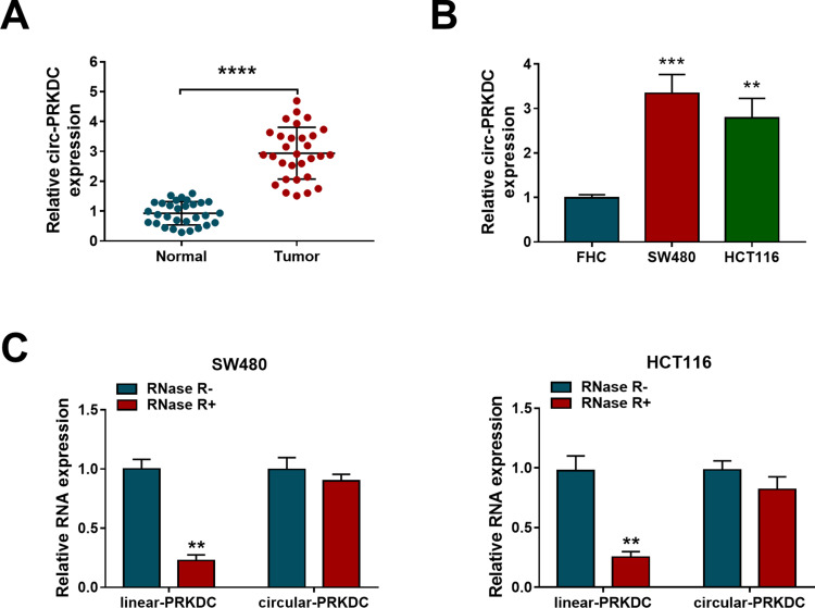
According to previous studies, we first examined the expression of circ-PRKDC in CRC tissues and adjacent non-tumor tissues. The results illustrated that circ-PRKDC level in CRC tissues was significantly increased in comparison with adjacent normal tissues (Figure 1A). Simultaneously, we detected circ-PRKDC expression in human normal colonic epithelial cell line FHC and CRC cells (SW480 and HCT116). qRT-PCR analysis showed a marked elevation in circ-PRKDC level in SW480 and HCT116 cells compared with FHC cells (Figure 1B). In addition, RNase R digestion assay revealed that circ-PRKDC was more resistant to RNase R than linear PRKDC in CRC cells (Figure 1C). As shown in Table 1, circ-PRKDC expression was not associated with age, gender and microsatellite instability (MSI) status, but was related to TNM stage. These data indicated that circ-PRKDC might play a carcinogenic role in CRC.
Figure 1.
Circ-PRKDC level is increased in CRC tissues and cells. (A) Expression of circ-PRKDC was examined in CRC tissues (n=30) and adjacent normal tissues (n=30). (B) Circ-PRKDC level was measured in CRC cells (SW480 and HCT116) and human normal colonic epithelial cells (FHC). (C) The levels of circ-PRKDC and linear PRKDC were detected in SW480 and HCT116 cells treated with or without RNase R. **P < 0.01, ***P < 0.001, ****P < 0.0001.
Circ-PRKDC Depletion Inhibits Proliferation, Migration and Invasion and Promotes Apoptosis in CRC Cells
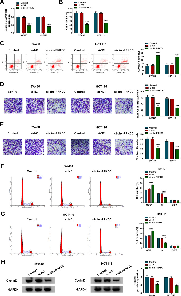
To investigate whether circ-PRKDC played a carcinogenic role in CRC, si-NC or si-circ-PRKDC was introduced into SW480 and HCT116 cells. First, qRT-PCR assay confirmed the significant inhibition efficiency of si-circ-PRKDC (Figure 2A). Next, CCK-8 analysis revealed that transfection with si-circ-PRKDC significantly reduced the viability of SW480 and HCT116 cells (Figure 2B). Flow cytometry suggested that the apoptosis rate in the si-circ-PRKDC group was significantly increased compared to the control group (Figure 2C). Moreover, introduction of si-circ-PRKDC significantly decreased the migrated and invaded cells compared with the control group (Figure 2D and E). Besides, circ-PRKDC silencing led to a striking increase in the percentage of cells at G0/G1 phase and a marked decrease in the percentage of cells at S phase, indicating silence of circ-PRKDC induced cell cycle arrest at G0/G1 phase (Figure 2F and G). Synchronously, Western blot assay displayed a marked reduction of CyclinD1 protein level in SW480 and HCT116 cells transfected with si-circ-PRKDC (Figure 2H). Overall, these data indicated that knockdown of circ-PRKDC suppressed CRC cell proliferation, migration and invasion, and induced apoptosis.
Figure 2.
Circ-PRKDC depletion inhibits proliferation, migration and invasion and promotes apoptosis in CRC cells. SW480 and HCT116 cells were introduced with si-NC or si-circ-PRKDC. (A) Circ-PRKDC expression was determined using qRT-PCR. (B) Cell viability was assessed using CCK-8 assay. (C) The apoptosis rate was measured via flow cytometry. (D and E) Cell migration and invasion were evaluated using transwell analysis. (F and G) Cell cycle was monitored using flow cytometry. (H) The protein level of CyclinD1 was examined using Western blot. ***P < 0.001, ****P < 0.0001.
Circ-PRKDC Acts as a Sponge for miR-198
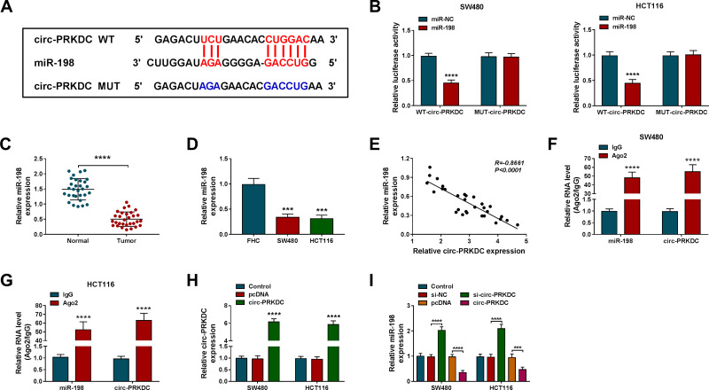
We speculated that circ-PRKDC and miR-198 had a putative binding sequence based on Circular RNA Interactome database (https://circinteractome.nia.nih.gov/) (Figure 3A). To verify this prediction, we conducted a dual-luciferase reporter assay. As displayed in Figure 3B, miR-198 mimics significantly decreased the luciferase activity of WT-circ-PRKDC reporter, while the effect was not significant when the binding site was mutated. Compared with adjacent non-tumor tissues, miR-198 level was significantly reduced in CRC tissues (Figure 3C). Similarly, miR-198 level in SW480 and HCT116 cells was markedly lower than that in FHC cells (Figure 3D). Additionally, circ-PRKDC and miR-198 levels in CRC tissues were negatively correlated (Figure 3E). Next, RIP analysis was performed to validate the binding relationship between circ-PRKDC and miR-198, and the results showed that miR-198 and circ-PRKDC were enriched in Ago2 group instead of lgG group (Figure 3F and G). Then, the overexpression efficiency of circ-PRKDC was verified via qRT-PCR assay (Figure 3H). Moreover, knockdown of circ-PRKDC expedited miR-198 expression, and up-regulation of circ-PRKDC restrained miR-198 expression (Figure 3I). Collectively, these results indicated that circ-PRKDC directly interacted with miR-198.
Figure 3.
Circ-PRKDC acts as a sponge for miR-198. (A) Circular RNA Interactome predicted that circ-PRKDC and miR-198 had targeted binding sites. (B) Luciferase activity was detected in SW480 and HCT116 cells co-transfected with WT-circ-PRKDC or MUT-circ-PRKDC and miR-NC or miR-198. (C) MiR-198 level was measured in CRC tissues and adjacent non-cancer tissues. (D) MiR-198 level was detected in FHC, SW480 and HCT116 cells. (E) The correlation between circ-PRKDC and miR-198 in CRC tissues was assessed using Spearman correlation coefficient. (F and G) The binding relationship between circ-PRKDC and miR-198 was verified via RIP analysis. (H) Circ-PRKDC level was examined in SW480 and HCT116 cells transfected with pcDNA or circ-PRKDC. (I) MiR-198 expression was measured in SW480 and HCT116 cells introduced with si-NC, si-circ-PRKDC, pcDNA or circ-PRKDC. ***P < 0.001, ****P < 0.0001.
Inhibition of miR-198 Alleviates the Effect of Circ-PRKDC Silencing on CRC Cell Progression
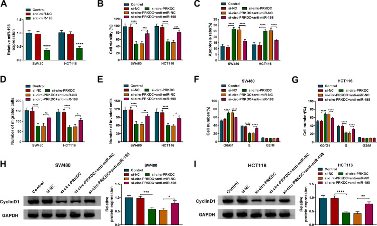
To illuminate whether circ-PRKDC targets miR-198 to affect CRC cell progression, a series of rescue experiments were performed in SW480 and HCT116 cells transfected with si-NC, si-circ-PRKDC, si-circ-PRKDC+anti-miR-NC or si-circ-PRKDC+anti-miR-198. First of all, the knockdown efficiency of miR-198 was confirmed in CRC cells introduced with anti-miR-NC or anti-miR-198 (Figure 4A). CCK-8 analysis and flow cytometry illustrated that inhibition of circ-PRKDC reduced cell viability and increased apoptosis of SW480 and HCT116 cells, while these changes were reversed by repressing miR-198 (Figure 4B and C). Transwell analysis depicted that silencing of circ-PRKDC inhibited the migration and invasion ability of CRC cells, whereas co-transfection of si-circ-PRKDC and anti-miR-198 partially alleviated these effects (Figure 4D and E). In addition, down-regulation of circ-PRKDC triggered G0/G1 phase arrest, which was abolished by inhibiting miR-198 (Figure 4F and G). Also, introduction of anti-miR-198 partially alleviated the decrease in CyclinD1 protein level caused by circ-PRKDC depletion (Figure 4H and I). Additionally, down-regulation of miR-198 markedly promoted the protein expression of CyclinD1 compared with the control group (Figure S1). Thus, these results evidenced that circ-PRKDCknockdown impeded CRC cell progression by regulating miR-198.
Figure 4.
Inhibition of miR-198 alleviates the effect of circ-PRKDC silencing on CRC cell progression. (A) The expression of miR-198 was measured in SW480 and HCT116 cells transduced with anti-miR-NC or anti-miR-198. Cell viability (B), apoptosis rate (C), migration (D), invasion (E), cell cycle (F and G) and CyclinD1 protein expression (H and I) were examined in SW480 and HCT116 cells transfected with si-NC, si-circ-PRKDC, si-circ-PRKDC+anti-miR-NC or si-circ-PRKDC+anti-miR-198. *P < 0.05, **P < 0.01, ***P < 0.001, ****P < 0.0001.
DDR1 is a Target of miR-198
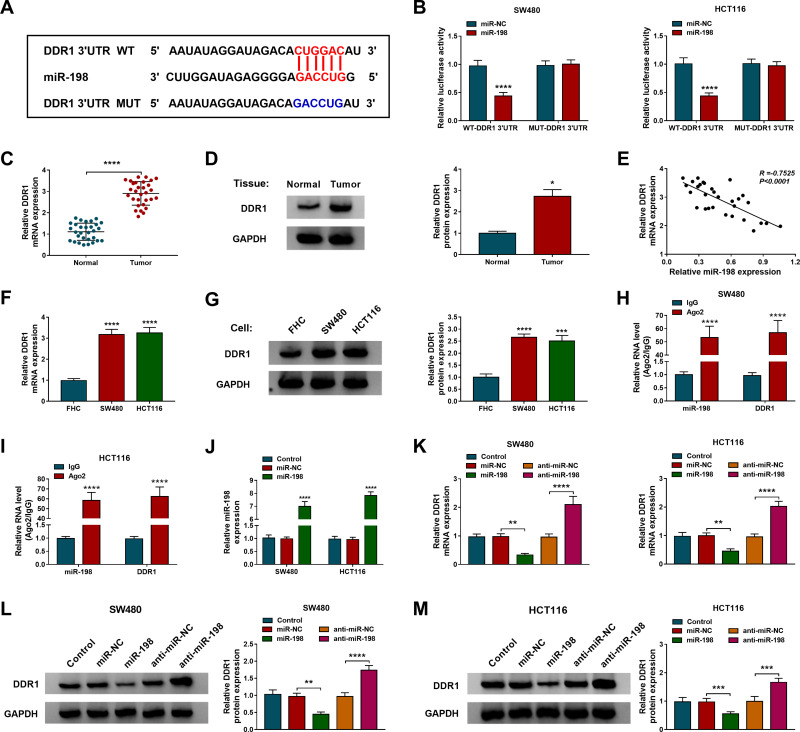
We predicted miRNAs that might bind to circ-PRKDC and screened four miRNAs (miR-198, miR-326, miR-330-5p and miR-485-3p) that were down-regulated in CRC. Next, the expression levels of four miRNAs were detected by qRT-PCR after circ-PRKDC knockdown. The results showed that the up-regulation of miR-198 was the most significant, so we chose miR-198 as the possible target of circ-PRKDC (Figure S2). The online database TargetScan suggested a shared sequence between miR-198 and DDR1 3ʹUTR (Figure 5A). Dual-luciferase reporter assay showed that mature miR-198 overtly decreased the luciferase activity of WT-DDR1 3ʹUTR reporter (Figure 5B). Compared to normal tissues, DDR1 mRNA and protein levels were significantly elevated in CRC tissues (Figure 5C and D). Spearman correlation coefficient revealed that miR-198 and DDR1 had a negative correlation in CRC tissues (Figure 5E). Additionally, the mRNA and protein levels of DDR1 in SW480 and HCT116 cells were remarkably higher than those in FHC cells (Figure 5F and G). Moreover, the binding relationship between miR-198 and DDR1 was verified by RIP assay, and the results exhibited that miR-198 and DDR1 were enriched in Ago2 group (Figure 5H and I). As shown in Figure 5J, the overexpression efficiency of miR-198 was examined using qRT-PCR assay. Besides, miR-198 up-regulation restrained DDR1 mRNA and protein expression, whereas miR-198 knockdown had the opposite effect (Figure 5K–M). Thus, these data reflected that miR-198 negatively targeted DDR1.
Figure 5.
DDR1 is a target of miR-198. (A) The predicted binding sites of miR-198 and DDR1 3ʹUTR were exhibited. (B) Luciferase activity was detected in SW480 and HCT116 cells introduced with WT-DDR1 3ʹUTR or MUT-DDR1 3ʹUTR and miR-NC or miR-198. (C and D) The mRNA and protein levels of DDR1 were measured in CRC tissues and normal tissues. (E) The correlation between miR-198 and DDR1 in CRC tissues was tested via Spearman correlation analysis. (F and G) DDR1 mRNA and protein levels were examined in FHC, SW480 and HCT116 cells. (H and I) RIP assay was used to confirm the relationship between miR-198 and DDR1. (J) Expression of miR-198 was detected in SW480 and HCT116 cells transduced with miR-NC or miR-198. (K–M) DDR1 mRNA and protein levels were measured in SW480 and HCT116 cells transfected with miR-NC, miR-198, anti-miR-NC or anti-miR-198. *P < 0.05, **P < 0.01, ***P < 0.001, ****P < 0.0001.
DDR1 Up-Regulation Attenuates the Effect of miR-198 Overexpression on CRC Cell Progression
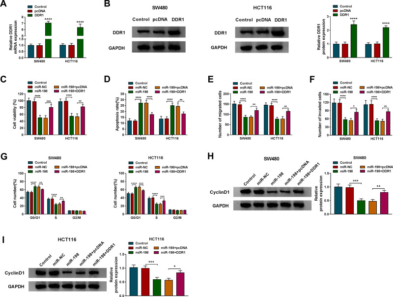
Firstly, DDR1 expression was markedly increased in the DDR1 group relative to the pcDNA group, indicating a significant overexpression efficiency (Figure 6A and B). To explore the effects of miR-198 and DDR1 on CRC cell growth, SW480 and HCT116 cells were transduced with miR-NC, miR-198, miR-198+pcDNA or miR-198+DDR1, respectively. As displayed in Figure 6C–F, augmentation of miR-198 suppressed cell viability, migration and invasion, and triggered apoptosis in SW480 and HCT116 cells, while introduction of miR-198+DDR1 partially abrogated these effects. Furthermore, overexpression of DDR1 partially abated the promotion of miR-198 up-regulation on cell cycle arrest (Figure 6G). Also, co-transfection with miR-198 and DDR1 partially alleviated the reduction of CyclinD1 level caused by miR-198 overexpression (Figure 6H and I). These results indicated that miR-198 hindered CRC cell progression via targeting DDR1.
Figure 6.
DDR1 up-regulation attenuates the effect of miR-198 overexpression on CRC cell progression. (A and B) After SW480 and HCT116 cells were introduced with pcDNA or DDR1, DDR1 mRNA and protein levels were determined using qRT-PCR and Western blot. Cell viability (C), apoptosis rate (D), migration (E), invasion (F), cell cycle (G) and CyclinD1 protein level (H and I) were detected in SW480 and HCT116 cells transfected with miR-NC, miR-198, miR-198+pcDNA or miR-198+DDR1. *P < 0.05, **P < 0.01, ***P < 0.001, ****P < 0.0001.
Circ-PRKDC Regulates DDR1 Expression via Sponging miR-198
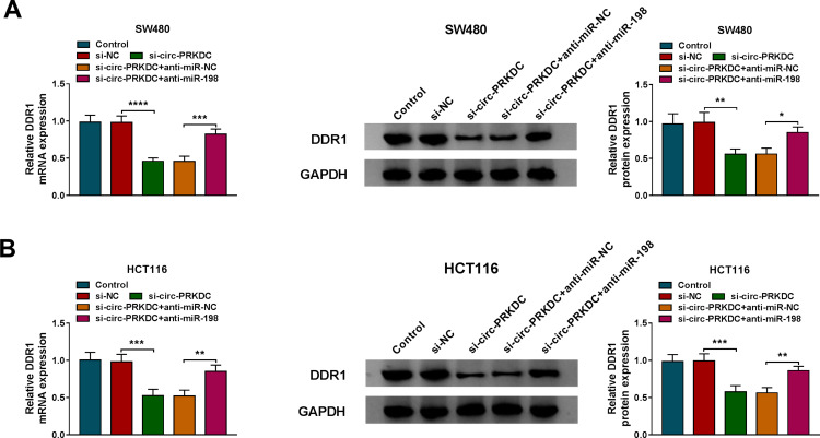
We also investigated the interaction among circ-PRKDC, miR-198 and DDR1 by detecting DDR1 expression in SW480 and HCT116 cells after transfection. The results depicted that DDR1 mRNA and protein levels were significantly reduced in the si-circ-PRKDC group compared with the si-NC group, whereas introduction of si-circ-PRKDC and anti-miR-198 undermined circ-PRKDC knockdown-induced decrease in DDR1 expression (Figure 7A and B). These results indicated that circ-PRKDC sponged miR-198 to elevate DDR1 expression.
Figure 7.
Circ-PRKDC regulates DDR1 expression via sponging miR-198. (A and B) The mRNA and protein levels of DDR1 were measured in SW480 and HCT116 cells introduced with si-NC, si-circ-PRKDC, si-circ-PRKDC+anti-miR-NC or si-circ-PRKDC+anti-miR-198. *P < 0.05, **P < 0.01, ***P < 0.001, ****P < 0.0001.
Silencing of Circ-PRKDC Inhibits Tumor Growth in vivo
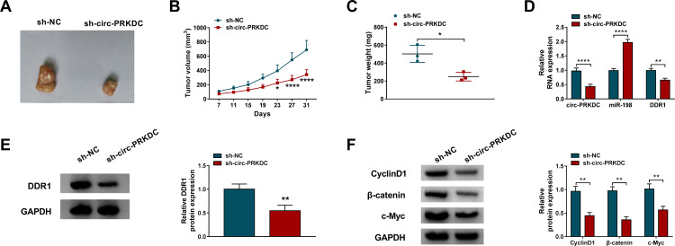
We used a xenograft mouse model to explore the role of circ-PRKDC in tumorigenesis in vivo. As exhibited in Figure 8A and B, tumor volume was markedly reduced in the sh-circ-PRKDC group compared with the sh-NC group. Also, circ-PRKDC silencing significantly decreased tumor weight (Figure 8C). Moreover, the expression of circ-PRKDC and DDR1 was significantly decreased, and miR-198 level was significantly elevated in the sh-circ-PRKDC group relative to the sh-NC group (Figure 8D and E). In addition, silencing of circ-PRKDC significantly reduced the expression of CyclinD1, β-catenin and c-Myc in xenograft tumors (Figure 8F). These data indicated that circ-PRKDC knockdown blocked tumor growth in vivo.
Figure 8.
Silencing of circ-PRKDC inhibits tumor growth in vivo. SW480 cells transfected with sh-NC or sh-circ-PRKDC were subcutaneously injected into nude mice. (A and B) Tumor volume was measured every 4 days. (C) After the mice were killed, the xenograft tumors were weighed. (D and E) The levels of circ-PRKDC, miR-198 and DDR1 in xenograft tumors were tested using qRT-PCR or Western blot. (F) The levels of CyclinD1, β-catenin and c-Myc in xenograft tumors were measured by Western blot. *P < 0.05, **P < 0.01, ****P < 0.0001.
Discussion
In recent years, circRNAs with high stability and tissue specificity are receiving increasing attention. Emerging evidence has manifested that circRNAs exert regulatory effects on biological functions, including proliferation, apoptosis and metastasis.28 A growing number of abnormally expressed circRNAs occupy a vital position in the regulation of CRC development.10 For example, circ5615 triggered CRC cell proliferation and restrained cell cycle arrest via interacting with miR-149-5p to up-regulate TNKS.29 CircVAPA contributed to CRC cell growth and glycolysis by directly combining with miR-125a to release CREB5.30 In this research, we determined that silenced circ-PRKDC suppressed CRC cell proliferation, migration and invasion, and triggered apoptosis. Subsequently, we selected miR-198 as a possible target for circ-PRKDC based on bioinformatics analysis.
Compelling evidence has illuminated that circRNAs serve as a competing endogenous RNA (ceRNA) to modulate gene expression, thereby affecting a range of biological processes.31 A previous report suggested that miR-198 is one of the top 10 miRNAs down-regulated in CRC tumor stroma with synchronized liver metastasis.32 Besides, more and more studies have confirmed that miR-198 is a tumor-inhibiting factor in various tumors. In breast cancer, miR-198 attenuated the malignancy of tumor by decreasing CDCP1 expression.33 In gastric cancer, miR-198 restrained tumorigenesis via targeting FGFR1 and had better efficacy in combination with cisplatin.34 Furthermore, Wang et al unveiled that low miR-198 expression was closely related to poor overall survival and up-regulation of miR-198 blocked CRC cell growth by repressing FUT8.20 In the present research, we first evidenced that circ-PRKDC might be a ceRNA for miR-198 and expedited CRC development via sponging miR-198.
To further clarify the potential mechanism of circ-PRKDC, we predicted the possible target genes of miR-198. Mounting evidence has manifested that miRNAs influence gene expression by pairing with 3ʹUTR of mRNAs.35 Plenty of investigations have revealed that DDR1 acts as an oncogene by regulating cell processes such as growth and metastasis in various tumors.36 For example, Zhong et al disclosed that DDR1 augmentation induced tumorigenesis via hindering antitumor immunity in breast cancer.37 Chou et al found that suppression of DDR1 restrained cell growth and accelerated apoptosis in oral cancer.38 Moreover, the previous study indicated that DDR1 silencing impeded tumor progression in CRC by interacting with miR-199a-5p.39 Our research unearthed that miR-198 targeted DDR1 to inhibit CRC cell progression.
In short, these findings demonstrated that circ-PRKDC aggravated the malignancy of CRC via modulating miR-198/DDR1 regulatory axis. We revealed a new ceRNA mechanism, which might provide a novel therapeutic strategy for CRC. However, the limitation of this study is that the sample size is too small. It is necessary to expand the sample size and further study the clinical significance of circ-PRKDC in CRC.
Funding Statement
This study was support by National Science Foundation for Young Scientists of China Grant No. 81903268.
Highlights
Circ-PRKDC level was enhanced in CRC tissues and cells.
Circ-PRKDC silencing inhibited CRC cell progression.
Circ-PRKDC promoted CRC progression via miR-198/DDR1 axis.
Circ-PRKDC knockdown blocked tumor growth in vivo.
Disclosure
The authors declare that they have no financial or non-financial conflicts of interest for this work.
References
- 1.Dekker E, Tanis PJ, Vleugels JLA, Kasi PM, Wallace MB. Colorectal cancer. Lancet. 2019;394(10207):1467–1480. doi: 10.1016/S0140-6736(19)32319-0 [DOI] [PubMed] [Google Scholar]
- 2.Bray F, Ferlay J, Soerjomataram I, Siegel RL, Torre LA, Jemal A. Global cancer statistics 2018: GLOBOCAN estimates of incidence and mortality worldwide for 36 cancers in 185 countries. CA Cancer J Clin. 2018;68(6):394–424. doi: 10.3322/caac.21492 [DOI] [PubMed] [Google Scholar]
- 3.Brenner H, Kloor M, Pox CP. Colorectal cancer. Lancet. 2014;383(9927):1490–1502. doi: 10.1016/S0140-6736(13)61649-9 [DOI] [PubMed] [Google Scholar]
- 4.Katona BW, Weiss JM. Chemoprevention of colorectal cancer. Gastroenterology. 2020;158(2):368–388. doi: 10.1053/j.gastro.2019.06.047 [DOI] [PMC free article] [PubMed] [Google Scholar]
- 5.Wolpin BM, Mayer RJ. Systemic treatment of colorectal cancer. Gastroenterology. 2008;134(5):1296–1310. doi: 10.1053/j.gastro.2008.02.098 [DOI] [PMC free article] [PubMed] [Google Scholar]
- 6.Qian L, Yu S, Chen Z, Meng Z, Huang S, Wang P. The emerging role of circRNAs and their clinical significance in human cancers. Biochim Biophys Acta Rev Cancer. 2018;1870(2):247–260. [DOI] [PubMed] [Google Scholar]
- 7.Cui X, Wang J, Guo Z, et al. Emerging function and potential diagnostic value of circular RNAs in cancer. Mol Cancer. 2018;17(1):123. doi: 10.1186/s12943-018-0877-y [DOI] [PMC free article] [PubMed] [Google Scholar]
- 8.Wang N, Lu K, Qu H, et al. CircRBM33 regulates IL-6 to promote gastric cancer progression through targeting miR-149. Biomed Pharmacother. 2020;125:109876. doi: 10.1016/j.biopha.2020.109876 [DOI] [PubMed] [Google Scholar]
- 9.Liu T, Ye P, Ye Y, Lu S, Han B. Circular RNA hsa_circRNA_002178 silencing retards breast cancer progression via microRNA-328-3p-mediated inhibition of COL1A1. J Cell Mol Med. 2020;24(3):2189–2201. doi: 10.1111/jcmm.14875 [DOI] [PMC free article] [PubMed] [Google Scholar]
- 10.Hao S, Cong L, Qu R, Liu R, Zhang G, Li Y. Emerging roles of circular RNAs in colorectal cancer. Onco Targets Ther. 2019;12:4765–4777. doi: 10.2147/OTT.S208235 [DOI] [PMC free article] [PubMed] [Google Scholar]
- 11.Jin C, Wang A, Liu L, Wang G, Li G. Hsa_circ_0136666 promotes the proliferation and invasion of colorectal cancer through miR-136/SH2B1 axis. J Cell Physiol. 2019;234(5):7247–7256. doi: 10.1002/jcp.27482 [DOI] [PubMed] [Google Scholar]
- 12.Chen H, Pei L, Xie P, Guo G. Circ-PRKDC contributes to 5-fluorouracil resistance of colorectal cancer cells by regulating miR-375/FOXM1 axis and Wnt/beta-catenin pathway. Onco Targets Ther. 2020;13:5939–5953. doi: 10.2147/OTT.S253468 [DOI] [PMC free article] [PubMed] [Google Scholar]
- 13.Liu LH, Tian QQ, Liu J, Zhou Y, Yong H. Upregulation of hsa_circ_0136666 contributes to breast cancer progression by sponging miR-1299 and targeting CDK6. J Cell Biochem. 2019;120(8):12684–12693. doi: 10.1002/jcb.28536 [DOI] [PubMed] [Google Scholar]
- 14.Chen Y, Gao DY, Huang L. In vivo delivery of miRNAs for cancer therapy: challenges and strategies. Adv Drug Deliv Rev. 2015;81:128–141. doi: 10.1016/j.addr.2014.05.009 [DOI] [PMC free article] [PubMed] [Google Scholar]
- 15.Di Leva G, Garofalo M, Croce CM. MicroRNAs in cancer. Annu Rev Pathol. 2014;9:287–314. doi: 10.1146/annurev-pathol-012513-104715 [DOI] [PMC free article] [PubMed] [Google Scholar]
- 16.Shirmohamadi M, Eghbali E, Najjary S, et al. Regulatory mechanisms of microRNAs in colorectal cancer and colorectal cancer stem cells. J Cell Physiol. 2020;235(2):776–789. doi: 10.1002/jcp.29042 [DOI] [PubMed] [Google Scholar]
- 17.Hong YG, Xin C, Zheng H, et al. miR-365a-3p regulates ADAM10-JAK-STAT signaling to suppress the growth and metastasis of colorectal cancer cells. J Cancer. 2020;11(12):3634–3644. doi: 10.7150/jca.42731 [DOI] [PMC free article] [PubMed] [Google Scholar]
- 18.Li J, Peng W, Yang P, et al. MicroRNA-1224-5p inhibits metastasis and epithelial-mesenchymal transition in colorectal cancer by targeting SP1-mediated NF-kappaB signaling pathways. Front Oncol. 2020;10:294. doi: 10.3389/fonc.2020.00294 [DOI] [PMC free article] [PubMed] [Google Scholar]
- 19.Sun N, Zhang L, Zhang C, Yuan Y. miR-144-3p inhibits cell proliferation of colorectal cancer cells by targeting BCL6 via inhibition of Wnt/beta-catenin signaling. Cell Mol Biol Lett. 2020;25:19. doi: 10.1186/s11658-020-00210-3 [DOI] [PMC free article] [PubMed] [Google Scholar]
- 20.Wang M, Wang J, Kong X, et al. MiR-198 represses tumor growth and metastasis in colorectal cancer by targeting fucosyl transferase 8. Sci Rep. 2014;4:6145. doi: 10.1038/srep06145 [DOI] [PMC free article] [PubMed] [Google Scholar]
- 21.Li LX, Lam IH, Liang FF, et al. MiR-198 affects the proliferation and apoptosis of colorectal cancer through regulation of ADAM28/JAK-STAT signaling pathway. Eur Rev Med Pharmacol Sci. 2019;23(4):1487–1493. [DOI] [PubMed] [Google Scholar]
- 22.Leitinger B. Discoidin domain receptor functions in physiological and pathological conditions. Int Rev Cell Mol Biol. 2014;310:39–87. [DOI] [PMC free article] [PubMed] [Google Scholar]
- 23.Yeh YC, Lin HH, Tang MJ. Dichotomy of the function of DDR1 in cells and disease progression. Biochim Biophys Acta Mol Cell Res. 2019;1866(11):118473. doi: 10.1016/j.bbamcr.2019.04.003 [DOI] [PubMed] [Google Scholar]
- 24.Chen YL, Tsai WH, Ko YC, et al. Discoidin domain receptor-1 (DDR1) is involved in angiolymphatic invasion in oral cancer. Cancers (Basel). 2020;12(4):841. doi: 10.3390/cancers12040841 [DOI] [PMC free article] [PubMed] [Google Scholar]
- 25.Wu A, Chen Y, Liu Y, Lai Y, Liu D. miR-199b-5p inhibits triple negative breast cancer cell proliferation, migration and invasion by targeting DDR1. Oncol Lett. 2018;16(4):4889–4896. [DOI] [PMC free article] [PubMed] [Google Scholar]
- 26.Vella V, Nicolosi ML, Cantafio P, et al. DDR1 regulates thyroid cancer cell differentiation via IGF-2/IR-A autocrine signaling loop. Endocr Relat Cancer. 2019;26(1):197–214. doi: 10.1530/ERC-18-0310 [DOI] [PubMed] [Google Scholar]
- 27.Sirvent A, Lafitte M, Roche S. DDR1 inhibition as a new therapeutic strategy for colorectal cancer. Mol Cell Oncol. 2018;5(4):e1465882. doi: 10.1080/23723556.2018.1465882 [DOI] [PMC free article] [PubMed] [Google Scholar]
- 28.Qu S, Liu Z, Yang X, et al. The emerging functions and roles of circular RNAs in cancer. Cancer Lett. 2018;414:301–309. doi: 10.1016/j.canlet.2017.11.022 [DOI] [PubMed] [Google Scholar]
- 29.Ma Z, Han C, Xia W, et al. circ5615 functions as a ceRNA to promote colorectal cancer progression by upregulating TNKS. Cell Death Dis. 2020;11(5):356. doi: 10.1038/s41419-020-2514-0 [DOI] [PMC free article] [PubMed] [Google Scholar]
- 30.Zhang X, Xu Y, Yamaguchi K, et al. Circular RNA circVAPA knockdown suppresses colorectal cancer cell growth process by regulating miR-125a/CREB5 axis. Cancer Cell Int. 2020;20:103. [DOI] [PMC free article] [PubMed] [Google Scholar]
- 31.Zhong Y, Du Y, Yang X, et al. Circular RNAs function as ceRNAs to regulate and control human cancer progression. Mol Cancer. 2018;17(1):79. [DOI] [PMC free article] [PubMed] [Google Scholar]
- 32.Murakami T, Kikuchi H, Ishimatsu H, et al. Tenascin C in colorectal cancer stroma is a predictive marker for liver metastasis and is a potent target of miR-198 as identified by microRNA analysis. Br J Cancer. 2017;117(9):1360–1370. doi: 10.1038/bjc.2017.291 [DOI] [PMC free article] [PubMed] [Google Scholar]
- 33.Hu Y, Tang Z, Jiang B, Chen J, Fu Z. miR-198 functions as a tumor suppressor in breast cancer by targeting CUB domain-containing protein 1. Oncol Lett. 2017;13(3):1753–1760. doi: 10.3892/ol.2017.5673 [DOI] [PMC free article] [PubMed] [Google Scholar]
- 34.Gu J, Li X, Li H, Jin Z, Jin J. MicroRNA-198 inhibits proliferation and induces apoptosis by directly suppressing FGFR1 in gastric cancer. Biosci Rep. 2019;39(6). doi: 10.1042/BSR20181258 [DOI] [PMC free article] [PubMed] [Google Scholar]
- 35.Zhang Y, Wang Z, Gemeinhart RA. Progress in microRNA delivery. J Control Release. 2013;172(3):962–974. doi: 10.1016/j.jconrel.2013.09.015 [DOI] [PMC free article] [PubMed] [Google Scholar]
- 36.Borza CM, Pozzi A. Discoidin domain receptors in disease. Matrix Biol. 2014;34:185–192. doi: 10.1016/j.matbio.2013.12.002 [DOI] [PMC free article] [PubMed] [Google Scholar]
- 37.Zhong X, Zhang W, Sun T. DDR1 promotes breast tumor growth by suppressing antitumor immunity. Oncol Rep. 2019;42(6):2844–2854. [DOI] [PubMed] [Google Scholar]
- 38.Chou ST, Peng HY, Mo KC, et al. MicroRNA-486-3p functions as a tumor suppressor in oral cancer by targeting DDR1. J Exp Clin Cancer Res. 2019;38(1):281. doi: 10.1186/s13046-019-1283-z [DOI] [PMC free article] [PubMed] [Google Scholar]
- 39.Hu Y, Liu J, Jiang B, et al. MiR-199a-5p loss up-regulated DDR1 aggravated colorectal cancer by activating epithelial-to-mesenchymal transition related signaling. Dig Dis Sci. 2014;59(9):2163–2172. doi: 10.1007/s10620-014-3136-0 [DOI] [PubMed] [Google Scholar]


