ABSTRACT
Liver is the central organ responsible for whole-body metabolism, and its constituent hepatocytes are the major players that carry out liver functions. Although they are highly differentiated and rarely divide, hepatocytes re-enter the cell cycle following hepatic loss due to liver damage or injury. However, the exact molecular mechanisms underlying cell cycle re-entry remain undefined. Gdown1 is an RNA polymerase II (Pol II)-associated protein that has been linked to the function of the Mediator transcriptional coactivator complex. We recently found that Gdown1 ablation in mouse liver leads to down-regulation of highly expressed liver-specific genes and a concomitant cell cycle re-entry associated with the induction of cell cycle-related genes. Unexpectedly, in view of a previously documented inhibitory effect on transcription initiation by Pol II in vitro, we found that Gdown1 is associated with elongating Pol II on the highly expressed genes and that its ablation leads to a reduced Pol II occupancy that correlates with the reduced expression of these genes. Based on these observations, we discuss the in vitro and in vivo functions of Gdown1 and consider mechanisms by which the dysregulated Pol II recruitment associated with Gdown1 loss might induce quiescent cell re-entry into the cell cycle.
KEYWORDS: Gdown1, RNA polymerase II, Mediator; transcription, elongation, liver regeneration
Introduction
Cell division, which is crucial to cellular proliferation, development, and self-renewal, is tightly regulated by multiple factors that are coordinately expressed during the cell cycle [1–3]. Controlled cell cycle progression involves precisely regulated gene expression that is governed by gene-specific transcription factors [4,5]. As demonstrated by the ability of small numbers of such factors to genetically reprogram and convert one cell type to another, of increasing importance for regenerative medicine [6,7], some transcription factors (TFs) can be master regulators of cell fate. On the other hand, transcriptional dysregulation caused by mutations or deletions in TFs such as p53 can lead to malignant tumors [8,9]. In response to signals that result in the expression of specific sets of genes, TFs bind selectively to cognate binding sites in the transcriptional regulatory regions that include promoter and enhancer regions. These bound TFs in turn interact with diverse transcriptional coactivators, leading ultimately to the recruitment of Pol II and corresponding general transcription factors (GTFs) to cognate gene core promoters [below]. To initiate accurate gene transcription, Pol II must be precisely positioned at the transcription start site within core promoters where the GTFs (TFIIA, -B, -D, -E, -F, and – H) play essential roles, through interactions with core promoter elements and Pol II, in facilitating pre-initiation complex (PIC) formation [10–12]. The TBP subunit of TFIID, TFIIB, and TFIIF are especially important for initial interactions with core promoters. TFIIF is composed of two subunits (Rap74 and Rap30) that directly bind to the second largest subunit (RPB2) of Pol II [13]. TFIIF binding to Pol II facilitates Pol II recruitment, stabilizes the PIC, and promotes open complex formation through interactions with other GTFs (including TFIIB and TFIIE) [14]. While the TFIIF requirement can be partially bypassed with a pre-melted promoter DNA template in vitro [15], TFIIB is absolutely essential for PIC formation through its pivotal role in positioning promoter DNA and determining the site of initial strand opening through interactions with TBP and Pol II [10,16,17].
Another factor that facilitates Pol II recruitment to core promoters is the large (1-MDa) 25–30 subunit Mediator complex that is essential for activator-dependent transcription by Pol II [18,19]. Mediator acts principally in the regulation of transcription through joint enhancer-bound activator and Pol II interactions that facilitate both Pol II recruitment and enhancer–promoter interactions [20–22]. In addition to direct interactions of Mediator with Pol II, recent structural studies of the PIC have revealed Mediator interactions with TFIIB, suggesting a multi-valent role for Mediator in PIC assembly [23,24]. However, Mediator function is not limited to the transcription initiation step through these interactions, but rather has expanding roles in various steps in transcription, chromatin regulation, and mRNA processing [24,25].
As discussed below, one of the newly discovered functions of Mediator in transcription is to convert an inactive form of Pol II, containing a tightly associated inhibitory protein (Gdown1) and designated Pol II(G), to an active form. However, whereas biochemical and structural studies led to an understanding of the mechanism by which Gdown1 inhibits initiation by Pol II, there had been no understanding of biological functions of Gdown1. Toward that goal, and most importantly, our Gdown1 knockout liver study revealed an important and unexpected role for Gdown1 in the transcription of highly expressed liver-specific genes and, further, that Gdown1 ablation induces hepatocyte cell cycle re-entry by down-regulating these genes. Hence, transcriptional regulation by Gdown1 is directly coupled to cell cycle control. As a prelude to a discussion of these newer findings on the biological functions of Gdown1, we first discuss mechanistic aspects of the Gdown1-mediated inhibition of Pol II initiation and its reversal by Mediator–which must ultimately be linked to the newly discovered functions. A subsequent discussion of the new functions, especially in relation to the control of cell cycle re-entry, is then followed by perspectives on current and future studies.
Mediator-dependent transcriptional activation by Pol II(G)
During purification of Pol II from animal tissues (calf thymus or pig liver) by methods that included affinity chromatography, a previously unidentified form of Pol II, designated Pol II(G), was discovered [26]. Beyond the conserved 12 subunits that comprise Pol II, this biochemically distinct Pol II(G) contained an additional 43-kDa polypeptide that was tightly associated with Pol II. Protein sequencing analysis identified the polypeptide as Gdown1, which was originally recognized as one of the multiple proteins encoded in the GRINL1A (glutamine receptor-like gene) locus of the human genome [27]. Studies with biochemically defined in vitro transcription systems (Pol II, GTFs, Mediator, and PC4) that support accurate transcription initiation from naked DNA templates [28] revealed a very low activity of Pol II(G) relative to Pol II in the absence of Mediator and, remarkably, an ability of Mediator to reverse the repressed activity of Pol II(G) [26] – thus indicating a Gdown1-elicited Mediator function for the regulation of transcription initiation. At the same time, no inhibitory effect of Gdown1 on elongation by Pol II was observed.
The mechanisms underlying Gdown1-mediated repression of Pol II initiation activity were subsequently uncovered in studies with an in vitro transcription system containing a minimal set of factors (Pol II, TFIIF, TFIIB, TBP) that suffice for basal (activator-independent) transcription from supercoiled TATA-containing DNA templates [29]. These analyses revealed that Pol II-bound Gdown1 blocks a Pol II-TFIIF interaction that is critical for PIC assembly [30] – thus indicating a repressive effect of Gdown1 at an early step in initiation and suggesting a potentially simple mechanism for reversal of the inhibition by Mediator-facilitated removal of Gdown1 from Pol II.
Architecture of Pol II(G) and molecular mechanism of transcription repression by Gdown1
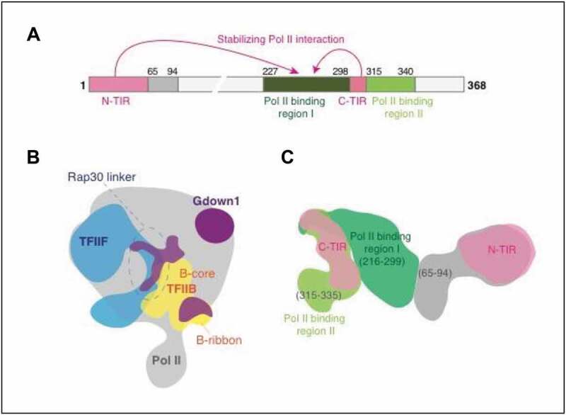
The 368 amino-acid human Gdown1 protein is composed of highly conserved regions at the N- and C-termini. However, there is no identifiable domain that might suggest biological functions. Protein folding analysis predicts that Gdown1 is mostly intrinsically disordered. Interestingly, however, a limited proteolysis analysis suggests that there might be a folded region at the N-terminus [31]. Biochemical assays and CX-MS analysis revealed two functionally distinct regions containing sub-regions: two Pol II binding regions and two transcription inhibitory regions designated N-TIR and C-TIR Figure 1A [32]. The Pol II binding regions of Gdown1 are the major sites of interaction with the RPB2 subunit protrusion domain. TIRs only weakly bind to Pol II, although two mutations, L303/ 304A and an NΔ33 deletion in TIRs, cause a loss of the inhibitory activity of Gdown1. Importantly, the combined TIR interactions with Pol II stabilize the binding of the Gown1 C-terminal domain to the RPB2 protrusion domain, which explains the robust binding of Gdown1 to Pol II [26].
Figure 1.

Architecture of Pol II(G). (A) Schematic of Gdown1 functional domains. (B) Positions of TFIIF (blue) and TFIIB (yellow) relative to Gdown1 (purple) on Pol II (gray). (C) Location of Gdown1 functional domains determined by integrative structure modeling. Models based on data in [32]
In more direct structural studies, a cryo-EM map of Pol II(G) at ~4 Å resolution confirmed the presence of Gdown1 on the RPB2 protrusion domain as well as RPB3 and RPB10 localizations. The density that was observed on the RPB2 protrusion domain perfectly matched the interaction site of the linker domain of the Rap30 subunit of initiation factor TFIIF Figure 1B. The Rap30 linker is an unstructured region that is essential both for growth in yeast [33] and for transcription initiation in vitro [34]. Recent cryo-EM studies of the PIC showed that the Rap30 linker contacts multiple sites that include RPB2 (albeit not the protrusion), TFIIB, TBP, and a downstream promoter region [35]. As these sites are essential for PIC formation, it was proposed that the linker interaction with the PIC might position other essential TFIIF domains [35]. The linker interaction may thereby be critical for TFIIF binding to Pol II, which would be perfectly consistent with Pol II(G)-mediated transcription repression via targeting TFIIF. The cryo-EM analyses also revealed that the Gdown1 density overlaps the Pol II interaction sites (RPB1 dock and RPB2 wall domains) of the TFIIB B-core and B-ribbon domains Figure 1B [36,37]. Consistent with this overlapping interaction, Gdown1 was found to interfere with the TFIIB-Pol II interaction and to inhibit transcription initiation. Therefore, the mechanism of transcription repression by Gdown1 is to prevent TFIIF and TFIIB from binding to Pol II, which contributes to the maintenance of Pol II(G) an inactive state in the absence of Mediator.
Molecular mechanism of the reversal of Gdown1-mediated repression by Mediator
Further mechanistic studies showed that mutations in both TIRs can bypass the Mediator requirement for transcriptional activation. These results suggest that TIRs play essential roles in the Gdown1 inhibitory activity [32] and, further, that disruption of the TIR interactions might be involved in the natural process by which Mediator reverses Gdown1-mediated repression. Although the cryo-EM study could not reveal the location of functional domains of Gdown1, likely because of the high degree of disorder, crosslinking-based integrative structure modeling [38,39] showed that N-TIR localizes near Pol II subunits RPB10 and RPB3 and that the connected N-terminal region (residues 65–94) localizes to the Pol II binding region I Figure 1C. Thus, N-TIR interacts with the Pol II binding region I through the (65–94) region, explaining how N-TIR could stabilize the Gdown1-Pol II interaction. The Pol II binding region I covers the RPB2 protrusion surface and the Pol II binding region II also localizes to the RPB2 protrusion. The location of C-TIR was identified as an overlapping region between regions (216–314) and (300–335). Hence, TIRs are located near the Pol II binding regions. Notably, N-TIR is positioned near the Pol II RPB3 and RPB10 subunits that (in yeast) may interact with Mediator [24]. This raises the possibility that a Mediator tail domain interaction with RPB3 may lead to a concomitant N-TIR dissociation and an associated de-stabilization of the Gdown1-Pol II interaction, which could be at least part of the mechanism by which Mediator reverses Gdown1-mediated transcription initiation inhibition.
Biological functions of Gdown1/Pol II(G)
While Mediator and Pol II are highly conserved from yeast to human, Gdown1 is metazoan-specific [26]. Genomic sequence comparisons have revealed orthologs of Gdown1 in all mammalian species, and further identified an ortholog of Gdown1 in Drosophila melanogaster but not in Caenorhabditis elegans. The obligatory Mediator requirement for Pol II(G)-mediated transcription in vitro clearly shows that Pol II(G) has unique properties relative to Pol II, and the ubiquitous expression of Gdown1 [40,41] suggests a global role in transcriptional regulation. However, the actual biological roles of Gdown1, and why higher metazoans need two distinct forms of Pol II to regulate gene transcription, remains largely unknown.
Gdown1 is essential for early embryonic development
Although it is present at all life cycle stages in Drosophila, Gdown1 is most abundant at the embryonic stage [32]. Consistent with this observation, Gdown1 nuclear localization is more evident in the embryo but not in the larval salivary gland. More interestingly, Gdown1 co-localizes with Pol II in nuclei at the transcriptionally silent syncytial blastoderm stage, but is detected only in the cytoplasm and not in the nuclei that retain Pol II at the later cellular blastoderm stage at which global transcription is initiated. Also, the pole cells that are transcriptionally silent at this early embryonic stage retain nuclear Gdown1, suggesting a role for Gdown1, through the formation of Pol II(G), in transcriptional repression at an early embryonic stage. More importantly, in genetic studies, adult flies were never obtained with Gdown1 mutation or siRNA-mediated Gdown1 knockdown in the embryo. Similarly, maternal Gdown1 knockout (KO) embryos also were not detected. These observations indicate that Gdown1 plays a critical role in early embryonic development in the fly.
Consistent with this demonstration of embryonic lethality in the fly, Gdown1 was also found to be essential for early embryonic development in mice [42]. In the mouse study, the number of Gdown1 null embryos began to decrease at embryonic day 3.5 (E3.5) and no nullizygous embryos were evident at E10.5. Although Gdown1 null blastocysts were observed, a Gdown1 KO embryonic stem cell line was not obtained. These data clearly show that Gdown1 is critical for early embryonic development in mammals, although underlying mechanisms remain to be elucidated.
Gdown1 ablation induces quiescent hepatocyte re-entry into the cell cycle
Unlike embryonic stem cells that proliferate rapidly, hepatocytes rarely divide (a few times a year). Hepatocytes are major players in carrying out liver functions, such as nutrient metabolism and synthesis of plasma proteins. Therefore, in normal hepatocytes, activation of transcription is mostly focused on liver-specific genes [43]. Although they are highly differentiated, hepatocytes possess a unique ability to re-enter the cell cycle upon liver injury or loss to restore liver mass [44]. Toward elucidation of the molecular mechanism of liver regeneration, numerous studies over the past decades have sought to identify factors that regulate this process [44–48]. However, what triggers hepatocytes to re-enter the cell cycle remains undefined.
Despite the Gdown1 KO embryonic lethality, Gdown1 is not essential for hepatocyte viability as hepatocyte KO mice display whole body and liver weights comparable to those of control mice [42]. However, Gdown1 hepatocyte KO leads to Cyclin D1 induction and cell cycle re-entry in 5-week (W) old liver in the absence of obvious hepatic injury and is followed by p21 induction through activation of a p53 signaling pathway. At 8 W, in addition to the continuous expression of Cyclin D1, p21, and p53, the KO liver shows histological abnormalities that include Ki67-positive hepatocytes, apoptotic hepatocytes, and the proliferation of SMA (smooth muscle actin)-positive cells that are associated with collagen deposition, whose emergence is often triggered by hepatic injury [49]. Consistent with the down-regulation of genes involved in lipid metabolism, Gdown1 KO mice show significantly lowered serum triglyceride levels. It is possible that the up-regulation of both p21 and Cyclin D1 for a long period (~ 3-weeks) causes the injury-like reaction in the KO liver.
It appears that the initial impact of Gdown1 KO on hepatocytes is to induce cell cycle re-entry, although progression appears to be prevented by p53-dependent activation of p21. In fact, Gdown1 and p53 double KO (DKO) promotes the cell cycle progression that eventually leads to dysplastic cell nuclei, incomplete mitoses, distortion of normal liver architecture, and human hepatocellular carcinoma-like RNA expression profile in DKO liver. Although the precise mechanisms by which Gdown1 KO causes cell cycle re-entry in hepatocytes, the down-regulation of highly transcribed genes that are involved in liver-specific functions and lipid metabolism appears to be implicated as a cause of cell cycle re-entry.
Gdown1 is associated with elongating Pol II in the liver
Consistent with the original discovery of Pol II(G) in porcine liver [26], most of the Gdown1 in mouse liver is associated with Pol II. This further suggests that Gdown1 plays a direct role in transcription as a component of Pol II(G) [42]. Because of the mutually exclusive Pol II interactions with Gdown1 and TFIIF observed in the in vitro studies, it was predicted that Gdown1 should be dissociated from Pol II when the PIC is assembled. However, ChIP-seq studies in mouse liver revealed a strong association of Gdown1 with elongating Pol II on coding regions of highly expressed liver genes such as those (e.g., Alb, Cyp2e1, and Serpina3k) encoding plasma proteins. These genes play major roles in the maintenance of normal liver functions. The number (222) of identified Gdown1 target genes seems low in view of the dramatic effect of the KO. However, the RNA transcripts of these Gdown1-targeted genes account for approximately 30% of total liver transcripts, which may explain the observed phenotype in KO liver. Although the primary effect of Gdown1 KO is an induction of cell cycle-related genes, including Cyclin D1 and p21, Gdown1 occupancy on these genes was not observed. Therefore, the cell cycle re-entry by Gdown1 KO must be induced through secondary (indirect) effects.
In view of earlier ChIP-seq results for Gdown1 in a cancer cell line [30] and an in vitro study indicating TFIIF displacement by Gdown1 in early elongation complexes [50], it was suggested that Gdown1 might be involved in stabilizing paused Pol II at proximal-promoter regions [51]. However, in mouse liver, Gdown1 occupancy was not observed on genes, such as immediate early genes Jun, Fos, and Btg2 [42], that contain paused Pol II. The expression of these immediate early genes, which in normal liver are regulated by paused Pol II at their proximal-promoter regions [52], is induced within a few hours upon liver injury or partial hepatectomy [53,54]. Whereas promoter-proximal Pol II pausing is an important transcription regulation strategy [55], it appears unlikely that Gdown1 plays a major role in Pol II pausing, at least in the mouse liver.
Potential mechanism of Gdown1 KO-induced cell cycle re-entry
The loss of Gdown1 in the liver leads to substantial decreases in elongating Pol II on the coding regions of genes that are highly transcribed in the liver, resulting in the down-regulation of RNA expression. In the absence of p53, the down-regulation of Gdown1 target genes immediately up-regulates genes encoding proteins involved in poly(A) binding, spliceosome function, and mitochondrion function, which are essential cellular components. The transcription of Cyclin D1 also is activated in an unknown and apparently non-canonical manner [42].
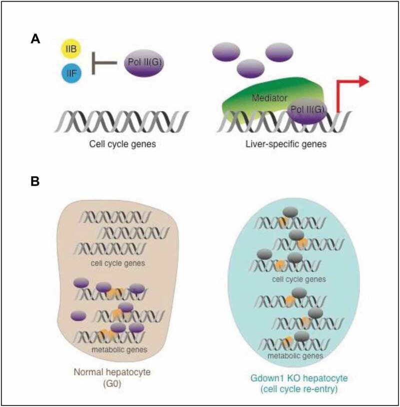
The exact mechanism leading to the activation of cell cycle-related genes remains unclear. However, a reduction of Pol II recruitment to the liver-specific genes in the absence of Gdown1 may lead to hepatocyte re-entry into the cell cycle. In normal liver, hepatocytes are committed to the expression of highly selected genes that are involved in the synthesis of plasma proteins or in metabolism, while other genes that include cell cycle-related genes are not expressed. Since it prevents Pol II recruitment to promoters in the absence of Mediator, Gdown1 plays a role in restricting Mediator/activator-independent transcription (at selected genes) and may thereby establish a more favorable situation for active transcription of liver-specific genes in normal liver Figure 2A. In the absence of Gdown1 where the restriction no longer exists, Pol II recruitment to promoters could be less competitive, which could make Pol II available for other genes. Also, the liver-specialized genes may heavily depend on Mediator/activator for their active transcription. The albumin gene is the most highly expressed gene in the liver, and its transcriptional activation depends on an enhancer region that lies around 10 kb upstream from the transcription start site [56] and closely resembles super-enhancers. Super-enhancers are extremely sensitive to perturbation of associated components [57], such that loss of Gdown1 might contribute to the prompt reduction of Pol II recruitment to the albumin gene. Consequently, the expression of heavily Mediator/activator-dependent genes would be decreased, while genes whose transcription is maintained at the basal level might be activated. We speculate that this Pol II re-allocation to other genes might underlie hepatocyte re-entry into the cell cycle Figure 2B.
Figure 2.

Model for Gdown1 KO-induced cell cycle re-entry in liver. (A) A potential role of Gdown1 for gene transcription in normal liver. (B) A model for Gdown1 KO-induced cell cycle re-entry. Models based on data in [42]
The above model also provides a basis for how tumorigenesis might be initiated in the liver. Genomic mutations in the human ALB gene, which may down-regulate its expression, are frequently observed in hepatocellular carcinoma [58–61]. Thus, dysregulated gene expression resulting from loss of transcriptional control of Pol II by Gdown1 could result in Pol II reallocation and induce quiescent cells to re-enter into the cell cycle, thereby triggering tumorigenesis.
Perspective
Following the discovery of Gdown1 as a Pol II-associated inhibitor of transcription initiation in vitro over a decade ago, further biochemical studies have provided a basic understanding of the initiation inhibition mechanism. More recent genetic analyses have provided evidence for physiological functions – including potentially direct functions in transcription elongation on highly expressed liver-specific genes as well as functions in restricting cell cycle progression and cell proliferation in quiescent cells. The combined studies raise many questions regarding the relationship of the in vitro results to the in vivo results and the potential generality of the genetic analyses indicating gene-specific effects. In particular, since Gdown1 inhibits PIC assembly and must be dissociated from Pol II, at least partially, for initiation by Pol II, it will be important to understand: the fate and dynamics of Pol II-associated Gdown1 during initiation and the transition to productive elongation, how the initiated Pol II regains its association with Gdown1 following promoter clearance, and the role of the Mediator in these processes. Related, the presumed role of Gdown1 in transcription elongation on occupied genes will need to be confirmed and associated mechanisms, including potential interactions with other elongation factors, established. Beyond this mechanistic question, another key question relates to the link between the loss of Gdown1 on highly expressed liver genes and the ultimate reactivation of cell cycle-related genes leading to cell cycle re-entry – including, especially, identification of intermediary pathways involving other genes/gene products. It will also be important to investigate potentially more general effects of Gdown1 in regulating cell cycle progression in other cell types as well as possible gene specificity for Pol II(G) recruitment and function. Answers to these open questions will help us understand the biological role(s) of Gdown1, including its role in cell cycle control, as well as metazoan-specific and cancer-related aspects of transcriptional regulation related to Gdown1.
Acknowledgments
We thank Dr. Sohail Malik for critical comments on the manuscript. This study was supported by National Institutes of Health grant CA202245.
Funding Statement
This work was supported by National Institutes of Health [CA202245].
Disclosure statement
No potential conflict of interest was reported by the authors.
References
- 1.Budirahardja Y, Gonczy P.. Coupling the cell cycle to development. Development. 2009;136:2861–2872. [DOI] [PubMed] [Google Scholar]
- 2.Murray AW. Recycling the cell cycle: cyclins revisited. Cell. 2004;116:221–234. [DOI] [PubMed] [Google Scholar]
- 3.He S, Nakada D, Morrison SJ. Mechanisms of stem cell self-renewal. Annu Rev Cell Dev Biol. 2009;25:377–406. [DOI] [PubMed] [Google Scholar]
- 4.Bahler J. Cell-cycle control of gene expression in budding and fission yeast. Annu Rev Genet. 2005;39:69–94. [DOI] [PubMed] [Google Scholar]
- 5.Bertoli C, Skotheim JM, de Bruin RA. Control of cell cycle transcription during G1 and S phases. Nat Rev Mol Cell Biol. 2013;14:518–528. [DOI] [PMC free article] [PubMed] [Google Scholar]
- 6.Smith ZD, Sindhu C, Meissner A. Molecular features of cellular reprogramming and development. Nat Rev Mol Cell Biol. 2016;17:139–154. [DOI] [PubMed] [Google Scholar]
- 7.Hirschi KK, Li S, Roy K. Induced pluripotent stem cells for regenerative medicine. Annu Rev Biomed Eng. 2014;16:277–294. [DOI] [PMC free article] [PubMed] [Google Scholar]
- 8.Bradner JE, Hnisz D, Young RA. Transcriptional addiction in cancer. Cell. 2017;168:629–643. [DOI] [PMC free article] [PubMed] [Google Scholar]
- 9.Kastenhuber ER, Lowe SW. Putting p53 in context. Cell. 2017;170:1062–1078. [DOI] [PMC free article] [PubMed] [Google Scholar]
- 10.Grunberg S, Hahn S. Structural insights into transcription initiation by RNA polymerase II. Trends Biochem Sci. 2013;38:603–611. [DOI] [PMC free article] [PubMed] [Google Scholar]
- 11.Cramer P. Organization and regulation of gene transcription. Nature. 2019;573:45–54. [DOI] [PubMed] [Google Scholar]
- 12.Haberle V, Stark A. Eukaryotic core promoters and the functional basis of transcription initiation. Nat Rev Mol Cell Biol. 2018;19:621–637. [DOI] [PMC free article] [PubMed] [Google Scholar]
- 13.Chen HT, Warfield L, Hahn S. The positions of TFIIF and TFIIE in the RNA polymerase II transcription preinitiation complex. Nat Struct Mol Biol. 2007;14:696–703. [DOI] [PMC free article] [PubMed] [Google Scholar]
- 14.He Y, Fang J, Taatjes DJ, et al. Structural visualization of key steps in human transcription initiation. Nature. 2013;495:481–486. [DOI] [PMC free article] [PubMed] [Google Scholar]
- 15.Pan G, Greenblatt J. Initiation of transcription by RNA polymerase II is limited by melting of the promoter DNA in the region immediately upstream of the initiation site. J Biol Chem. 1994;269:30101–30104. [PubMed] [Google Scholar]
- 16.Chen HT, Hahn S. Mapping the location of TFIIB within the RNA polymerase II transcription preinitiation complex: a model for the structure of the PIC. Cell. 2004;119:169–180. [DOI] [PubMed] [Google Scholar]
- 17.Hahn S. Structure and mechanism of the RNA polymerase II transcription machinery. Nat Struct Mol Biol. 2004;11:394–403. [DOI] [PMC free article] [PubMed] [Google Scholar]
- 18.Bjorklund S, Kim YJ. Mediator of transcriptional regulation. Trends Biochem Sci. 1996;21:335–337. [DOI] [PubMed] [Google Scholar]
- 19.Soutourina J. Transcription regulation by the Mediator complex. Nat Rev Mol Cell Biol. 2018;19:262–274. [DOI] [PubMed] [Google Scholar]
- 20.Malik S, Roeder RG. The metazoan mediator co-activator complex as an integrative hub for transcriptional regulation. Nat Rev Genet. 2010;11:761–772. [DOI] [PMC free article] [PubMed] [Google Scholar]
- 21.Allen BL, Taatjes DJ. The Mediator complex: a central integrator of transcription. Nat Rev Mol Cell Biol. 2015;16:155–166. [DOI] [PMC free article] [PubMed] [Google Scholar]
- 22.Malik S, Roeder RG. Mediator: a drawbridge across the enhancer-promoter divide. Mol Cell. 2016;64:433–434. [DOI] [PMC free article] [PubMed] [Google Scholar]
- 23.Plaschka C, Lariviere L, Wenzeck L, et al. Architecture of the RNA polymerase II-mediator core initiation complex. Nature. 2015;518:376–380. [DOI] [PubMed] [Google Scholar]
- 24.Robinson PJ, Trnka MJ, Bushnell DA, et al. Structure of a complete mediator-RNA polymerase II pre-initiation complex. Cell. 2016;166:1411–1422 e1416. [DOI] [PMC free article] [PubMed] [Google Scholar]
- 25.Jeronimo C, Robert F. The mediator complex: at the nexus of RNA polymerase II transcription. Trends Cell Biol. 2017;27:765–783. [DOI] [PubMed] [Google Scholar]
- 26.Hu X, Malik S, Negroiu CC, et al. A Mediator-responsive form of metazoan RNA polymerase II. Proc Natl Acad Sci U S A. 2006;103:9506–9511. [DOI] [PMC free article] [PubMed] [Google Scholar]
- 27.Roginski RS, Mohan Raj BK, Birditt B, et al. The human GRINL1A gene defines a complex transcription unit, an unusual form of gene organization in eukaryotes. Genomics. 2004;84:265–276. [DOI] [PubMed] [Google Scholar]
- 28.Malik S, Roeder RG. Isolation and functional characterization of the TRAP/mediator complex. Methods Enzymol. 2003;364:257–284. [DOI] [PubMed] [Google Scholar]
- 29.Tyree CM, George CP, Lira-DeVito LM, et al. Identification of a minimal set of proteins that is sufficient for accurate initiation of transcription by RNA polymerase II. Genes Dev. 1993;7:1254–1265. [DOI] [PubMed] [Google Scholar]
- 30.Cheng B, Li T, Rahl PB, et al. Functional association of Gdown1 with RNA polymerase II poised on human genes. Mol Cell. 2012;45:38–50. [DOI] [PMC free article] [PubMed] [Google Scholar]
- 31.Wu YM, Chang JW, Wang CH, et al. Regulation of mammalian transcription by Gdown1 through a novel steric crosstalk revealed by cryo-EM. Embo J. 2012;31:3575–3587. [DOI] [PMC free article] [PubMed] [Google Scholar]
- 32.Jishage M, Yu X, Shi Y, et al. Architecture of Pol II(G) and molecular mechanism of transcription regulation by Gdown1. Nat Struct Mol Biol. 2018;25:859–867. [DOI] [PMC free article] [PubMed] [Google Scholar]
- 33.Eichner J, Chen HT, Warfield L, et al. Position of the general transcription factor TFIIF within the RNA polymerase II transcription preinitiation complex. Embo J. 2010;29:706–716. [DOI] [PMC free article] [PubMed] [Google Scholar]
- 34.Tan S, Conaway RC, Conaway JW. Dissection of transcription factor TFIIF functional domains required for initiation and elongation. Proc Natl Acad Sci U S A. 1995;92:6042–6046. [DOI] [PMC free article] [PubMed] [Google Scholar]
- 35.He Y, Yan C, Fang J, et al. Near-atomic resolution visualization of human transcription promoter opening. Nature. 2016;533:359–365. [DOI] [PMC free article] [PubMed] [Google Scholar]
- 36.Sainsbury S, Niesser J, Cramer P. Structure and function of the initially transcribing RNA polymerase II-TFIIB complex. Nature. 2013;493:437–440. [DOI] [PubMed] [Google Scholar]
- 37.Kostrewa D, Zeller ME, Armache KJ, et al. RNA polymerase II-TFIIB structure and mechanism of transcription initiation. Nature. 2009;462:323–330. [DOI] [PubMed] [Google Scholar]
- 38.Sali A, Berman HM, Schwede T, et al. Outcome of the first wwPDB hybrid/integrative methods task force workshop. Structure. 2015;23:1156–1167. [DOI] [PMC free article] [PubMed] [Google Scholar]
- 39.Russel D, Lasker K, Webb B, et al. Putting the pieces together: integrative modeling platform software for structure determination of macromolecular assemblies. PLoS Biol. 2012;10:e1001244. [DOI] [PMC free article] [PubMed] [Google Scholar]
- 40.Fagerberg L, Hallstrom BM, Oksvold P, et al. Analysis of the human tissue-specific expression by genome-wide integration of transcriptomics and antibody-based proteomics. Mol Cell Proteomics. 2014;13:397–406. [DOI] [PMC free article] [PubMed] [Google Scholar]
- 41.Yue F, Cheng Y, Breschi A, et al. A comparative encyclopedia of DNA elements in the mouse genome. Nature. 2014;515:355–364. [DOI] [PMC free article] [PubMed] [Google Scholar]
- 42.Jishage M, Ito K, Chu CS, et al. Transcriptional down-regulation of metabolic genes by Gdown1 ablation induces quiescent cell re-entry into the cell cycle. Genes Dev. 2020;34:767–784. [DOI] [PMC free article] [PubMed] [Google Scholar]
- 43.Hirota K, Fukamizu A. Transcriptional regulation of energy metabolism in the liver. J Recept Signal Transduct Res. 2010;30:403–409. [DOI] [PubMed] [Google Scholar]
- 44.Michalopoulos GK. Hepatostat: liver regeneration and normal liver tissue maintenance. Hepatology. 2017;65:1384–1392. [DOI] [PubMed] [Google Scholar]
- 45.Michalopoulos GK. Advances in liver regeneration. Expert Rev Gastroenterol Hepatol. 2014;8:897–907. [DOI] [PubMed] [Google Scholar]
- 46.Fausto N, Campbell JS, Riehle KJ. Liver regeneration. Hepatology. 2006;43:S45–S53. [DOI] [PubMed] [Google Scholar]
- 47.Taub R. Liver regeneration: from myth to mechanism. Nat Rev Mol Cell Bio. 2004;5:836–847. [DOI] [PubMed] [Google Scholar]
- 48.Forbes SJ, Newsome PN. Liver regeneration - mechanisms and models to clinical application. Nat Rev Gastroenterol Hepatol. 2016;13:473–485. [DOI] [PubMed] [Google Scholar]
- 49.Yin C, Evason KJ, Asahina K, et al. Hepatic stellate cells in liver development, regeneration, and cancer. J Clin Invest. 2013;123:1902–1910. [DOI] [PMC free article] [PubMed] [Google Scholar]
- 50.DeLaney E, Luse DS, Huang X. Gdown1 associates efficiently with RNA polymerase II after promoter clearance and displaces TFIIF during transcript elongation. PLoS One. 2016;11:e0163649. [DOI] [PMC free article] [PubMed] [Google Scholar]
- 51.Chen FX, Smith ER, Shilatifard A. Born to run: control of transcription elongation by RNA polymerase II. Nat Rev Mol Cell Biol. 2018;19:464–478. [DOI] [PubMed] [Google Scholar]
- 52.Liu XL, Kraus WL, Bai XY. Ready, pause, go: regulation of RNA polymerase II pausing and release by cellular signaling pathways. Trends Biochem Sci. 2015;40:516–525. [DOI] [PMC free article] [PubMed] [Google Scholar]
- 53.Thompson NL, Mead JE, Braun L, et al. Sequential protooncogene expression during rat liver regeneration. Cancer Res. 1986;46:3111–3117. [PubMed] [Google Scholar]
- 54.Mohn KL, Laz TM, Melby AE, et al. Immediate-early gene expression differs between regenerating liver, insulin-stimulated H-35 cells, and mitogen-stimulated Balb/c 3T3 cells. Liver-specific induction patterns of gene 33, phosphoenolpyruvate carboxykinase, and the jun, fos, and egr families. J Biol Chem. 1990;265:21914–21921. [PubMed] [Google Scholar]
- 55.Core L, Adelman K. Promoter-proximal pausing of RNA polymerase II: a nexus of gene regulation. Genes Dev. 2019;33:960–982. [DOI] [PMC free article] [PubMed] [Google Scholar]
- 56.Pinkert CA, Ornitz DM, Brinster RL, et al. An albumin enhancer located 10 kb upstream functions along with its promoter to direct efficient, liver-specific expression in transgenic mice. Genes Dev. 1987;1:268–276. [DOI] [PubMed] [Google Scholar]
- 57.Hnisz D, Shrinivas K, Young RA, et al. A phase separation model for transcriptional control. Cell. 2017;169:13–23. [DOI] [PMC free article] [PubMed] [Google Scholar]
- 58.Fujimoto A, Furuta M, Totoki Y, et al. Whole-genome mutational landscape and characterization of noncoding and structural mutations in liver cancer. Nat Genet. 2016;48:500–+. [DOI] [PubMed] [Google Scholar]
- 59.Cancer Genome Atlas Research Network. Electronic address, w. b. e., and Cancer Genome Atlas Research, N. Ally A, Balasundaram M, Carlsen R. Comprehensive and integrative genomic characterization of hepatocellular Carcinoma. Cell. 2017;169(7):1327–1341 e1323. DOI: 10.1016/j.cell.2017.05.046. [DOI] [PMC free article] [PubMed] [Google Scholar]
- 60.Schulze K, Imbeaud S, Letouze E, et al. Exome sequencing of hepatocellular carcinomas identifies new mutational signatures and potential therapeutic targets. Nat Genet. 2015;47:505–U106. [DOI] [PMC free article] [PubMed] [Google Scholar]
- 61.Fernandez-Banet J, Lee NP, Chan KT, et al. Decoding complex patterns of genomic rearrangement in hepatocellular carcinoma. Genomics. 2014;103:189–203. [DOI] [PubMed] [Google Scholar]


