Abstract
Pure erythroid leukemia is a rare and aggressive form of acute leukemia with a deleterious clinical course. It is of erythroid lineage without myeloblastic component, representing >80% of marrow cellularity, with ≥30% proerythroblasts.
Keywords: AML, bone marrow pathology, hematological oncology, pancytopenia, pure erythroid leukemia
Pure erythroid leukemia is a rare and aggressive form of acute leukemia with a deleterious clinical course. It is of erythroid lineage without myeloblastic component, representing >80% of marrow cellularity, with ≥30% proerythroblasts.

A 60‐year‐old woman presented with lower gastrointestinal bleeding to the emergency department. She was a smoker, known for Crohn's disease and antiphospholipid syndrome treated with warfarin and mercaptopurine. Upon hospitalization, pancytopenia was discovered (leukocytes 1.3 × 10⁹/L, neutrophils 0.72 × 10⁹/L, hemoglobin 64 × 109/L, reticulocytes 4.4 × 10⁹/L, and platelets 57 × 109/L). Peripheral smear was interpreted as reactive changes with no myelodysplastic syndrome. Infectious and occult bleeding work‐ups were negative. A bone marrow biopsy was deemed unsafe as patient was febrile and had desaturation episodes. Thoracic CT scan showed mild bilateral pleural effusion, without pneumonia or lymphadenopathy. She died 10 days after hospitalization from respiratory insufficiency and severe lactic acidosis.
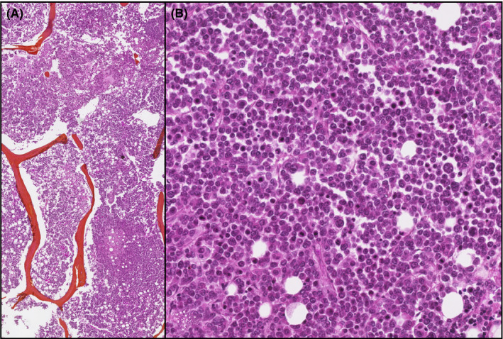
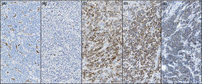
Autopsy findings included splenomegaly (484 g), hemorrhagic foci in lungs, heart, and gastrointestinal tract, and severe pulmonary edema and emphysema. Microscopic examination revealed infiltration of lungs, spleen, kidneys, liver, and lymph nodes by undifferentiated neoplastic cells. The bone marrow was hypercellular (90%) with >80% neoplastic erythroid precursors (Figure 1). Neoplastic cells expressed CD117, CD71, and E‐cadherin, but not CD45, CD34, MPO, TdT, pancytokeratin, and S100 (Figure 2). Diffuse TP53 immunohistochemical staining was demonstrated. A diagnosis of pure erythroid leukemia was made.
FIGURE 1.

Bone marrow shows a markedly hypercellular marrow (90%) (A, HE, ×20 objective), infiltrated by numerous monomorphous neoplastic cells of erythroid lineage (B, HE, ×200 objective)
FIGURE 2.

The neoplastic erythroblasts in the spleen were negative for CD34 (A, CD34, ×200 objective) and myeloperoxidase (B, myeloperoxidase, ×200 objective) and positive for CD117 (C, CD117, ×200 objective), CD71 (D, CD71, ×200 objective), and E‐cadherin (E, E‐cadherin, ×200 objective)
Pure erythroid leukemia (PEL) is a rare and aggressive form of acute leukemia with deleterious clinical course and median survival of <3 months. 1 PEL is of erythroid lineage without myeloblastic component, representing >80% of marrow cellularity, with ≥30% proerythroblasts. 2 It remains a challenging diagnosis that requires correlating clinical, pathological, and cytogenetic results.
CONFLICT OF INTEREST
The authors declare no potential conflicts of interest with respect to the research, authorship, and/or publication of this article. Informed consent was obtained for this publication.
AUTHOR CONTRIBUTIONS
MF: collected data and wrote the manuscript. SFR: wrote the manuscript. MH and AM: supervised the study and reviewed the manuscript.
ACKNOWLEDGMENTS
None. Written consent was obtained from next of kin, following institutional guidelines.
Forest M, Roy SF, Houde M, Maietta A. Pure erythroid leukemia. Clin Case Rep. 2020;8:3598–3599. 10.1002/ccr3.3316
Contributor Information
Méghan Forest, Email: meghan.forest@umontreal.ca.
Antonio Maietta, Email: antonio.maietta.chum@ssss.gouv.qc.ca.
REFERENCES
- 1. Reinig EF, Greipp PT, Chiu A, Howard MT, Reichard KK. De novo pure erythroid leukemia: refining the clinicopathologic and cytogenetic characteristics of a rare entity. Mod Pathol. 2018;31(5):705‐717. [DOI] [PubMed] [Google Scholar]
- 2. Swerdlow SH, Campo E, Pileri SA, et al. WHO Classification of Tumours of Haematopoietic and Lymphoid Tissues. Revised, 4th ed Lyon, France: IARC; 2017. [Google Scholar]


