
FIG. 2.
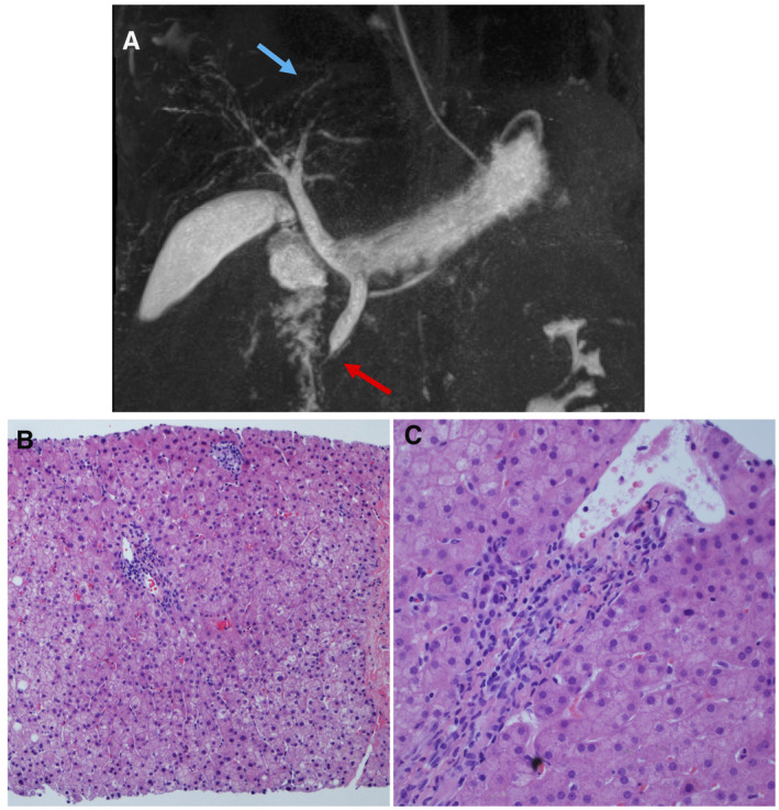
MR cholangiopancreatography and liver biopsy findings demonstrating sclerosing cholangitis. (A) MR cholangiopancreatography findings of KISC as noted by blue arrow highlighting intrahepatic biliary dilatation with a beaded appearance and red arrow pointing to a dilated common bile duct with distal narrowing. (B) Liver biopsy showing one small portal tract with mild bile duct injury/reactive changes, ductular proliferation, and one adjacent small lobular non‐necrotizing granuloma (HE, ×100). (C) Liver biopsy showing a portal tract with bile duct injury and minimal infiltration of neutrophils in the portal tract and lobule without marked cholestasis (HE, 40×).