Dear Editor,
Coronavirus disease 2019 (COVID‐19) is a pandemic infection owing to the Severe Respiratory Syndrome Coronavirus‐2 (SARS‐CoV‐2) that, in the worst case, can lead to fatal acute respiratory distress syndrome. It was first identified in hospitalized patients in Wuhan, China, in December 2019 and January 2020. 1 Since the start of the COVID‐19 epidemic, many clinical manifestations have been described outside of the respiratory tract, most of which are thought to be caused by inflammation and deleterious immune responses to the virus.
To date, several dermatological manifestations have been described during COVID‐19: petechial rash, erythematous rash, varicella‐like vesicles, urticarial eruption, 2 , 3 and livedo reticularis. Other dermatological manifestations of acral involvement have been published such as red‐purple papules mimicking chilblains disease, acro‐ischemia, and recently atypical erythema multiforme palmar plaques. 4 Cebeci F. et al. 5 first described the mucosal damage in COVID‐19.
Herein, we report on a case of severe and pure mucous damages in a COVID‐19‐positive inpatient. This case brings additional data implementing the knowledge on the dermatological signs during COVID‐19.
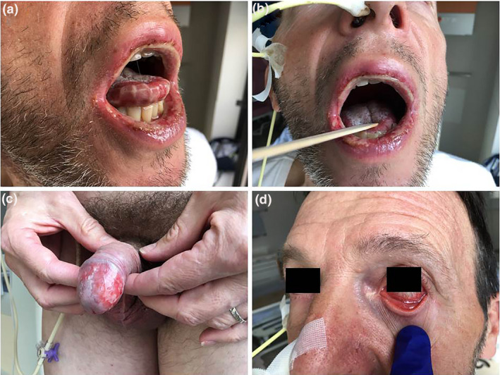
A 57‐year‐old man with no medical history had first complained of cough, headache, myalgia, and fever for 3 days, before all symptoms resolved spontaneously. Five days later, he presented to the emergency department with painful gingivostomatitis, fever (39°C), ageusia, and anosmia and was hospitalized as a result of complete inability to eat or drink. Physical examination found bilateral ulcers of the mouth (a and b) and glans (c), erythematous conjunctivitis (d), but no other lesion on the whole tegument: these findings were consistent with erythema multiforme major (Fig. 1). Biologically, there was a marked inflammatory syndrome (CRP = 237 mg/l, elevated ferritinemia = 20 times normal) associated with hepatic cytolysis (3–5 times normal) without jaundice. COVID‐19 infection was proven by amplifying the virus from a nasopharyngeal swab using polymerase chain reaction (PCR) at admission day (Day 0). A chest CT scan was found normal at this time. Swabs of the mouth ulcers, glans, and conjunctiva were negative for herpes simplex virus, varicella‐zoster virus, and COVID‐19. Human immunodeficiency virus antigen and antibodies were negative, cytomegalovirus and Epstein‐Barr virus serologies only found IgG, and mycoplasma pneumoniae nasopharyngeal polymerase chain reaction was negative. Research of antinuclear factors was not contributory. Amoxicillin/clavulanic acid and antiviral drugs were started at Day 1 resulting in a rapid improvement of the general condition, resumption of a liquid then mixed diet at Day 3 and normal diet at Day 6. Fever resolved at Day 5, whereas C‐reactive protein had fallen to 25 mg/l. The patient returned home at Day 8 with no treatment afterwards.
Figure 1.

Bilateral ulcers of the mouth (a and b) and glans (c), and erythematous conjunctivitis (d)
This case report provides an additional case of mucosal involvement during the course of COVID‐19; this is an erythema multiforme major case with the most widespread mucosal involvement we have seen so far. The search for other infections and diseases conventionally linked to erythema multiforme were negative. Noteworthy, mucosal damages appeared at a distance similar to flu‐like and respiratory symptoms. Moreover, there was no detectable SARS‐CoV‐2 in mucosal lesions (mouth, glans, and conjunctiva), although the virus was amplified from a nasopharyngeal swab (which test can be found positive weeks after COVID symptoms have stopped). In front of clinical history and semiology, the most obvious diagnosis was an erythema multiforme major with multiple mucosal involvements. Altogether, these findings suggest mucosal damages were more likely to result from deleterious immune responses towards self‐tissues rather than a cytopathic effect directly caused by the virus. Given the location of the lesions, we gave up doing biopsies to decipher the mechanism and eliminate differential diagnoses.
Physicians should be aware that erythema multiforme major can represent a skin manifestation of COVID‐19 and a circumstance where COVID‐19 should be suspected.
Acknowledgment
The patient in this manuscript has given written permission for the publication of their case details.
Conflict of interest: None.
Funding source: None.
References
- 1. Zhu N, Zhang D, Wang W, et al. A novel coronavirus from patients with pneumonia in China, 2019. N Engl J Med 2020; 382: 727–733. [DOI] [PMC free article] [PubMed] [Google Scholar]
- 2. Marzano AV, Genovese G, Fabbrocini G, et al. Varicella‐like exanthem as a specific COVID‐19‐associated skin manifestation: multicenter case series of 22 patients. J Am Acad Dermatol 2020; 83: 280–285. [DOI] [PMC free article] [PubMed] [Google Scholar]
- 3. Recalcati S. Cutaneous manifestations in COVID‐19: a first perspective. J Eur Acad Dermatol Venereol 2020; 34: e212–e213. [DOI] [PubMed] [Google Scholar]
- 4. Janah H, Zinebi A, Elbenaye J. Atypical erythema multiforme palmar plaques lesions due to Sars‐Cov‐2. J Eur Acad Dermatol Venereol 2020; 34: e373–e375. [DOI] [PMC free article] [PubMed] [Google Scholar]
- 5. Cebeci Kahraman F, Çaşkurlu H. Mucosal involvement in a COVID‐19‐positive patient: A case report. Dermatol Ther 2020; 33: e13797. 10.1111/dth.13797 [DOI] [PMC free article] [PubMed] [Google Scholar]


