Abstract
The antimicrobial capability of chitosan from Tenebrio molitor as compared with chitosan from crustacean (Penaeus monodon) on different pathogenic microorganisms of concern in food safety was studied. The antimicrobial effect was tested at pH 5 and pH 6.2 and at two different initial concentrations (103 or 106 CFU/mL). Results indicated that chitosan from both sources have antimicrobial activity, although the effect depended on the microorganism considered (Salmonella Typhimurium, Listeria monocytogenes and Escherichia coli O157:H7). Our results indicated that Salmonella was the most resistant bacteria, and that chitosan from insect was less active than chitosan from crustacean, especially against Salmonella. Another important factor on antimicrobial activity was the pH of the sample. When chitosan was added to a solution with a pH of 6.2 it was more active against Listeria and Escherichia coli, than at pH 5.00. Besides, the effect of chitosan appears to decrease with the incubation time, since some increases in counts were observed on E. coli and Salmonella after the 24 and 49 hours of incubation.
Introduction
Chitosan is a natural and biodegradable biopolymer that has been used in different industrial applications as flocculating and chelating agent, permeability control agent or antimicrobial substance, among others [1–3]. Today, chitosan is mainly produced on an industrial scale from deacetylation of chitin, chis is present in the exoskeleton of crustaceans and insects and in the cell walls of most fungi and some algae [4]. Although residues from crab, prawns, crayfish and shrimps are the main source of chitin [4], the importance of insect chitosan is due to the role that insects play as a sustainable source of protein. Insects are seen as an alternative to the proteins that are conventionally consumed and that come mainly from traditional livestock (cows, chickens or pigs fundamentally) and fish. Besides, the use of insect as protein source will produce two by-side products of undoubted industrial interest, lipids (30–40% total dry weight) [5], that could be used as biofuel and a residual material composed fundamentally by chitin which have some bioactive properties and from which chitosan can be obtained.
Chitin and chitosan have interesting physicochemical, biological and mechanical properties [6]. One of those properties of chitosan is related with its antimicrobial activity. There are diverse studies in which the antimicrobial and antifungal properties of chitosan and several of its derivatives have been revealed [7, 8]. More recently, the effect of the physical form of chitosan on its antibacterial activity against pathogenic bacteria has been studied [9]. Serio et al. [10] studied the chitosan coating as inhibitor of Listeria monocytogenes (L. monocytogenes) on vacuum-packed pork loins and Brown et al. [11] on fresh cheese. It has been also reported that the antibacterial action is usually rapid and eliminates bacteria in few hours [12].
As far as physical properties of chitosan, they are governed, fundamentally, by two factors; the degree of deacetylation and the molecular weight. Moreover, the natural origin as well as the variability on their chemical structure can affect the properties of chitosan and could impact on their industrial utilization [13]. Some studies have revealed that the degree of deacetylation has been associated with the antimicrobial activity of chitosan [14].
Pathogenic microorganisms as Listeria monocytogenes, Salmonella Typhimurium or Escherichia coli (E. coli) O157:H7 are of concern under the point of view of food safety. Those microorganisms are present in a broad range of foodstuffs contributing every year to a huge amount of food transmitted illnesses. In 2018 (the latest year for which EU data is available), a total of 5.146 disease outbreaks were identified, with bacteria and their toxins being the main causative agent. Campylobacter, Salmonella spp., Escherichia coli and Listeria monocytogenes are among the main pathogens linked to foodborne outbreaks in Europe (2011–2020) [15]. Salmonella enterica is generally acquired from contaminated food and is a common cause of human gastroenteritis and bacteremia worldwide [15–17]. The common reservoir of Salmonella is the intestinal tract of a wide range of domestic and wild animals, which results in a variety of foodstuffs covering both food of animal and plant origin as sources of infection. Transmission often occurs when organisms are introduced in food preparation areas and are allowed to multiply in food, e.g., owing to inadequate storage temperatures, inadequate cooking, or cross-contamination of ready-to-eat (RTE) food [18, 19]. Salmonella enterica subsp. enterica serovar Typhimurium (S. Typhimurium) is one of the most common serovars associated with clinically reported salmonellosis in humans, accounting for at least 15% of infections [20].
E. coli O157:H7 produces verotoxins. This bacterium is the major serotype that was recognized as a cause of human illness, but not in cattle, its primary host [21, 22]. The source of infection is the contamination of food such as raw or undercooked meat products and raw milk by human and animal feces [23]. The most common sources of Shiga toxin-producing E. coli (STEC) O157 infection are beef and leafy vegetables [24], but fresh-pressed apple juice or cider, yoghurt, cheese, salad vegetables, and cooked maize have also been implicated [25].
L. monocytogenes is an opportunistic pathogen that has been recognized as an important foodborne pathogen since the early 1980s [26]. It is resistant to diverse environmental conditions and can grow at temperatures as low as 3°C [27, 28]. L. monocytogenes can cause invasive disease in livestock, mainly sheep, goats, and cattle [29]. It is found in a wide variety of raw and processed foods such as milk and cheeses, meat (including poultry) and meat products, and seafood and fish products where it can survive and multiply rapidly during storage [30, 31].
Therefore, control measures should be taken to avoid foodborne outbreaks. Synthetic antimicrobials are each time more refused by the consumer worried by the toxicity of those products. In consequence, people are looking for new natural antimicrobials to help in controlling those pathogenic microorganisms. The control of those microorganisms can be carried out by using natural antimicrobials as chitosan to be used in different foodstuffs reducing in this way the annual cases of illnesses transmitted by foods [32–34].
Considering the previously exposed antecedents, a comparative study of the antimicrobial activity of chitosan from Tenebrio molitor as an alternative to crustacean chitosan at different pH levels against E. coli, L. monocytogenes and Salmonella enterica subsp. Enterica serovar Typhimurium at different inoculation sizes was carried out.
Materials and methods
Microbial strains
Pure cultures of Listeria monocytogenes serovar 4b (Spanish Type Culture Collection (CECT) 4032) (L. monocytogenes), Escherichia coli O157:H7 (CECT 5947) (E. coli) and Salmonella enterica serovar Typhimurium (CECT 443) (S. Typhimurium) were provided by the Spanish Type Culture Collection. Following the procedure described by Sanz-Puig et al. [35], lyophilized samples (0.5 g lyophil) were rehydrated in Tryptone Soy Broth (TSB) (Scharlab S.A., Barcelona, Spain) (L. monocytogenes and S. Typhimurium) or in Luria Broth (LB) (Scharlab S.A., Barcelona, Spain) (E. coli). Rehydrated cultures were transferred to 500 mL of its corresponding media and incubated in a bath shaker (200 rpm) for 14 h at 37°C. After centrifugation process, glycerinated bacteria cells were maintained in frozen cryovials at -80°C. The final inoculums concentration was determined by plate count and was of 108−109 colony forming units/mL (CFU/mL).
Chitosan samples preparation
Crustacean chitosan (chitosan from shrimp shells, practical grade) was purchased from Merck (MerckKGaA, Darmstadt, Germany) and insect chitosan (Tenebrio molitor) was acquired from MealFood Europe S.L. (Doñinos de Salamanca, Salamanca, Spain). For both chitosans, a 1% (w/v) chitosan sample stock solution was prepared in a 1% (v/v) acetic acid solution. Chitosan solutions at 0.15% (w/v) were prepared from the 1% (w/v) chitosan stock solution (crustacean or insect) in 1% (v/v) acetic acid. Each stock solution was sterilized by 0.45 μm pore size membrane filters.
Evaluation of antimicrobial activity of chitosan
The antimicrobial activity was tested at pH 5 and pH 6.2 by acidifying with acetic acid (1% w/v) LB for E. coli and TSB for L. monocytogenes and S. Typhimurium studies and at two different initial concentrations, 103 or 106 CFU/mL (3.2x10³ and 2.7x10⁶ CFU/mL, respectively). Microbial growth at these conditions was compared with the one of control samples (without chitosan). Acetic acid controls (LB and TSB at pH 5 and pH 6.2) were also tested to evaluate the possible antimicrobial effect of acidification.
In order to carry out the experiment, each microorganism maintained in cryovials at -80°C was thawed and serial dilutions in peptone water (0.1% (w/v)) were performed until the desired initial inoculum concentration was achieved (103 or 106 CFU/mL). Inoculated media (LB or TSB) were incubated in a bath shaker at 37° C for 49h and sampled at t0 = 0h, t1 = 3h, t2 = 6h, t3 = 8h, t4 = 24h and t5 = 49h. Samples removed at each time were serially diluted in peptone water (0.1%), plated by duplicate and incubated in Luria Agar (LA) (E. coli) or Tryptone Soy Agar (TSA) (S. Typhimurium and L. monocytogenes) at 37°C for 24h (E. coli and S. Typhimurium) or 48h (L. monocytogenes). After the incubation period, bacteria cells were counted (CFU/mL). Experimental results were shown as log10 of survival fraction (log S) calculated as (Eq 1):
| (1) |
where N is bacterial concentration (CFU/mL) at time t (h) and N0 initial bacterial concentration (CFU/mL) (t0). Therefore, positive values indicate microbial growth and negative values microbial inactivation respect the initial inoculation value (N0).
Statistical analysis
For each condition, three independent repetitions were performed and for each sampling time, two duplicates were taken, plating each one in duplicate (4 plates/sampling time). The analysis of experimental data was carried out by using Statgraphics Centurion XVI (Statpoint Technologies, Inc., USA). Outliers were identified and removed prior data analysis. The statistical significance of data was determined by an Analysis of Variance (ANOVA) (p < 0.05) and differences between groups were determined by Tukey test.
Results
Factors affecting antimicrobial activity of chitosan solutions
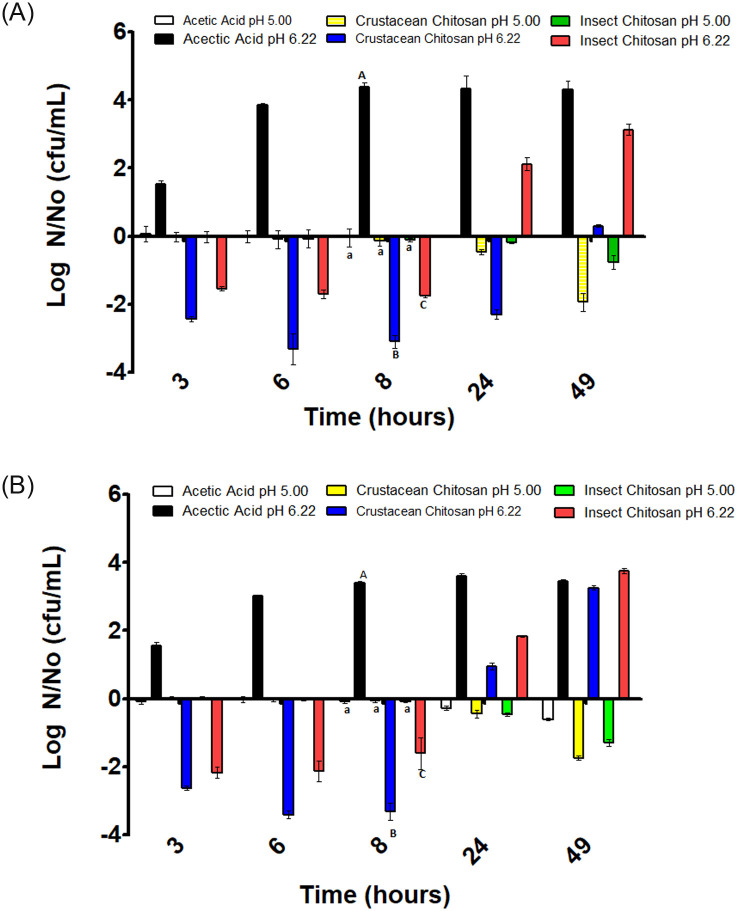
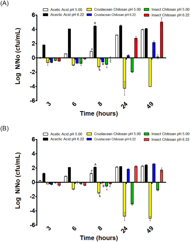
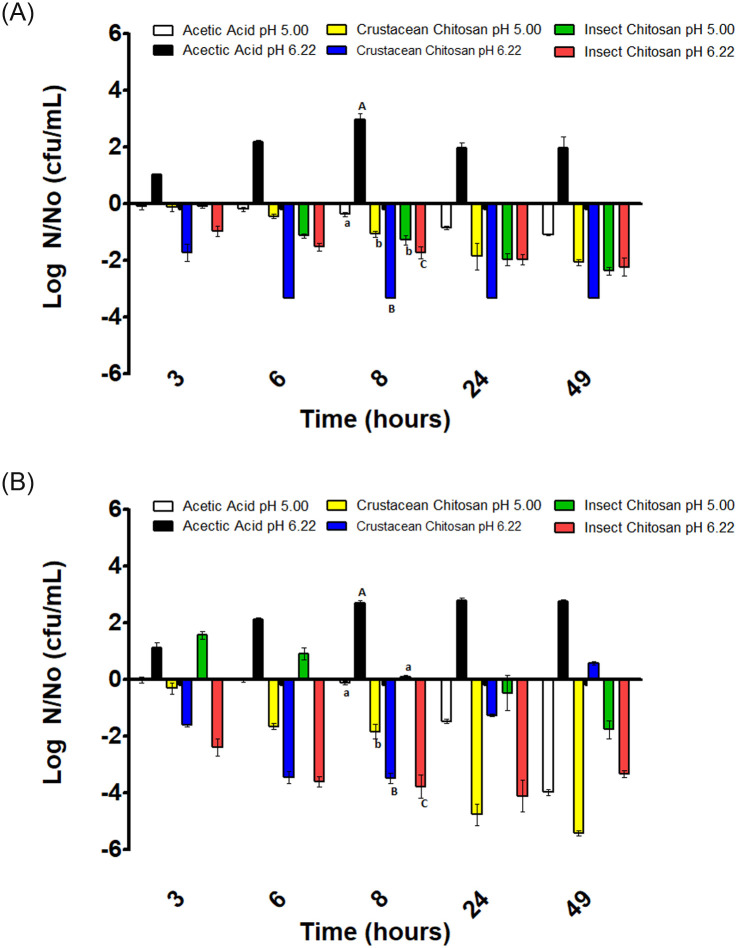
Results on the effect of acetic acid and chitosan from different sources on the behavior of E. coli, L. monocytogenes and S. Typhimurium can be seen in Figs 1–3. In general, the response of tested microorganisms to the different antimicrobials and pH conditions was microorganism dependent. To determine the possible antimicrobial effect of chitosan from different sources dissolved in acetic acid, acetic acid control solutions at pH 5 and 6.2 were tested as antimicrobials for comparison purposes and to define the impact of chitosan independently of the antimicrobial activity of acetic acid used to dissolve chitosan.
Fig 1. Variation of E. coli concentration in substrates inoculated with 103 CFU/mL (A) and 106 CFU/mL (B).
Positive values indicate growth and negative values inactivation respect the initial inoculation value (N0). Different letters mean significant differences between samples of the same pH.
Fig 3. Variation of S. Typhimurium concentration in substrates inoculated with 103 CFU/mL (A) and 106 CFU/mL (B).
Positive values indicate growth and negative values inactivation respect the initial inoculation value (N0). Different letters mean significant differences between samples of the same pH.
Antimicrobial effects of chitosan solutions on E. coli
For E. coli studies with an initial contamination level of 103 CFU/mL, the acetic acid solution at pH 5 resulted in a non-substantial variation on microbial counts during the whole incubation period, 49 h (Fig 1A). A similar result was observed when the inoculum size was 106 CFU/mL (Fig 1B). As for pH 6.2, E. coli cells grew until reach a plateau at the 8 hour of incubation; probably due to the pH 6.2 represent a more favorable environment for the microorganism (Fig 1A). For an initial inoculum size of 106 CFU/mL the behavior of cells was similar than that at the lower initial microbial concentration. Cells grew until reach the stationary phase at the 8 hours of incubation (Fig 1B).
Regarding the effect of chitosan from crustacean and insect sources at pH 5 against E. coli, non-appreciable variation on the microbial counts was observed along the incubation period, except at the 49 hours of incubation a light reduction on the microbial counts was observed, being that reduction highest for crustacean chitosan than for insect chitosan (Fig 1A and 1B). Although, this reduction could be due to the decline phase after a stationary phase and not to the presence of chitosan. As for the effect at pH 6.2, the addition of chitosan produced a decrease in the number of cells that was highest in the case of chitosan from crustacean than from insect. However, after 8 hours of incubation, an increase in the microbial counts was observed until the 49 hours of incubation, being the level of microbial concentration higher than one at time zero.
In general, considering both pH levels, the behavior of E. coli was independently of the initial inoculum size. In consequence, it can be considered that the inoculum size did not affect the microbial pattern along the incubation time.
Antimicrobial effects of chitosan solutions on L. monocytogenes
Results for L. monocytogenes can be seen in Fig 2A and 2B for a 103 and 106 CFU/mL initial concentration level, respectively.
Fig 2. Variation of L. monocytogenes concentration in substrates inoculated with 103 CFU/mL (A) and 106 CFU/mL (B).
Positive values indicate growth and negatives inactivation respect the initial inoculation value (N0). Different letters mean significant differences between samples of the same pH.
Acetic acid solution at pH 5 produced a decrease in the microbial counts during the incubation period regardless the initial concentration level of L. monocytogenes. Nevertheless, the number of log reductions was higher when the inoculum size was 106 CFU/mL (Fig 2A and 2B). This result appears to indicate that there is a clear bactericidal effect of the acetic acid against this microorganism. On the other hand, when L. monocytogenes cells were treated with acetic acid at a pH of 6.2, cells grew reaching the stationary phase after 8 hours of incubation. Results indicated that no bactericidal effect of acetic acid at pH 6.2 took place as compared with the bactericidal effect observed at pH 5.
When L. monocytogenes cells were subjected to the action of crustacean chitosan in acetic acid at pH 5, a decrease in bacterial counts was observed throughout the incubation period with respect to the initial inoculum, regardless of inoculum size. Nevertheless, the inactivation effect was stronger for the inoculum size of 106 CFU/mL, and no viable cells were observed at the incubation time of 49 hours (Fig 2B).
The effect of crustacean chitosan in acetic acid at pH 6.2 can also be observed in Fig 2A and 2B. For the inoculum size of 103 CFU/mL a rapid reduction in the number of viable cells was observed such that at the 6 hour of incubation no viable cells were observed in the culture. This decrease in cells counts was highest than in acetic acid alone at pH 5. For the inoculum size of 106 CFU/mL, the effect was similar although there is a slight decreasing of the antimicrobial effect that produced a small growth in the number of cells at the 49 hour of incubation.
In relation to insect chitosan in acetic acid, the behavior was similar to that observed for crustacean chitosan at both pH and inoculum size values. There was a decrease in the number of cells in comparison with the initial inoculum concentration. Nevertheless, according to the results that can be seen in Fig 2A and 2B, the antimicrobial activity of the crustacean chitosan is somewhat greater than showed by the insect chitosan.
Antimicrobial effects of chitosan solutions on S. Typhimurium
Results of the evolution of S. Typhimurium in the different substrates with initial inoculation levels of 103 CFU/mL and 106 CFU/mL are shown in Fig 3A and 3B respectively.
As can be seen in Fig 3 for both inoculum sizes, there is a cell growth in the acetic acid substrate at pH values of 5 and 6.2. This means that acetic acid at these pH values of the study does not seem to have bacteriostatic or bactericidal effect against Salmonella Typhimurium.
The crustacean chitosan in acetic acid at pH 5 produced a reduction of the microbial load along the incubation period of 49 hours for the two initial inoculation levels. It appears that there was a synergistic effect among acetic acid and crustacean chitosan in acetic acid at this pH level. At pH 6.2, a slight decrease followed by a latency period was observed that lasted about 8 hours followed of an increase in the number of cells at hours 24 and 49 regardless of inoculum size.
The insect chitosan in acetic acid at pH 5 and with an inoculation level of 103 CFU/mL produced a reduction in the number of viable cells at 24 hours of incubation period followed by a slight growth at 49 hours of incubation period. In the case of the inoculum level of 106 CFU/mL a reduction of 3 log cycles was observed at 24 hours of incubation, but some growth appears to take place at 49 hours of incubation period manifested as a decrease of the log reduction cycles (Fig 3B).
At pH 6.2 a latency phase was observed that lasted until 8 hours of incubation, followed by an increase in the number of cells exceeding the level of initial inoculation. This behavior was similar for both initial inocula concentrations.
Antimicrobial effects of chitosan solutions on E. coli, L. monocytogenes and S. Typhimurium after 8 hours of incubation
Tables 1–3 show the concentrations of microorganisms after 8 hour of incubation period at different pH values and inoculum concentrations for each microorganism considered in the study. The time of 8 hours of incubation was considered a good check point for comparison because it was the moment at which the stationary phase was reached with acetic acid at pH 6.2 used as control for comparison purpose.
Table 1. E. coli behaviour after 8 hours of incubation in different substrates (mean ± standard deviation).
| Substrate | Log N/N0 (CFU/mL) | |||
|---|---|---|---|---|
| Inoculum size 103 | Inoculum size106 | |||
| Control (LB pH = 7) | 5.47± 0.075 A | 3.31±0.018 A | ||
| pH 5.0 | pH 6.2 | pH 5.0 | pH 6.2 | |
| Acetic Acid Control | -0.11±0.126 B | 4.39±0.041 B | -0.09±0.004 B | 3.34±0.033 A |
| Crustacean chitosan | -0.12±0.053 B | -3.09±0.062 C | -0.06±0.026 B | -3.3±0.080 B |
| Insect chitosan | -0.11±0.017 B | -1.76±0.012 D | -0.07±0.0236 B | -1.46±0.045 C |
Figures with the same letter are not significantly different by columns. Different letters indicate significant differences (p value≤0.05). Negative figures mean inactivation and positive figures mean growth.
Table 3. Salmonella Typhimurium behaviour after 8 hours of incubation in different substrates (mean ± standard deviation).
| Substrate | Log N/N0 (CFU/mL) | |||
|---|---|---|---|---|
| Inoculum size 103 | Inoculum size106 | |||
| Control (TSB pH = 7) | 4.47±0.0566 A | 2.68±0.05 A | ||
| pH 5.0 | pH 6.2 | pH 5.0 | pH 6.2 | |
| Acetic Acid Control | 0.95±0.0236 B | 4.42±0.07 A | 1.29±0.018 B | 2.12±0.046 B |
| Crustacean chitosan | -1.28±0.045 D | -0.57±0.012 C | -1.51±0.0804 D | -0.02±0.105 C |
| Insect chitosan acid | -0.90±0.028 C | -0.04±0.073 B | -0.55±0.043 C | -0.22±0.018 D |
Figures with the same letter are not significantly different by columns. Different letters indicate significant differences (p value≤0.05). Negative figures mean inactivation and positive figures mean growth.
Table 1 shows the E. coli microbial counts after 8h of incubation in blank control, acid control and chitosan treatments. According to the table, significant differences (p≤0.05) on bacterial counts were found between the blank control and the rests of substrates except for the inoculation level 106 and pH 6.2. Besides, at pH 6.2 there were significant differences (p≤0.05) on counts among acetic acid, crustacean chitosan and insect chitosan substrates in the way that all chitosan treatments reduced the microbial load at pH 6.2 compared to acetic acid or blank controls (p≤0.05). In addition, crustacean chitosan showed a greater microbial reduction than insect chitosan.
As for L. monocytogenes, Table 2 shows bacterial counts after 8h of incubation in blank control and the studied substrates. Acetic acid control produced a small reduction of bacteria cells independent of inoculum size at pH 5 as compared with the blank control. Similarly to E. coli behaviour (Table 1), chitosan treatments at pH 6.2 showed greater Listeria cell reductions than at pH 5, being the maximum reduction achieved with insect chitosan at pH 6.2 for an inoculum size of 106 CFU/mL (3.78 log reductions).
Table 2. Listeria monocytogenes behaviour after 8 hours of incubation in different substrates (mean ± standard deviation).
| Substrate | Log N/N0 (CFU/mL) | |||
|---|---|---|---|---|
| Inoculum size 103 | Inoculum size106 | |||
| Control (TSB pH = 7) | 4.27±0.0269 A | 2.98±0.0170 A | ||
| pH 5.0 | pH 6.2 | pH 5.0 | pH 6.2 | |
| Acetic Acid Control | -0.37±0.023 B | 2.96±0.11 B | -0.11±0.020 B | 2.73±0.046 B |
| Crustacean chitosan | -1.07±0.036 C | ND | -1.85±0.020 C | -3.48±0.062 C |
| Insect chitosan | -1.28±0.057 C | -1.73±0.0698 C | -0.013±0.049 B | -3.78±0.136 D |
ND = Non Detectable. Figures with the same letter are not significantly different by columns. Different letters indicate significant differences (p value≤0.05).
Negative figures mean inactivation and positive figures mean growth.
Table 3 shows the S. Typhimurium bacterial counts after 8h of incubation in blank control and the studied substrates. As can be seen, acetic acid control media resulted in an increase of microbial counts at all pH and inoculum concentrations. Regarding Salmonella cells exposed to chitosan, small reductions were observed in all cases, at 8 hour of incubation, showing significantly differences (p≤0.05) among them. According to the table, those reductions in Salmonella cells were dependent on the source of chitosan, pH and inoculation level. In general, crustacean chitosan was more effective against Salmonella than insect chitosan excepting at an inoculum size of 106 CFU/mL and pH 6.2 (Table 3).
Discussion
The effect of chitosan as an antimicrobial in agriculture and the food industry has been studied for some time ago. Allan et al. [36] already studied the effect of chitosan on E. coli and Staphylococcus aureus (S. aureus). However recently, and as a consequence of the use of minimum preservation procedures, the interest for the antimicrobial capability of this compound obtained from chitin has been accentuated. According to previous studies carried out by other authors, the antimicrobial activity of chitosan is influenced by several extrinsic and intrinsic factors (type of chitosan, molecular weight, degree of deacetylation, solvent and concentration) as well as some environmental factors such as microorganism specie, its physiological state, pH, temperature, ionic strength, metal ions, the presence of ethylenediaminetetraacetic acid (EDTA) or organic matter [37–40].
Chitosan is a biopolymer primarily commercially produced from crabs and shrimp residues. The physicochemical characteristics of chitosan influence its functional properties, which differ between species of crustaceans and methods of preparation [41]. In the present work, chitosan from crustaceans and insects have been studied. The objective was to compare the functionality as antimicrobials of commercial products of crustacean and insect chitosan. Results pointed out differences between the antimicrobial capacity of commercial crustacean chitosan and commercial insect chitosan. In general, at 49 hours of incubation period, the crustacean chitosan at pH 5.0 showed greater antimicrobial capacity than the insect chitosan at the same pH value. This behavior was mainly seen in the case of Salmonella where the crustacean chitosan produced more than 4 logarithmic reductions while the insect chitosan was bacteriostatic or produced around 1 logarithmic reduction. The same behavior is observed for E. coli although the differences in the antimicrobial activity are smaller than in the case of Salmonella. As indicated above, some previous studies have indicated that there may be differences between functional capacity and physical characteristics of chitosan from different crustaceans. This behavior could be more evident among chitosan from sources as diverse as crustaceans and insects.
The pH plays an important role in the antimicrobial capability of chitosan in such a way that the antimicrobial activity is inversely affected by the pH value; at low pH the chitosan seems more antimicrobial than at high pH values [38, 42–44].
Wang [45] conducted a study on the effect of different chitosan concentrations at two levels of pH 6.5 and 5.5 on different pathogenic microorganisms including S. Typhimurium, E. coli and L. monocytogenes. The author concluded that chitosan at pH 6.5 had a very weak effect on pathogenic microorganisms; in fact, there was no inhibition of L. monocytogenes. At pH 5.5 there was inhibition of the microorganisms tested between 24 and 72 hours of storage at 30°C. The author concluded that chitosan acts better at pH 5.5 than at pH 6.5. More recently, Gücükoğlu [46] also studied the antibacterial activity of chitosan of different molecular weights at various pH levels (pH 4, 4.5 and 5) on L. monocytogenes strains. Results indicated that the pH 5 showed also the greatest bacterial reduction effect at 24 hours of the incubation period excepting for two L. monocytogenes strains.
In the present work two pH levels were tested at a 0.15% (w/v) concentration of chitosan. After an incubation period of 8 hours, the antimicrobial effect was higher at pH 5.00 than at pH 6.2 for Salmonella Typhimurium, while for E coli and Listeria occurred the contrary, chitosan at pH 6.2 had a stronger antimicrobial effect than at pH 5.00. It appears that the effect of chitosan at both pH levels was microorganism dependant.
In our study, all microorganisms grew in the control at pH 7 reaching the stationary phase in many cases at 8 hours of the incubation period. The effect of control (acetic acid) at pH 5.0 appears to be microorganism dependent, it was bacteriostatic for E. coli, bactericidal for L. monocytogenes and in the case of S. Typhimurium there was a growth similar to that achieved in the control (acetic acid) at pH 6.2 after 49 hours of incubation. These results seem to indicate that the acetic acid control at pH 5.0 has an antimicrobial effect only on some microorganisms. Regarding the chitosan at the two pH levels, differences were observed regarding the control in acetic acid. Chitosan showed a clear antimicrobial activity, especially in L. monocytogenes at both pH values. Chitosan from both sources, crustacean and insect, at pH 5.0 were bacteriostatic or bactericidal for the three pathogenic microorganisms studied at the 49 hours of the incubation period, while for the same period of time, growth was observed on chitosan at pH 6.2, except on L. monocytogenes were still chitosan at pH 6.2 had a bactericidal effect. These results are in general in agreement with those obtained by Wang [45] who indicated that all chitosan concentrations tested at pH 5.5 had a greater antimicrobial effect than at pH 6.5 excepting some chitosan concentrations against Samonella. Gücükoğlu [46] also observed a general greater antimicrobial effect on the pH 5 tested on L. monocytogenes strains in spite of a different inoculum concentration was used in his study (2.0x10⁵ CFU/mL) compared with our study.
According to the results, it appears that the effect of chitosan was microorganism dependent; maybe differences in the membrane of the different microorganisms could affect the antimicrobial activity of chitosan. There are some hypotheses about the antimicrobial mechanism of chitosan. The ionic interactions between positive charges of amino groups and negative bacteria surface molecules in acid conditions alter the membrane permeability leading to cellular lysis [40, 47]. Another mechanism could be the interaction with essential nutrients for bacteria [48].
The bacterial inoculum size could also affect the bactericidal effect of chitosan [49]. Those authors indicated that after 4 hours of incubation for an inoculum size of 103 cells/mL, all tested compounds proved to be bactericidal at any tested chitosan concentration; while for higher concentration of initial inoculum, 0.1% (w/v) of chitosan was only bacteriostatic; they also concluded that, independently of the inoculum level, 0.25% (w/v) of any chito-oligosaccharide mixture was sufficient to reduce the E. coli initial population by at least 3 log cycles. However, results of the present work, as indicated above in results section, are not conclusive regarding the effect of the inoculum size because it varied according to pH and microorganism specie; in consequence it is not possible to establish that in all cases there is a higher antimicrobial activity at one particular inoculum size.
Conclusions
In the present study, both chitosans showed antimicrobial activity, although their effect depended on the pH and the microorganism studied. L. monocytogenes was the most sensitive at both pH values. Crustacean chitosan seemed more active than insect for the microorganisms studied and at the pHs tested, specially against Salmonella. However, more studies on the characterization of both chitosan compounds would be necessary to interpret these differences.
For the pHs studies, after 8h of incubation antimicrobial activity seems to be microorganisms’ dependent; the antimicrobial effect was higher at pH 5.00 than at pH 6.2 for Salmonella, while for E. coli and Listeria occurred the contrary. As for inoculum size, no clear conclusions can be drawn on the antimicrobial effect of the chitosan compounds studied.
Supporting information
(XLSX)
Acknowledgments
The authors would like to thank Mr. Karel Clapshaw for his assistance in correcting the manuscript’s English.
Data Availability
All relevant data are within the manuscript and its Supporting information files.
Funding Statement
The authors are grateful to the Spanish Ministry of Science for funding through project AGL2017-86840-C2-2-R, and to FEDER funds from EU. The funders had no role in study design, data collection and analysis decision to publish, or preparation of the manuscript.
References
- 1.Rajoka MSR, Mehwish HM, Wu Y, Zhao L, Arfat Y, Majeed K, et al. Chitin/chitosan derivatives and their interactions with microorganisms: a comprehensive review and future perspectives. Crit Rev Biotechnol. 2020;40(3):365–79. 10.1080/07388551.2020.1713719 [DOI] [PubMed] [Google Scholar]
- 2.Rehman A, Jafari SM, Aadil RM, Assadpour E, Randhawa MA, Mahmood S. Development of active food packaging via incorporation of biopolymeric nanocarriers containing essential oils. Trends Food Sci Technol. 2020;101:106–21. [Google Scholar]
- 3.Tian B, Liu Y. Chitosan-based biomaterials: From discovery to food application. Polym Adv Technol [Internet]. n/a(n/a). https://onlinelibrary.wiley.com/doi/abs/10.1002/pat.5010 [Google Scholar]
- 4.Hu Z. Challenges and opportunities related to the use of chitosan as food preservative. J Appl Microbiol. 2018;126 10.1111/jam.14131 [DOI] [PubMed] [Google Scholar]
- 5.Barroso FG, de Haro C, Sánchez Muros M-J, Venegas E, Martínez-Sánchez A, Pérez-Bañón C. The potential of various insect species for use as food for fish. Aquac. 2014;422–423:193–201. [Google Scholar]
- 6.Kasaai MR. A review of several reported procedures to determine the degree of N-acetylation for chitin and chitosan using infrared spectroscopy. Carbohydr Polym. 2008;71(4):497–508. [Google Scholar]
- 7.Du Y, Zhao Y, Dai S, Yang B. Preparation of water-soluble chitosan from shrimp shell and its antibacterial activity. Innov Food Sci Emerg Technol. 2009;10(1):103–7. [Google Scholar]
- 8.Kulawik P, Jamróz E, Ozogul F. Chitosan role for shelf-life extension of seafood. Environ Chem Lett. 2019; [Google Scholar]
- 9.Ardila N, Daigle F, Heuzey M-C, Ajji A. Effect of Chitosan Physical Form on Its Antibacterial Activity Against Pathogenic Bacteria. J Food Sci. 2017;82 10.1111/1750-3841.13635 [DOI] [PubMed] [Google Scholar]
- 10.Serio A, Lòpez C, Sacchetti G, Rossi C, Paparella A. Chitosan Coating Inhibits the Growth of Listeria monocytogenes and Extends the Shelf Life of Vacuum-Packed Pork Loins at 4 • C. Foods. 2018;7. [DOI] [PMC free article] [PubMed] [Google Scholar]
- 11.Brown SRB, Kozak SM, D’Amico DJ. Applications of Edible Coatings Formulated with Antimicrobials Inhibit Listeria monocytogenes Growth on Queso Fresco. Front Sustain Food Syst. 2018;2:1. [Google Scholar]
- 12.Gerasimenko D, Avdienko I, Bannikova G, Zueva O, Varlamov V. Antibacterial Effects of Water-Soluble Low-Molecular-Weight Chitosans on Different Microorganisms. Prikl Biokhim Mikrobiol. 2004;40:301–6. [PubMed] [Google Scholar]
- 13.Ibitoye E, Idris L, Mohd Noor MH, Goh Y, Abu Bakar MZ, Jimoh A. Extraction and Physicochemical Characterization of Chitin and Chitosan Isolated from House Cricket. Biomed Mater. 2018;13 10.1088/1748-605X/aa9dde [DOI] [PubMed] [Google Scholar]
- 14.Shin C-S, Kim D-Y, Shin W-S. Characterization of chitosan extracted from Mealworm Beetle (Tenebrio molitor, Zophobas morio) and Rhinoceros Beetle (Allomyrina dichotoma) and their antibacterial activities. Int J Biol Macromol. 2019. March;125:72–77. 10.1016/j.ijbiomac.2018.11.242 [DOI] [PubMed] [Google Scholar]
- 15.European Food Safety Authority and European Centre for Disease Prevention and Control (EFSA and ECDC). The European Union One Health 2018 Zoonoses Report. EFSA J. 2019;17(12):e05926 10.2903/j.efsa.2019.5926 [DOI] [PMC free article] [PubMed] [Google Scholar]
- 16.Xu X, Chen Y, Pan H, Pang Z, Li F, Peng X, et al. Genomic characterization of Salmonella Uzaramo for human invasive infection. Microb Genomics. 2020;6 10.1099/mgen.0.000401 [DOI] [PMC free article] [PubMed] [Google Scholar]
- 17.Ed-Dra A, Rhazi Filali F, Karraouan B, el Allaoui A, Aboulkacem A, Bouchrif B. Prevalence, molecular and antimicrobial resistance of Salmonella isolated from sausages in Meknes, Morocco. Microb Pathog. 2017;105:340–5. 10.1016/j.micpath.2017.02.042 [DOI] [PubMed] [Google Scholar]
- 18.Liu Q, Chen W, Elbediwi M, Pan H, Wang L, Zhou C, et al. Characterization of Salmonella Resistome and Plasmidome in Pork Production System in Jiangsu, China. Front Vet Sci. 2020;7:617 10.3389/fvets.2020.00617 [DOI] [PMC free article] [PubMed] [Google Scholar]
- 19.Yang X, Huang J, Wu Q, Zhang J, Liu S, Guo W, et al. Prevalence, antimicrobial resistance and genetic diversity of Salmonella isolated from retail ready-to-eat foods in China. Food Control. 2016;60:50–6. [Google Scholar]
- 20.Scallan E, Hoekstra M, Angulo F, Tauxe R, Widdowson M-A, Roy S, et al. Foodborne Illness Acquired in the United States—Major Pathogens. Emerg Infect Dis. 2011;17:7–15. 10.3201/eid1701.p11101 [DOI] [PMC free article] [PubMed] [Google Scholar]
- 21.European Food Safety Authority (EFSA). Monitoring of verotoxigenic Escherichia coli (VTEC) and identification of human pathogenic VTEC types. The Efsa Journal; 2007 p. 1–61. Report No.: 579.
- 22.Mora A, Herrrera A, López C, Dahbi G, Mamani R, Pita J, et al. Characteristics of the Shiga-toxin-producing enteroaggregative Escherichia coli O104:H4 German outbreak strain and of STEC strains isolated in Spain. Int Microbiol Off J Span Soc Microbiol. 2011;14:121–41. 10.2436/20.1501.01.142 [DOI] [PubMed] [Google Scholar]
- 23.Buchanan R, Doyle MP. Foodborne disease significance of Escherichia coli O157:H7 and other enterohemorrhagic E. coli. Food Technol. 1997;51:69–76. [Google Scholar]
- 24.Painter J, Hoekstra M, Ayers T, Tauxe R, Braden C, Angulo F, et al. Attribution of Foodborne Illnesses, Hospitalizations, and Deaths to Food Commodities by using Outbreak Data, United States, 1998–2008. Emerg Infect Dis. 2013;19:407–15. 10.3201/eid1903.111866 [DOI] [PMC free article] [PubMed] [Google Scholar]
- 25.Van de Venter T. Emerging foodborne diseases: a global responsibility. Food Nutrit Agric. 2000;26:4–13. [Google Scholar]
- 26.Farber J, Peterkin PI. Listeria monocytogenes, a food-borne pathogen. Microbiol Rev. 1991;55: 476–511. [DOI] [PMC free article] [PubMed] [Google Scholar]
- 27.Agencia Española de Seguridad Alimentaria y Nutrición (AESAN). Informe del Comité Científico de la Agencia Española de Seguridad Alimentaria y Nutrición (AESAN) en relación a los estudios de vida útil para Listeria monocytogenes en determinados productos alimenticios. 2011 p. 43–63. Report No.: 14.
- 28.Luber P, Crerar S, Dufour C, Farber J, Datta A, Todd E. Controlling Listeria monocytogenes in ready-to-eat foods: Working towards global scientific consensus and harmonization—Recommendations for improved prevention and control. Food Control. 2011;22: 1535–49. [Google Scholar]
- 29.Muñoz ÁB, Chaves JA, Rodríguez EC, Realpe ME. Listeria monocytogenes en manipuladores de alimentos: un nuevo enfoque para tener en cuenta en los peligros de la industria alimentaria. Biomédica. 2013;33:283–91. [PubMed] [Google Scholar]
- 30.Andreoletti O, Budka H, Buncic S, Colin P, Collins J, De A. Request for updating the former SCVPH opinion on Listeria monocytogenes risk related to ready-to-eat foods and scientific advice on different levels of Listeria monocytogenes in ready-to-eat foods and the related risk for human. EFSA J. 2007;599:1–42. [Google Scholar]
- 31.WHO (World Health Organization) /Food and Agriculture Organization (FAO). Risk assessment of Listeria monocytogenes in ready-to-eat Foods. Microbiol Risk Assess Ser. 2004;4.
- 32.Ibáñez-Peinado D, García-Carrión G, Martínez A, Rodrigo D. In vivo Antimicrobial Activity Assessment of a Cauliflower By-Product Extract Against Salmonella Typhimurium. Front Sustain Food Syst. 2020;4. [Google Scholar]
- 33.Yilmaz Atay H. Antibacterial Activity of Chitosan-Based Systems In: Jana S, Jana S, editors. Functional Chitosan: Drug Delivery and Biomedical Applications [Internet]. Singapore: Springer Singapore; 2019. p. 457–89. 10.1007/978-981-15-0263-7_15 [DOI] [Google Scholar]
- 34.Goy RC, de Britto D, Assis OBG. A review of the antimicrobial activity of chitosan. Polímeros. 2009;19: 241–7. [Google Scholar]
- 35.Sanz-Puig M, Moreira A, Torres C, Beltran J, Cunha L, Martínez A, et al. Resistance changes in Salmonella enterica serovar Typhimurium treated by High Hydrostatic Pressure and Pulsed Electric Fields and assessment of virulence changes by using Caenorhabditis elegans as a test organism. Innov Food Sci Emerg Technol. 2019;51:51–6. [Google Scholar]
- 36.Allan G. G., Altman L. C., Bensinger R. E., Ghosh D. K., Hirabayshi Y., Neogi S. Biomedical application of chitin and chitosan In: Chitin, chitosan and related enzymes. Orlando, Florida: Academic Press, Inc; 1984. p. 119–33. (Zikakis, J. P.; vol. 1). [Google Scholar]
- 37.Chaudhary S, Kumar S, Kumar V, Sharma R. Chitosan nanoemulsions as advanced edible coatings for fruits and vegetables: Composition, fabrication and developments in last decade. Int J Biol Macromol. 2020;152:154–70. 10.1016/j.ijbiomac.2020.02.276 [DOI] [PubMed] [Google Scholar]
- 38.No HK, Park NY, Lee SH, Meyers SP. Antibacterial activity of chitosans and chitosan oligomers with different molecular weights. Int J Food Microbiol. 2002;74(1):65–72. 10.1016/s0168-1605(01)00717-6 [DOI] [PubMed] [Google Scholar]
- 39.Benbettaïeb N, Karbowiak T, Debeaufort F. Bioactive edible films for food applications: Inluence of the bioactive compounds on film structure and properties. Crit Rev Food Sci Nutr. 2017;59:00–00. [DOI] [PubMed] [Google Scholar]
- 40.Rabea E, Badawy M, Stevens C, Smagghe G, Steurbaut W. Chitosan as Antimicrobial Agent: Applications and Mode of Action. Biomacromolecules. 2003;4:1457–65. 10.1021/bm034130m [DOI] [PubMed] [Google Scholar]
- 41.Cho YI, No HK, Meyers SP. Physicochemical Characteristics and Functional Properties of various Commercial Chitin and Chitosan Products. Products J of Agr.l and Food Chemi. 1998;46(9): 3839–43. [Google Scholar]
- 42.Tsai GJ, Su WH. Antibacterial Activity of Shrimp Chitosan against Escherichia coli. J Food Prot. 1999;62(3): 239–43. 10.4315/0362-028x-62.3.239 [DOI] [PubMed] [Google Scholar]
- 43.Jumaa M, Furkert FH, Müller BW. A new lipid emulsion formulation with high antimicrobial efficacy using chitosan. Eur J Pharm Biopharm. 2002;53(1):115–23. 10.1016/s0939-6411(01)00191-6 [DOI] [PubMed] [Google Scholar]
- 44.Raafat, D. Evaluation of the effect of chitosan on the activity of some antimicrobial agents. [Master Thesis]. [Alexandria, Egypt]: University of Alexandria; 2004.
- 45.Wang GH. Inhibition and inactivation of five species of foodborne pathogens by chitosan. J Food Prot. 1992. November;55(11):916–919. 10.4315/0362-028X-55.11.916 [DOI] [PubMed] [Google Scholar]
- 46.Gücükoğlu A, Yildirim Y, Terzi G, Erdem, Sireli. In vitro effects of chitosan on the survival of Listeria monocytogenes. Vet Med. 2016;13:11–8. [Google Scholar]
- 47.Goy RC, Assis OBG. Antimicrobial analysis of films processed from chitosan and N,N,N-trimethylchitosan. Braz J Chem Eng. 2014;31:643–8. [Google Scholar]
- 48.Jia Z, shen D, Xu W. Synthesis and antibacterial activities of quaternary ammonium salt of chitosan. Carbohydr Res. 2001;333(1):1–6. 10.1016/s0008-6215(01)00112-4 [DOI] [PubMed] [Google Scholar]
- 49.Fernandes J, Tavaria F, Soares J, Ramos O, Monteiro M, Pintado M, et al. Antimicrobial effects of chitosans and chitooligosaccharides, upon Staphylococcus aureus and Escherichia coli, in food model systems. Food Microbiol. 2008;25:922–8. 10.1016/j.fm.2008.05.003 [DOI] [PubMed] [Google Scholar]
Associated Data
This section collects any data citations, data availability statements, or supplementary materials included in this article.
Supplementary Materials
(XLSX)
Data Availability Statement
All relevant data are within the manuscript and its Supporting information files.


