PLOS ONE logoLink to PLOS ONE
. 2020 Dec 22;15(12):e0244145. doi: 10.1371/journal.pone.0244145

Age influences on Propofol estimated brain concentration and entropy during maintenance and at return of consciousness during total intravenous anesthesia with target-controlled infusion in unparalyzed patients: An observational prospective trial

Federico Linassi 1,2,*, Matthias Kreuzer 2, Eleonora Maran 1, Antonio Farnia 3, Paolo Zanatta 4, Paolo Navalesi 1, Michele Carron 1
Editor: JianJun Yang5
PMCID: PMC7755218  PMID: 33351856

Abstract

Purpose

Aging affects pharmacodynamics/pharmacokinetics of anesthetics, but age effects on Entropy-guided total intravenous anesthesia with target-controlled infusions (TIVA-TCI) are not fully characterized. We compared aging effects on effective estimated brain concentration of Propofol (CeP) during TIVA-TCI Entropy-guided anesthesia, without neuromuscular blockade (NMB).

Methods

We performed an observational, prospective, single-center study enrolling 75 adult women undergoing Entropy-guided Propofol-Remifentanil TIVA-TCI for breast surgery. Primary endpoint was the relationship between age and CeP at maintenance of anesthesia (MA) during Entropy-guided anesthesia. Secondary endpoints were relationships between age and CeP at arousal reaction (AR), return of consciousness (ROC) and explicit recall evenience. We calculated a linear model to evaluate the age’s impact on observational variable and performed pairwise tests to compare old (≥65 years, n = 50) and young (<65 years, n = 25) patients or patients with and without an AR.

Results

We did not observe age-related differences in CeP during MA, but CeP significantly (p = 0,01) decreased with age at ROC. Entropy values during MA increased with age and were significantly higher in the elderly (RE: median 56[IQR49.3–61] vs 47.5[42–52.5],p = 0.001; SE: 51.6[45–55.5] vs 44[IQR40-50],p = 0.005). 18 patients had an AR, having higher maximum RE (92.5[78–96.3] vs 65[56.5–80.5],p<0.001), SE (79[64.8–84] vs 61[52.5–69],p = 0.03, RE-SE (12.5[9.5–16.5] vs 6 [3–9],p<0.001.

Conclusion

Older age was associated with lower CeP at ROC, but not during MA in unparalysed patients undergoing breast surgery. Although RE and SE during MA, at comparable CeP, were higher in the elderly, Entropy, and in particular an increasing RE-SE, is a reliable index to detect an AR.

Introduction

Total intravenous anesthesia with target-controlled infusions (TIVA-TCI) allows anesthetists to achieve a stable plasma or estimated brain concentration of Propofol and Remifentanil (CeP and CeR, respectively) and to promptly respond to signs of inappropriate anesthetic/analgesic plans, being considered the “ideal” approach by some clinicians [1]. TIVA-TCI systems use multi-compartment pharmacodynamics/pharmacokinetic models to calculate the necessary infusion rates to reach and maintain CeP and CeR, based on patient age, sex, weight, and height [25]. CeP at loss of consciousness (LOC) and return of consciousness (ROC) during TIVA-TCI are lower in the elderly (age ≥65 years) than in younger patients (age <65 years) [6,7]. Further, it seems that lower CeP is necessary to maintain a similar anesthetic level in elderly using TIVA-TCI during cardiac surgery [8], suggesting that older patients are more sensitive to Propofol administered for anesthesia maintenance.

Commercial monitors using processed electroencephalographic (pEEG) parameters can be used to target the adequate range of general anesthesia, by titrating the anesthetic to a recommended index range. The Entropy module calculating the State (SE) and Response Entropy (RE) (Entropy Module, GE Healthcare, Chicago, IL, USA) [9] is one of these devices that translates the EEG activity into dimensionless indices inversely related to the anesthetic level by using specially designed algorithms [9]. The use of these indices seems to decrease the risk of Post-Operative Cognitive Dysfunction (POCD) or intraoperative awareness by helping to avoid excessively deep or light levels of anesthesia [10,11], but their usefulness for these purposes is matter of controversial discussion [1215]. Previous results describe an influence of age on these indices [16,17], because aging changes intraoperative EEG features [18,19].

Noxious stimulation during general anesthesia can cause arousal events that are accompanied by body movements in the absence of neuromuscular blocking agents. These events may reflect an increased probability of consciousness or insufficient general anesthesia without neuromuscular blockade [20]. To detect arousal reactions, non EEG-based parameters such as the Surgical Pleth Index (SPI; GE Healthcare, Helsinki, Finland) also exist. The SPI measures the nociception/anti-nociception balance during anesthesia and is a function of stimulation intensity and the analgesic component provided by an opiate infusion [21]. An increase in SPI or RE and RE-SE is indicative of an arousal reaction with movement [22]. Here, we present results from our investigation regarding the impact of age on pEEG-guided TIVA-TCI as well as the RE, SE and SPI reaction during intraoperative arousal events in the absence of neuromuscular blockade.

Methods

This observational, prospective study was approved by the Ethical Committee of Treviso Regional Hospital, Italy (N. 681/CE Marca) and registered with ClinicalTrials.gov (NCT04129112). All procedures in the study were in accordance with the ethical standards of our institutional and/or national research committees, as well as the 1964 Helsinki Declaration and its later amendments or comparable ethical standards.

After obtaining written informed consent, we recruited adult (age ≥18 years) females undergoing oncologic breast surgery (quadrantectomy or mastectomy) at the Treviso Regional Hospital from July 1, 2019 to December 15, 2019. We excluded patients with neurological, cerebrovascular, or psychiatric disease, with severe respiratory, cardiovascular, kidney, or liver disease, or patients with American Society of Anesthesiolgists (ASA) classification > II. Patients in continuous therapy with anti-depressive drugs, anxiolytics, or with a history of drugs or psychoactive drug abuse were excluded. We also excluded patients if they required preoperative anxiolysis, intraoperative vasoactive drugs for hemodynamic instability, or received neuromuscular blocking agents (NMB). S1 Fig presents the corresponding CONSORT chart.

2.1 General anesthesia

Routine monitoring included continuous electrocardiogram, pulse-oximetry, and non-invasive blood pressure (BP) measurements. We placed an EEG electrode strip on the patient’s forehead to monitor the brain electrical activity with the Entropy Module (GE Entropy™ Module, GE Healthcare). RE (display range, 0–100) and SE (display range, 0–91) were continuously monitored and displayed. SE reflects the spectral Entropy of the EEG signal calculated up to 32 Hz, whereas RE is the spectral Entropy including higher frequencies (up to 47 Hz) that includes the electromyography- (EMG)-dominant part of the spectrum [9]. Consequently, frontal EMG activity causes a fast response of RE and an increase in RE-SE. We used the SPI to monitor hemodynamic responses to surgical stimuli [22]. The unique SPI algorithm processes plethysmographic amplitude and pulse intervals to create a single index. SPI between 20 and 50 seem to present target values for Propofol-Remifentanil anesthesia [23].

We used A TIVA-TCI pump (Orchestra® Base Primea with two module DPS, Fresenius-Kabi, Brézins, France) to induce and maintain general anesthesia, targeting the CeP using the Schnider model [2,3] and the CeR using the Minto model [4,5]. According to good standard for routine clinical practice, we suggested anesthesiologist to target CeP values to the range of 2–4 μg ml-1 [1], then adjusted to achieve a target SE of 40–60 [25]. When SE deviated from this range, CeP was adjusted in 0.5 μg ml-1 increments at intervals of ≥1 min until SE returned to the suggested range. To avoid side effects (e.g., tachycardia, hypotension, and chest wall rigidity), fentanyl 1–2 μg kg-1 was administered to facilitate laryngeal mask airway (LMA) insertion. CeR was increased in 0.5 ng ml-1 increments every 2 min until the suggested range of 2–3 ng ml-1 [1], was reached. CeR was then adjusted to a target SPI between 20 and 50. When the SPI deviated from this range, CeR was adjusted in 0.5 μg kg-1 increments at intervals of ≥1 min until the SPI returned to the suggested range [23].

We gently placed a LMA following LOC and ventilated the lungs (Primus Anesthesia Workstation, Drager, Telford, PA, USA) at a tidal volume of 6–8 ml kg-1 using volume-control mode with an inspiration:expiration ratio of 1:2 and positive end-expiratory pressure of 5 cm H2O. The respiratory rate was initially 12 min-1 and subsequently adjusted to maintain an end-tidal CO2 of 35 to 40 mmHg.

After surgery, we targeted the TIVA-TCI to a CeP of 0 μg ml-1 and CeR of 0 ng ml-1. We removed the LMA at ROC, defined as spontaneous eye opening and execution of simple commands.

2.2 Clinical endpoints and variables

The aim of this study was to assess the effects of age on Entropy-guided TIVA-TCI anesthesia. Primary end-point was the relationship between age and TIVA-TCI’s effective estimated brain concentrations of Propofol (CeP) and Remifentanil (CeR) during anesthesia maintenance.

Secondary endpoints were the relationship between age and CeP at arousal, ROC and the occurrence of explicit recall.

For the primary endpoint, the following potential confounder variables were considered: body mass index (BMI), ASA physical status, number of previous general anesthetic expousure, years of schooling, surgery duration, fentanyl use (total quantity).

An anesthetist among the authors, who was not involved in delivering anesthesia to patients, recorded variables on a paper data-collection form and was in charge to record any arousal events, defined as any involuntary movement, inadequate ventilation because of vocal cord closure, or significant hemodynamic response [24]; these patients were defined arousable. Another author, blinded to the arousal events during anesthesia, assessed the Brice modified questionnaire [25] for explicit recall 15 minutes after return of consciousness (in the Post-Anesthesia Care unit) and on the first post-operative day, in both cases in the quietest and most comfortable condition as possible.

2.3 Statistical analysis

We based the sample size calculation on the difference in CeP at ROC. Therefore, we used a pilot sample of 10 patients, five younger (age 18–64 years) and five older (age ≥65 years). We used following parameters: 0.2 μg ml-1 difference in median CeP ROC between younger and older groups; 0.25 μg ml-1 SD; a type I error probability of 0.05 and a type II error probability of 0.2. At least 25 patients were required for each group. We recruited patients consecutively until the necessary number in both groups was reached.

We used the Shapiro-Wilk test to test for normality. We report continuous, normally distributed variables as mean ± SD and continuous non-normally distributed variables as median [IQR] or minimum to maximum. We tested for differences between groups using the two-tailed Student t-test or two-tailed Mann-Whitney U test for normally and non-normally distributed variables, respectively. We report categorical variables as number (percentage), and tested for differences between groups using the Chi-square test. We supplement the findings from the t-test/Mann-Whitney U test with the area under the curve as effect size, together with 10k-fold bootstrapped 95% confidence intervals (CI). Therefore, we used the MATLAB-based MES toolbox [26].

We calculated a linear model to assess the relationships between age and selected variables with the MATLAB fitlm function. The strength and direction of association between two continuous variables were determined using the Pearson’s correlation.

We calculated the Wilcoxon signed rank test to evaluate possible differences between CeP/CeR at ROC and arousal.

We used R version 3.4.0 (2017-04-21) and MATLAB R2017b (The Mathworks, Natick, MA) for statistical analyses. We considered P<0.05 statistically significant.

Results

3.1 Demographics

During the study period, 81 of 89 patients undergoing breast surgery provided written informed consent to join the study. Of these, 5 patients were excluded because of not meeting inclusion criteria, and 1 patient was excluded due to incomplete data. Data from 75 patients were therefore analyzed. Their median age, weight, height, BMI and schooling years were 60 years (IQR:50–70), 65 kg (IQR:57–78), 161 cm (IQR:158–168), 25.1 kg m-2 (IQR:20,6–29,6) and 13 years (IQR:8–13) respectively; ASA I/II was 37/38. Median surgery duration was 68.6 min (IQR:40–90). Table 1 contains the patient characteristics for the young and old patients. Older patients had significantly fewer schooling years and a higher number of previous general anesthesia.

Table 1. Patient, drug, and monitoring parameter characteristics according to age.

Variable Age (years) P value AUC value
<65 (n = 50) ≥65 (n = 25)
Patient characteristics
BMI (kg m-2) 25.1 [21.6 to 28.3], 16.8 to 35.3 25.6 [21.9 to 28], 17.4 to 32.8 0.996 1 [0.36–0.34]
ASA I/II (n) 28/22 (56%/44%) 9/16 (36%/84%) 0.102* N/A
Schooling (years) 13 [8 to 13], 5 to 23 5 [5 to 8], 5 to 18 <0.001 0.83 [0.72–0.93]
Previous GA (n) 1 [0.25 to 2], 0 to 5 3 [1 to 4], 0 to 7 0.001 0.27 [0.16–0.40]]
Duration of surgery (min) 60 [50 to 80], 20 to 180 45 [35 to 90], 25 to 200 0.154 0.60 [0.44–0.75]
Fentanyl (μg kg-1 min-1) 0.04 [0.02 to 0.06], 0.01 to 0.17 0.03 [0.02 to 0.07], 0.01 to 0.11 0.562 0.54 [0.39–0.69]
Arousal reaction Y/N (n) 13/37 (26%/74%) 5/20 (20%/80%) 0.566* N/A
Anesthesia maintenance
CeP (μg ml-1) 2.5 [2 to 3], 1.5 to 4.5 3 [2 to 3], 1.5 to 4.5 0.332 0.43 [0.29–0.58]
CeR (ng ml-1) 1.8 [1.5 to 2.0], 0.6 to 2.8 2.0 [1.5 to 2.2], 1 to 3 0.384 0.43 [0.28–0.59]
RE 47.5 [42 to 52.5], 22 to 85 55 [49.3 to 61], 33 to 95 0.003 0.29 [0.16–0.42]
SE 45 [40 to 50], 20 to 75 50 [45 to 55,5], 30 to 88 0.005 0.30 [0.18–0.43]
RE–SE 3 [2 to 5], 1 to 10 5 [2.8 to 5.5], 0 to 10 0.020 0.33 [0.21–0.49]
ROC
CeP (μg ml-1) 0.7 [0.5 to 0.9], 0.1 to 1.3 0.5 [0.4 to 0.6], 0.2 to 0.9 0.010 0.68 [0.56–0.80]
CeR (ng ml-1) 0.3 [0.2 to 0.5], 0.1 to 1.5 0.4 [0.2 to 0.5], 0 to 0.9 0.820 0.52 [0.36–0.67]
RE 92.5 [86 to 95.25], 28 to 100 87 [67 to 95], 34 to 99 0.153 0.60 [0.46–0.74]
SE 81.5 [71 to 86], 27 to 91 78 [61.5 to 85], 31 to 89 0.223 0.59 [0.45–0.72]
RE–SE 10 [8 to 15], 0 to 36 10 [7.5 to 12.5], 0 to 27 0.424 0.56 [0.42–0.69]

Notes. Data are median [IQR], minimum to maximum unless otherwise indicated. Arousal reaction was defined as ‘light anesthesia’ associated with involuntary movement, inadequate ventilation because of vocal cord closure, or a definite hemodynamic response (Sanders RD 2012).

*Chi-square p-value.

Abbreviations. BMI: Body mass index; CeP: Effective estimated brain concentration of propofol; CeR: Effective estimated brain concentration of remifentanil; GA: General anesthesia; RE: Response entropy (range, 0–100); SE: State entropy (range, 0–91); RE–SE: Difference between RE and SE; ROC: At return of consciousness.

3.2 Age effects on CeP and CeR

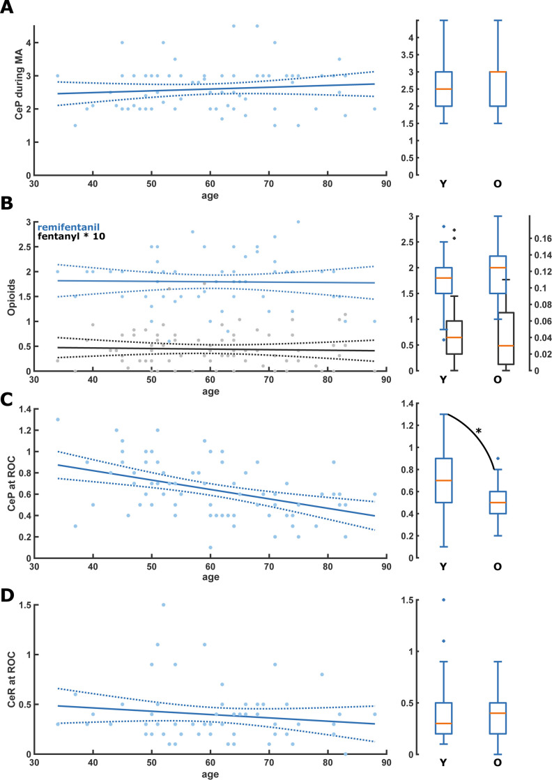
During anesthesia maintenance, we did not find any age-related difference or linear trend in median CeP or CeR concentrations: CeP was 3[IQR:2–3]μg ml-1 in the old vs 2.5[IQR:2–3]μg ml-1 in the young (p = 0,332) and CeR was 2[IQR:1.5–2.2]ng ml-1 in the old vs 1.8[IQR:1.5–2.0] ng ml-1 in the young (p = 0,384). The fentanyl doses did not differ significantly between the age groups. Fig 1A and 1B contain the linear model plots and the young vs old box plots. Table 1 contains the statistical details of the young versus old comparisons and Table 2 contains the details of the linear models.

Fig 1. Scatter plots with linear regression as well as box plots for the comparison of the young (Y: <65 yr) and old (O: ≥65 yr) for the different drugs during anesthesia maintenance and at return of consciousness (ROC).

Fig 1

A. There was no significant change or difference of CeP during anesthesia maintenance. B. There was no significant change or difference of CeR (blue) and fentanyl (black) during anesthesia maintenance. C. CeP at ROC was significantly lower in the old patients (p = 0.010; Area Under the Curve (AUC): 0.68 [0.56 0.80]); D. There was no significant change or difference of CeR at ROC. CeP: Effective estimated brain concentration of Propofol (μg ml-1); CeR: Effective estimated brain concentration of Remifentanil (ng ml-1); MA: Maintenance of anesthesia; ROC: Return of consciousness.

Table 2. Statistical parameters of the linear model.

Linear equation t-statistic P-value Corr. coeff
Anesthesia maintenance 0.88 0.382 0.10
CeP = 0.005*age+2.277 -0.14 0.887 -0.02
CeR = -0.001*age+1.846 -0.33 0.741 -0.04
fent = -0.001*age+0.494 3.61 <0.001 0.39
RE = 0.364*age+28.217 3.46 <0.001 0.38
SE = 0.313*age+27.389 2.58 0.012 0.29
RE-SE = 0.051*age+0.828 -1.35 0.181 -0.16
SPI = -0.140*age+42.066 2.80 0.007 0.31
max(RE-SE) = 0.116*max(SPI)+1.969
ROC -4.04 <0.001 -0.43
CeP = -0.009*age+1.174 -1.12 0.268 -0.15
CeR = -0.003*age+0.598 -0.40 0.694 -0.05
RE = -0.057*age+77.221 -0.84 0.406 -0.10
SE = -0.126*age+93.119 -1.11 0.272 -0.13
RE-SE = -0.070*age+15.898 0.88 0.382 0.10

Abbreviations: CeP: Effective estimated brain concentration of propofol; CeR: Effective estimated brain concentration of remifentanil; ROC: Return of consciousness.

At ROC, we found a statistically significant influence on age on CeP: CeP = -0.01*age+1.2 (p<0.001) (old vs. young: 0.5[IQR:0.4–0.6]μg ml-1 vs. 0.7[IQR:0.5–0.9]μg ml-1 p = 0.010). We did not find a statistical difference between old and young for CeR (old vs. young: 0,3 ng ml-1[IQR:0,2–0,5] vs 0,5 ng ml-1[IQR:0.2–0.5], p = 0.820). Fig 1C and 1D and Tables 1 and 2 present the details.

3.3 Age effects on processed EEG

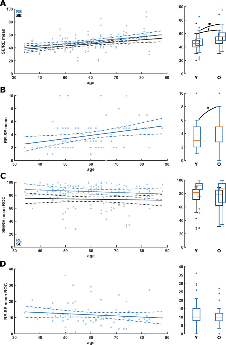
When looking at the Entropy values during maintenance, we found an age-related increase in mean RE and SE: RE = 0.36*age+28.22 (p<0.001); SE = 0.31*age+27.39 (p<0.001). Consequently, RE and SE were significantly higher in the old: RE: 55[IQR:49.3–61] vs 47.5[IQR:42–52.5], p = 0.003]; SE: 50[IQR:45–55.5] vs 45[IQR:40–50], p = 0.005). The RE-SE also increased with age, RE-SE = 0.05*age+0.83 (p = 0.012); Old vs. young: Median RE-SE (5[IQR:2.8–5.5] vs 3[25], p = 0.020). Fig 2A and 2B and Tables 1 and 2contain the detailed information. We did not observe significant differences in the entropies with age at ROC (Fig 2C and 2D and Tables 1 and 2). Further, there was no significant age effect on the mean SPI during maintenance (S2A Fig).

Fig 2. Scatter plots with linear regression as well as box plots for the comparison of the young (Y: <65 yr) and old (O: ≥65 yr) for the state (SE) and response Entropy (RE) during anesthesia maintenance and at return of consciousness (ROC).

Fig 2

A. RE (blue) and SE (black) were significantly higher in the old during anesthesia maintenance. SE: p = 0.005; AUC = 0.30 [0.18 0.43]; RE: p = 0.003; AUC = 0.29 [0.16 0.42]; B. RE-SE was significantly (p = 0.016; AUC = 0.33 [0.21 0.47]) higher in the old during anesthesia maintenance. C. There was no significant change or difference of RE (blue) and SE (black) at ROC. D. There was no significant change or difference of RE-SE at ROC. RE: Response Entropy; SE: State Entropy; MA: Maintenance of anesthesia; ROC: Return of consciousness.

3.4 PK/PD and processed EEG differences between arousers and non-arousers

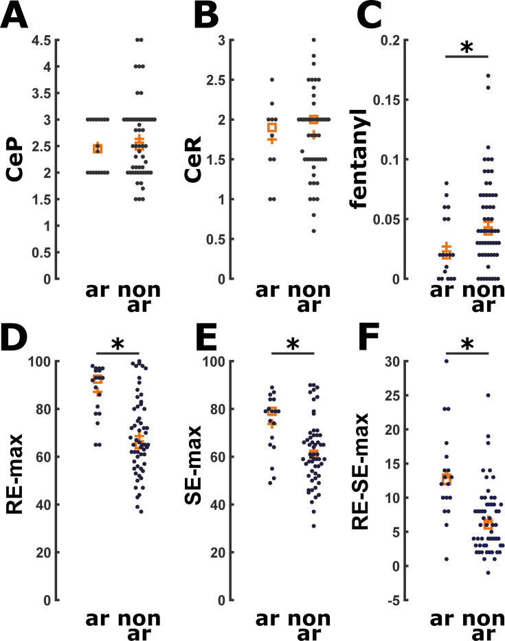
No patients exhibited explicit recall 15 minutes after ROC or at the first post-operative day after the interview with the Brice questionnaire. An arousal reaction maintenance occurred in 18 of 75 patients (24%), with no significant difference (p = 0.566) in occurrence rate between the young and old patient group. We did not observe a significant difference in CeP and CeR in patients with and without arousal reaction (Fig 3A and 3B). Median CeP during maintenance of the arousable patients was 2.45 μg ml-1[IQR:2–3μg ml-1] and it was 2 μg ml-1[IQR:1.7–2.8μg ml-1] at the arousal reaction (p = 0.069). This was not statistically different from the median CeP during maintenance of the non-arousable patients with 2.5 μg ml-1[IQR:2–3μg ml-1], p = 0.596. We found a significant difference (p<0.001) between CeP at the arousal reaction (2 μg ml-1[IQR:1.7–2.8μg ml-1]) and at ROC in the arousable patients (0.67 μg ml-1[IQR:0.4–0.9 0.67 μg ml-1]). Median CeR during maintenance in the arousable patients (1.9 ng ml-1[IQR:1.5-2ng ml-1]), as well as their CeR at the arousal reaction (2 ng ml-1[IQR:1.5-2ng ml-1]) was not statistically different if compared with median CeR during maintenance of the non-arousable patients (2 ng ml-1[IQR:1.1–2.4ng ml-1], p = 0.842 and p = 0.603, respectively) as presented in Fig 3B and Table 3. In addition, median CeR at arousal reaction was significantly different if compared with the CeR at ROC in the same arousable patients (2 ng ml-1[IQR:1.1–2.4 ng ml-1] vs 0.4 μg ml-1[IQR:0.3–0.5 ng ml-1], p = 0.005). Regarding the fentanyl dose, arousable patients received a significantly (p = 0.027) lower dosage of fentanyl 0.02 μg kg-1min-1[IQR:0.02–0.05μg kg-1 min-1] than non-arousable patients (0.04 μg kg-1min-1[IQR: 0.02–0.07μg kg-1 min-1]) (Fig 3C). When analyzing the pEEG indices, we found higher maximum (but not mean) RE (p<0.001), SE (p = 0.003), RE–SE (p<0.001; 0.81[0.68–0.92]) during maintenance in the arousable patients (Fig 3D–3F and Table 3). We also found higher maximum, but not mean, SPI values during maintenance (p = 0.030) in the arousable patients. (S3 Fig and Table 3). There was a significant positive (p = 0.006) correlation between maximum SPI and maximum RE–SE difference during surgery, i.e., max(RE-SE) = 0.116*max(SPI)+1.969. S2B Fig displays this relationship.

Fig 3. Scatter plots for drug and state (SE) and response (RE) values comparing arousers (ar) versus non-arousers (n-ar).

Fig 3

A. There was no significant difference in CeP (μg ml-1) during anesthesia maintenance. B. There was no significant difference in CeR (ng ml-1) during anesthesia maintenance. C. Arousers received significantly less fentanyl (mg/kg/min) (p = 0.026; AUC = 0.33 [0.19 0.47]). D. Arousers had significantly higher RE (p<0.001; AUC = 0.80 [0.70 0.90]). E. Arousers had significantly higher SE (p = 0.003; AUC = 0.73 [0.59 0.86]). F. Arousers had significantly higher RE-SE (<0.001; AUC = 0.81 [0.68 0.92]). CeP: Effective estimated brain concentration of Propofol (μg ml-1) during maintenance of anesthesia; CeR: Effective estimated brain concentration of Remifentanil (ng ml-1) during maintenance of anesthesia; ar: Arousers; n-ar: Non-arousers.

Table 3. Characteristics of patients according to presence of an arousal reaction.

Variable Arousal reaction P value AUC value
No (n = 57) Yes (n = 18)
Patient characteristics
Age (years) 61 [52 to 69], 34 to 83 55.5 [49 to 70.8], 39 to 88 0.572 0.46 [0.29–0.63]
BMI (kg m-2) 24.2 [21.6 to 27.5], 16.8 to 34.5 27.1 [24.4 to 29.6], 17.4 to 35.3 0.130 0.62 [0.45–0.77]
ASA I/II 30/27 (52.6%/47.4%) 7/11 (38.9%/61.1%) 0.310* N/A
Years of schooling (years) 13 [8 to 13], 5 to 23 8 [8 to 13], 5 to 18 0.675 0.47 [0.32–0.62]
Previous GA (n) 1 [1 to 3], 0 to 7 2 [1 to 2.75], 0 to 5 0.689 0.53 [0.39–0.67]
Duration of surgery (min) 60 [45 to 80], 20 to 200 62.5 [41.2 to 103.7], 35 to 180 0.426 0.56 [0.41–0.72]
Fentanyl (μg kg-1 min-1) 0.04 [0.02 to 0.07], 0.01 to 0.17 0.02 [0.02 to 0.05], 0.01 to 0.08 0.027 0.33 [0.19–0.47]
Anesthesia maintenance
CeP (μg ml-1) 2.5 [2 to 3], 1.50 to 4.5 2.45 [2 to 3], 2 to 3 0.596 0.46 [0.32–0.60]
CeR (ng ml-1) 2.0 [1.5 to 2.0], 0.6 to 3.0 1.9 [1.5 to 2], 1 to 2.5 0.842 0.48 [0.29–0.67]
RE 48 [42 to 55], 22 to 95 51.5 [47,3 to 56,3], 35 to 85 0.126 0.62 [0.48–0.75]
SE 45 [40 to 50], 20 to 88 49 [44.5 to 51.3], 33 to 75 0.091 0.63 [0.50–0.76]
RE–SE 3.9 [2 to 5], 1 to 10 4 [2 to 5], 1 to 10 0.409 0.49 [0.34–0.64]
RE max 65 [56.5 to 80.5], 37 to 100 92.5 [78 to 96.3], 65 to 98 <0.001 0.80 [0.70–0.90]
SE max 61.0 [52.5 to 69], 31 to 90 79 [64.8 to 84], 51 to 89 0.003 0.73 [0.59–0.86]
RE–SE max 6 [3 to 9], 1 to 25 12.5 [9.5 to 16.5], 1 to 30 <0.001 0.81 [0.68–0.92]
SPI 30 [25 to 40], 15 to 60 37.5 [30 to 44.2], 17 to 60 0.159 0.61 [0.46–0.75]
SPI max 52.5 [40 to 66.2], 24 to 83 63.5 [49.5 to 74.7], 42 to 89 0.030 0.67 [0.53–0.80]

Notes. Data are median (IQR), minimum to maximum unless otherwise indicated. Arousal reaction was defined as “light anesthesia” associated with involuntary movement, inadequate ventilation because of vocal cord closure, or a definite hemodynamic response (Sanders RD 2012).

*Chi-squared p-values.

Abbreviations. BMI: Body mass index; CeP: Effective estimated brain concentration of propofol; CeR: Effective estimated brain concentration of remifentanil; GA: General anesthetics; max: Maximum; RE: Response entropy (range, 0–100); SE: State entropy (range, 0–91); RE–SE: Difference between RE and SE; SPI: Surgical Pleth Index (range, 0–100).

Discussion

Our results confirm previous findings about age-influence on CeP at ROC: older patients regain consciousness at lower CeP [6,7]. We couldn’t find significant differences in the median CeP during Entropy-guided TIVA-TCI maintenance described by previous trials [8], but meanwhile we observed an age-induced change in the pEEG parameters: RE, SE, and RE-SE were significantly higher during maintenance in the elderly. Further, we couldn’t find age-related differences in CeP at arousal reactions. Arousable patients expressed significantly higher maximum RE–SE difference values.

Hence, we could reveal an age-influence of the pEEG-guidance on anesthesia navigation. Without targeting the anesthetic level to the recommended index range, CeP of our older patients might have been lower [3]. In our study, targeting the level to a SE of 40–60, it wasn’t. This is the case, because age-induced changes in the EEG seem to influence the pEEG indices.

4.1 Characteristic of patients according to age

Pharmacodynamics and pharmacokinetics of anesthetics are affected by age [26,16], and an increased Propofol-sensitivity in older patients may explain the lower CeP at ROC observed in our and previous studies [6,7]. However, we could not find the expected age-related difference in CeP during maintenance, probably because of the use of SE to target the anesthetic level. EEG amplitude under general anesthesia decreases with age and composition of recorded EEG changes towards a stronger contribution of higher frequencies [18,19]. EEG architecture’s changes could explain our higher Entropy values observation in elders during anesthesia maintenance, even in the case of actively targeting the range. Higher indices with age during maintenance were described for the Bispectral Index (BIS) and for the spectral Entropy [18], the underlying method for SE and RE calculation [9]. Here, we can confirm the impact of age for SE and RE. Further, the increase in RE-SE with age during maintenance is indicative of a stronger, age-related, contribution of higher frequencies in the 32 Hz to 47 Hz range processed by RE [9].

Age influence on SE and RE probably is the reason for our older patients having a higher CeP during maintenance, because anesthesiologists navigated anesthesia by the index. Hence, our results highlight a possible conflict between TCI and EEG-based indices for elders. Consequently, more hypnotics than required were possibly given to older patients, increasing the risk of hypnotics overdosage in this population. The case of one 82-year-old patient with an adequate CeP during maintenance (2.5 μg ml-1) showing median RE and SE of 95 and 88 during maintenance most probably highlights another issue: undetected EEG burst suppression (BSupp). BSupp indicate severe brain neuronal activity and metabolic rate reduction that may increase postoperative delirium and POCD risk [27,28]. pEEG indices seem to have limited ability to reliably detect BSupp patterns, underestimating their occurrence [29]. If undetected, the suppressed EEG low-amplitude and high frequency characteristics can cause high index values [30]. This may have been our patient’s case since she showed no arousal reaction during surgery and had no awareness with explicit recall. In order to generally identify BSupp, even if the indices do not, a visual identification of the raw EEG or its spectral representation was suggested [29]. This case also highlights that Propofol concentrations that are usually appropriate can cause persisting BSupp in certain cases. Hence, EEG-based patient monitoring can help to identify these cases and to individually adjust the anesthetic level. But, using processed EEG indices only to guide TCI anesthesia with Propofol may expose older patients to higher than necessary doses that could lead to a higher risk of adverse outcomes such as postoperative neurocognitive disorders [10].

4.2 Characteristic of patients according to arousal reaction

Response surface models revealed prominent synergistic effects between Propofol and Remifentanil in blunting the response to nociceptive stimuli and to avoid arousal reactions [21]. In our study, arousable patients had adequate CeP and CeR values during maintenance [1]. However, they received less fentanyl during surgery, and they had a higher median SPI during maintenance. This suggests that, even within the suggested TCI interval [23], analgesic management may have been inadequate, and SPI threshold of 30 suggested for inhalational anesthesia [21] should be adopted also for TCI anesthesia in order to prevent an arousal reaction’s risk.

Entropy monitoring may reduce the likelihood of a reaction to intraoperative nociceptive stimulus [31]. In our study, arousable patients had higher maximum RE-SE RE, SE, and SPI values than non-arousable patients, suggesting monitoring devices’ detection of arousal reaction. Because we didn’t see differences in median values between arousable and non-arousable patients, the predictive power for a possible arousal may be low.

Painful stimulation increases cardiovascular activity, detected by SPI [21]. However, muscle tension increases, another facet of the response to painful stimulation [32], also occur because of proprioceptive inputs to the reticulo-thalamic activating system [33]. SE, computed over the EEG-dominant frequency range (0.8–32 Hz), mainly reflects the patient’s cortical state. By contrast, RE is computed over the EEG- and EMG-dominant frequency ranges (0.8–47 Hz) and thereby at least partly serves as an indicator of upper facial EMG activation, which has been reported to represent nociception or impending awakening [31]. Increment in RE–SE, even if it occurs only transiently because of a subsequent increase in SE, may be useful for identifying an inadequate anesthetic/analgesic plan with arousal reaction’s risk following painful stimulation. So in case of increasing EMG the SE and RE will start to drift apart because of an increased influence of high frequency oscillatory activity picked up by RE, but not SE. Another case, more important to our investigations is the age-related flattening of the Power spectral density under general anesthesia [18]. This flattening also leads to an increased influence of higher frequencies that will also increase the difference between the RE and SE. So we are confident that the observed increase in the RE-SE with age under general anesthesia is not caused by increase EMG with age, but by the age-induced increased of higher EEG frequencies. Hence, the indices seem to appropriately respond to an arousal reaction after noxious stimulation independent of age.

4.3 Limitations

This study has some limitations. Firstly, it was conducted only on female patients undergoing breast surgery: future trials should confirm our observations also on males and other surgery-types; in addition, although all of our patients were in the menopausal period, we did not account for hormonal contribution in our analysis. Secondly, SE and RE trends, and the raw EEG tracings, weren’t electronically stored, so we couldn’t analyze RE and SE values during arousal reaction: we can only speculate that the observed maximum RE and SE in arousers were its consequence. Analysis of raw EEG data could have provided additional insights into the arousal reaction’s nature. Raw EEG data could have provided additional informationa also on BSupp, that can alter SE and RE values, however because the vast majority of our patients had indices in the index interval reflecting “adequate anesthesia”, i.e., between 40 and 60, we are very confident that BSupp is not an issue in our data.

Conclusion

Estimated concentrations of Propofol during anesthesia maintenance seem not to be age-related in patients during TIVA-TCI for breast surgery when anesthesia is navigated by the processed EEG index, highlighting the impact on age on general anesthesia. The SE and RE seem to increase with age during adequate levels of anesthesia.

The occurrence of an arousal reaction seems independent of age, but lower CeR during anesthesia maintenance might lead to an increased risk of an arousal reaction. Finally, additional information as for instance provided by the SPI could help to improve intraoperative patient monitoring and to optimize the hypnotic and analgesic component of anesthesia.

Supporting information

S1 Fig. CONSORT flow diagram showing selection of the enrolled patients.

(TIF)

S2 Fig

Scatter plots with linear regression for the A) mean SPI versus age (years) and B) max(SPI) versus max(RE-SE) relationship during anesthesia maintenance. There was no significant change of the mean SPI with age. There was a significant increase of the max (RE-SE) with increase of the maximum SPI observed during anesthesia maintenance. SPI: Surgical Pleth Index; RE: Response entropy; SE: State Entropy.

(TIF)

S3 Fig. Scatter plots for mean and maximum SPI values comparing arousers (ar) versus non-arousers (n-ar).

There was no significant difference of mean SPI between arousers and non-arousers during anesthesia maintenance. The maximum SPI was significantly higher in arouser during anesthesia maintenance (p = 0.03; AUC = 0.67 [0.53–0.80]). SPI: Surgical Pleth Index; ar: arousers; n-ar: non-arousers.

(TIF)

S1 Database

(TIF)

Acknowledgments

Authors would like to thank Paolo Burelli, MD, Chief of the Breast Unit of Treviso Regional Hospital, and Domenico Gentile, MD, Alessandro de Laurenzis, PsyD, Lisa Entilli, MD and Cristina Gioia, MD of the Department of Anesthesiology and Intensive Care of Treviso Regional Hospital for their assistance with the study.

Abbreviations

ASA

American Society of Anesthesiolgists

BIS

Bispectral Index (Medtronic, Dublin, Ireland)

BP

Blood Pressure

BSupp

Burst suppression

CeP

estimated brain concentration of Propofol

CeR

estimated brain concentration of Remifentanil

CI

Confidence Interval

EMG

electromyography

IQR

Interquartile range

LMA

laryngeal mask airway

LOC

loss of consciousness

MA

maintenance of anesthesia

NMB

neuromuscular blocking agents

pEEG

processed electroencephalographic

PK/PD

pharmacokinetics/pharmacodynamic

POCD

Post-Operative Cognitive Dysfunction

RE

Response Entropy (Entropy Module, GE Healthcare, Chicago, IL, USA)

ROC

return of consciousness

SD

Standard deviation

SE

State Entropy (Entropy Module, GE Healthcare, Chicago, IL, USA)

SPI

Surgical Pleth Index

TIVA-TCI

Total intravenous anesthesia with target-controlled infusions

Data Availability

All relevant data are within the manuscript and its Supporting Information files.

Funding Statement

Authors received no specific funding for this work

References

  • 1.Al-Rifai Z, Mulvey D. Principles of total intravenous anesthesia: practical aspects of using total intravenous anesthesia. BJA Education 2016; 16: 276–280. [Google Scholar]
  • 2.Schnider TW, Minto CF, Gambus PL, et al. The influence of method of administration and covariates on the pharmacokinetics of Propofol in adult volunteers. Anesthesiology 1998; 88:1170–1182. 10.1097/00000542-199805000-00006 [DOI] [PubMed] [Google Scholar]
  • 3.Schnider TW, Minto CF, Shafer SL, et al. The influence of age on Propofol pharmacodynamics. Anesthesiology 1999; 90:1502–1516. 10.1097/00000542-199906000-00003 [DOI] [PubMed] [Google Scholar]
  • 4.Minto CF, Schnider TW, Egan TD. Influence of age and gender on the pharmacokinetics and pharmacodynamics of remifentnil. I. Model development. Anesthesiology 1997; 86:10–23. 10.1097/00000542-199701000-00004 [DOI] [PubMed] [Google Scholar]
  • 5.Minto CF, Schnider TW, Shafer SL. Pharmacokinetics and pharmacodynamics of Remifentanil. II. Model application. Anesthesiology 1997; 86:24–33. 10.1097/00000542-199701000-00005 [DOI] [PubMed] [Google Scholar]
  • 6.Liu SH, Tian M. Factors affecting recovery from anesthesia with Propofol-Remifentanil target-controlled infusion in laparoscopic surgery. J Int Med Res 2017; 45:1007–1014. 10.1177/0300060517705539 [DOI] [PMC free article] [PubMed] [Google Scholar]
  • 7.Koo BN, Lee JR, Noh GJ, Lee JH, Kang YR, Han DW. A pharmacodynamic analysis of factors affecting recovery from anesthesia with Propofol-Remifentanil target controlled infusion. Acta Pharmacol Sin 2012; 33:1080–1084. 10.1038/aps.2012.85 [DOI] [PMC free article] [PubMed] [Google Scholar]
  • 8.Ouattara A, Boccara G, Lemaire S, et al. Target-controlled infusion of Propofol and Remifentanil in cardiac anesthesia: influence of age on predicted effect-site concentrations. Br J Anesth 2003; 90:617–622. [DOI] [PubMed] [Google Scholar]
  • 9.Viertio-Oja H, Maja V, Sarkela M, Talja P, et al. Description of the Entropy™ algorithm as applied in the Datex-Ohmeda S/5™ Entropy Module. Acta Anesthesiol. Scand.2004. 48:154–161. [DOI] [PubMed] [Google Scholar]
  • 10.Chan MT, Cheng BC, Lee TM, Gin T; CODA Trial Group. BIS-guided anesthesia decreases postoperative delirium and cognitive decline. J Neurosurg Anesthesiol 2013; 25:33–42. 10.1097/ANA.0b013e3182712fba [DOI] [PubMed] [Google Scholar]
  • 11.Absalom AR, Green D. NAP5: the tip of the iceberg, or all we need to know? Br J Anesth 2014; 113:527–530. 10.1093/bja/aeu349 [DOI] [PubMed] [Google Scholar]
  • 12.Shortal BP, Hickman LB, Mak-McCully RA, et al. Duration of EEG suppression does not predict recovery time or degree of cognitive impairment after general anesthesia in human volunteers. Br J Anesth 2019; 123(2):206–218. 10.1016/j.bja.2019.03.046 [DOI] [PMC free article] [PubMed] [Google Scholar]
  • 13.Wildes TS, Mickle AM, Avidian MS. Electroencephalography-Guided Anesthestic Administration and Postoperative Delirium-Reply. JAMA 2019, 321(24):2471–2472. 10.1001/jama.2019.5161 [DOI] [PubMed] [Google Scholar]
  • 14.Linassi F, Zanatta P, Tellaroli P, Ori C, Carron M. Isolated forearm technique: a meta-analysis of connected consciousness during different general anesthesia regimens. Br J Anesth. 2018; 121:198–209. [DOI] [PubMed] [Google Scholar]
  • 15.Sanders RD, Gaskell A, Sleigh J. Can use of frontal EEG monitoring increase intraoperative connected consciousness? Br. J. Anesth. 2018. 121, 191–193. 10.1016/j.bja.2018.03.019 [DOI] [PubMed] [Google Scholar]
  • 16.Ni K, Cooter M, Gupta DK, et al. Paradox of age: older patients receive higher age-adjusted minimum alveolar concentration fractions of volatile anesthetics yet display higher bispectral index values Br J Anesth, 2019. 123:288–297. 10.1016/j.bja.2019.05.040 [DOI] [PMC free article] [PubMed] [Google Scholar]
  • 17.Schults A, Grouven U, Zander I. Age-related effects in the EEG during Propofol anesthesia. Aceta Anesthesiol Scand 2004; 58:27–34. [DOI] [PubMed] [Google Scholar]
  • 18.Kreuzer M, Stern MA, Hight D, et al. , Spectral and Entropic Features are altered by age in the electroencephalogram in patients under sevoflurane anesthesia. Anesthesiology, 2020; [Epub ahead of print]. 10.1097/ALN.0000000000003182 [DOI] [PMC free article] [PubMed] [Google Scholar]
  • 19.Purdon PL, Pavone KJ, Akeju O, et al. The Ageing Brain: Age-dependent changes in the electroencephalogram during Propofol and sevoflurane general anesthesia. Br J Anesth 2015; 115 Suppl 1:i46–i57. [DOI] [PMC free article] [PubMed] [Google Scholar]
  • 20.Wang M, Messina AG, Russell IF. The topography of awareness: a classification of intra-operative cognitive states. Anesthesia 2012; 67:1197–1201. 10.1111/anae.12041 [DOI] [PubMed] [Google Scholar]
  • 21.Ledowski T, Burke J, Hruby J. Surgical pleth index: prediction of postoperative pain and influence of arousal. Br J Anesth 2016; 117:371–374. 10.1093/bja/aew226 [DOI] [PubMed] [Google Scholar]
  • 22.Struys MM, Vanpeteghem C, Huiku M, Uutela K, Blyaert NB, Mortier EP. Changes in a surgical stress index in response to standardized pain stimuli during Propofol-Remifentanil infusion. Br J Anesth. 2007; 99:359–367. [DOI] [PubMed] [Google Scholar]
  • 23.Bergmann I, Göhner A, Crozier TA,et al. Surgical pleth index-guided Remifentanil administration reduces Remifentanil andPropofol consumption and shortens recovery times in out-patient anesthesia. Br J Anesth 2013; 110: 622–8. [DOI] [PubMed] [Google Scholar]
  • 24.Sanders RD, Tononi G, Laureys S, Sleigh JW. Unresponsiveness ≠ unconsciousness. Anesthesiology. 2012; 116:946–959. 10.1097/ALN.0b013e318249d0a7 [DOI] [PMC free article] [PubMed] [Google Scholar]
  • 25.Pollard RJ, Coyle JP, Gilbert RL, Beck JE. Intraoperative awareness in a regional medical system: a review of 3 years' data. Anesthesiology. 2007; 106:269–274. 10.1097/00000542-200702000-00014 [DOI] [PubMed] [Google Scholar]
  • 26.Hentschke H, Stüttgen MC. Computation of measures of effect size for neuroscience data sets. Eur. J. Neurosci 2011. 34:1887–1894. 10.1111/j.1460-9568.2011.07902.x [DOI] [PubMed] [Google Scholar]
  • 27.Soehle M, Dittmann A, Ellerkmann RK, Baumgarten G, Putensen C, Guenther U. Intraoperative burst suppression is associated with postoperative delirium following cardiac surgery: a prospective, observational study. BMC Anesthesiol 2015; 15:61 10.1186/s12871-015-0051-7 [DOI] [PMC free article] [PubMed] [Google Scholar]
  • 28.Fritz BA, Kalarickal PL, Maybrier HR, et al. Intraoperative Electroencephalogram Suppression Predicts Postoperative Delirium. Anesth Analg 2016; 122:234–242. 10.1213/ANE.0000000000000989 [DOI] [PMC free article] [PubMed] [Google Scholar]
  • 29.Muhlhofer WG, Zak R, Kamal T, et al. Burst-suppression ratio underestimates absolute duration of electroencephalogram suppression compared with visual analysis of intraoperative electroencephalogram. Br J Anesth 2017; 118:755–761. 10.1093/bja/aex054 [DOI] [PMC free article] [PubMed] [Google Scholar]
  • 30.Hart SM, Buchannan CR, Sleigh JW. A failure of M-Entropy to correctly detect burst suppression leading to sevoflurane overdosage. Anesth Intensive Care 2009; 37:1002–1004. 10.1177/0310057X0903700619 [DOI] [PubMed] [Google Scholar]
  • 31.Aho AJ, Yli-Hankala A, Lyytikäinen LP, Jäntti V. Facial muscle activity, Response Entropy, and State Entropy indices during noxious stimuli in Propofol-nitrous oxide or Propofol-nitrous oxide-Remifentanil anesthesia without neuromuscular block. Br J Anesth. 2009; 102:227–233. 10.1093/bja/aen356 [DOI] [PMC free article] [PubMed] [Google Scholar]
  • 32.Brandon R, Howatson G, Strachan F, Hunter AM. Neuromuscular response differences to power vs strength back squat exercise in elite athletes. Scand J Med Sci Sports. 2015; 25:630–639. 10.1111/sms.12289 [DOI] [PubMed] [Google Scholar]
  • 33.Lanier WL, Iaizzo PA, Milde JH, Sharbrough FW. The cerebral and systemic effects of movement in response to a noxious stimulus in lightly anesthetized dogs. Possible modulation of cerebral function by muscle afferents. Anesthesiology 1994; 80:392–401. 10.1097/00000542-199402000-00019 [DOI] [PubMed] [Google Scholar]

Decision Letter 0

JianJun Yang

8 Sep 2020

PONE-D-20-16382

Age influences on Propofol estimated brain concentration and Entropy during maintenance and at return of consciousness during Total Intravenous Anesthesia With Target-Controlled Infusion in Unparalyzed Patients: an Observational Prospective Trial

PLOS ONE

Dear Dr. Linassi,

Thank you for submitting your manuscript to PLOS ONE. After careful consideration, we feel that it has merit but does not fully meet PLOS ONE’s publication criteria as it currently stands. Therefore, we invite you to submit a revised version of the manuscript that addresses the points raised during the review process.

The authors described an observational prospective trial to validate the aging effects on effective estimated brain concentration of propofol during TIVA-TCI Entropy-guided anesthesia. The manuscript has been assessed by three reviewers and their comments are available below. The reviewers both concerned more details regarding the results.

Please submit your revised manuscript by Oct 21 2020. If you will need more time than this to complete your revisions, please reply to this message or contact the journal office at plosone@plos.org. When you're ready to submit your revision, log on to https://www.editorialmanager.com/pone/ and select the 'Submissions Needing Revision' folder to locate your manuscript file.

Please include the following items when submitting your revised manuscript:

  • A rebuttal letter that responds to each point raised by the academic editor and reviewer(s). You should upload this letter as a separate file labeled 'Response to Reviewers'.

  • A marked-up copy of your manuscript that highlights changes made to the original version. You should upload this as a separate file labeled 'Revised Manuscript with Track Changes'.

  • An unmarked version of your revised paper without tracked changes. You should upload this as a separate file labeled 'Manuscript'.

If you would like to make changes to your financial disclosure, please include your updated statement in your cover letter. Guidelines for resubmitting your figure files are available below the reviewer comments at the end of this letter.

If applicable, we recommend that you deposit your laboratory protocols in protocols.io to enhance the reproducibility of your results. Protocols.io assigns your protocol its own identifier (DOI) so that it can be cited independently in the future. For instructions see: http://journals.plos.org/plosone/s/submission-guidelines#loc-laboratory-protocols

We look forward to receiving your revised manuscript.

Kind regards,

JianJun Yang, M.D., Ph.D.

Academic Editor

PLOS ONE

Journal Requirements:

When submitting your revision, we need you to address these additional requirements.

1. Please ensure that your manuscript meets PLOS ONE's style requirements, including those for file naming. The PLOS ONE style templates can be found at

https://journals.plos.org/plosone/s/file?id=wjVg/PLOSOne_formatting_sample_main_body.pdf and

https://journals.plos.org/plosone/s/file?id=ba62/PLOSOne_formatting_sample_title_authors_affiliations.pdf

2. "Please explain why the CT registration (NCT04129112) status is still 'recruiting

3.  We note that you have indicated that data from this study are available upon request. PLOS only allows data to be available upon request if there are legal or ethical restrictions on sharing data publicly. For information on unacceptable data access restrictions, please see http://journals.plos.org/plosone/s/data-availability#loc-unacceptable-data-access-restrictions.

In your revised cover letter, please address the following prompts:

a) If there are ethical or legal restrictions on sharing a de-identified data set, please explain them in detail (e.g., data contain potentially identifying or sensitive patient information) and who has imposed them (e.g., an ethics committee). Please also provide contact information for a data access committee, ethics committee, or other institutional body to which data requests may be sent.

b) If there are no restrictions, please upload the minimal anonymized data set necessary to replicate your study findings as either Supporting Information files or to a stable, public repository and provide us with the relevant URLs, DOIs, or accession numbers. Please see http://www.bmj.com/content/340/bmj.c181.long for guidelines on how to de-identify and prepare clinical data for publication. For a list of acceptable repositories, please see http://journals.plos.org/plosone/s/data-availability#loc-recommended-repositories.

We will update your Data Availability statement on your behalf to reflect the information you provide.

4. PLOS requires an ORCID iD for the corresponding author in Editorial Manager on papers submitted after December 6th, 2016. Please ensure that you have an ORCID iD and that it is validated in Editorial Manager. To do this, go to ‘Update my Information’ (in the upper left-hand corner of the main menu), and click on the Fetch/Validate link next to the ORCID field. This will take you to the ORCID site and allow you to create a new iD or authenticate a pre-existing iD in Editorial Manager. Please see the following video for instructions on linking an ORCID iD to your Editorial Manager account: https://www.youtube.com/watch?v=_xcclfuvtxQ

5. Please include your tables as part of your main manuscript and remove the individual files. Please note that supplementary tables (should remain/ be uploaded) as separate "supporting information" files

[Note: HTML markup is below. Please do not edit.]

Reviewers' comments:

Reviewer's Responses to Questions

Comments to the Author

1. Is the manuscript technically sound, and do the data support the conclusions?

The manuscript must describe a technically sound piece of scientific research with data that supports the conclusions. Experiments must have been conducted rigorously, with appropriate controls, replication, and sample sizes. The conclusions must be drawn appropriately based on the data presented.

Reviewer #1: Partly

Reviewer #2: Partly

Reviewer #3: Yes

**********

2. Has the statistical analysis been performed appropriately and rigorously?

Reviewer #1: I Don't Know

Reviewer #2: No

Reviewer #3: Yes

**********

3. Have the authors made all data underlying the findings in their manuscript fully available?

The PLOS Data policy requires authors to make all data underlying the findings described in their manuscript fully available without restriction, with rare exception (please refer to the Data Availability Statement in the manuscript PDF file). The data should be provided as part of the manuscript or its supporting information, or deposited to a public repository. For example, in addition to summary statistics, the data points behind means, medians and variance measures should be available. If there are restrictions on publicly sharing data—e.g. participant privacy or use of data from a third party—those must be specified.

Reviewer #1: Yes

Reviewer #2: Yes

Reviewer #3: Yes

**********

4. Is the manuscript presented in an intelligible fashion and written in standard English?

PLOS ONE does not copyedit accepted manuscripts, so the language in submitted articles must be clear, correct, and unambiguous. Any typographical or grammatical errors should be corrected at revision, so please note any specific errors here.

Reviewer #1: Yes

Reviewer #2: Yes

Reviewer #3: Yes

**********

5. Review Comments to the Author

Please use the space provided to explain your answers to the questions above. You may also include additional comments for the author, including concerns about dual publication, research ethics, or publication ethics. (Please upload your review as an attachment if it exceeds 20,000 characters)

Reviewer #1: In this manuscript, the authors examined the effect of aging on the pharmacodynamics/pharmacokinetics of propofol and remifentanil, in particular as they relate to

Entropy-guided total intravenous anesthesia with target-controlled infusions (TIVA-TCI) They compared aging effects on effective estimated brain concentration of Propofol (CeP) during TIVA-TCI Entropy-guided anesthesia, without neuromuscular blockade (NMB). Secondary endpoints included the relationships between age and CeP at arousal reaction (AR), return of consciousness (ROC) and explicit recall. They calculated a linear model to evaluate the impact of age on observational variables and performed pairwise tests to compare older (≥65 years, n=50) and younger (<65 years, n=25) patients as well as patients with and without an an arousal response. Their results suggest that there is no age difference in CeP during anesthesia maintenance, but the CeP significantly decreased with age for return of consciousness. Interestingly, entropy values during MA increased with age despite comparable CeP's. The authors are clearly experienced and well versed in the field, but their work seemingly contradicts their previous work as well as that of others regarding increased sensitivities of the elderly to anesthetics, especially during TIVA. As such, there are several questions that need to be addressed:

1. The authors only enrolled women in their cohort. Since we know that MAC is altered by hormonal contributions, most notably progesterone in the pregnant patient, did they account for any hormonal contributions related to menstrual cycle and/or menopause. If not, could they at least comment on this?

2. It makes sense that older age, with an implied increased sensitivity to anesthetics, would demonstrate a lower CeP at ROC, but why is this same sensitivity not found in higher doses required for true anesthesia in contradiction to their previous work and that of others?

3. Similarly, if older age is associated with increased sensitivity to anesthetics, why would the entropy values for wakefulness be higher in the elderly at comparable CeP? Doesn't this contradict the ROC data?

4. We have had significant experience with the BIS and entropy measures of consciousness and have always found a degree of EMG contamination, which the static entropy is "supposed" to filter out. Can the authors comment on this given that the patients were not paralyzed?

5. How does this study differ from those mentioned in the following and why are the results so different:"CeP at loss of consciousness (LOC) and return of consciousness (ROC) during TIVA-TCI are lower in the elderly (age ≥65 years) than in youngers (age <65 years).[6,7] Further, it seems that lower CeP is necessary to maintain a similar anesthetic level in elderly using TIVA-TCI during cardiac surgery,[8] suggesting that older patients are more sensitive to Propofol administered for anesthesia maintenance."

6. While formal PK/PD models for propofol and remifentanil have been established for quite some time, it is also well known that the variability in seeminly adequate effect site concentrations is quite high. Can the authors comment on this variability in their population?

7. Can the authors comment more on their fentanyl doses as a confounding factor? Differential dosing here could grossly effect their results if not properly controlled. For instance, is the lack of difference in CeP and CeR between old and young patients during maintance of anesthesia possibly due to the younger patients receiving more fentanyl? They already state that such is the case in arousable patients: "Regarding the fentanyl dose, arousable patients received a significantly (p=0.027,AUC:0.26[0.19-0.47]) lower dosage of fentanyl..."

8. As I have no expertise in the statistics used, a more formal review of such by a statistician is warranted, especially in light of the significant scatter noted in the figures.

9. I am also confused by the conclusion that there is do difference in the CeR and CeP during maintenance, yet the entropy is higher in the elderly. This makes the older patients sound like they were merely not as deeply anesthetized compared to the younger patients at similar predicted Ce's and would actually suggest a decreased sensitivity to the anesthetic in the elderly!

10. On page 10, without referring to the figures, it is not clear which groups are being compared in the statements "When analyzing the pEEG indices, we found higher maximum (but not mean) RE (p<0.001;AUC:0.80[0.70-0.90]), SE (p=0.003; AUC:0.73[0.59-0.86]), RE–SE (p<0.001; 0.81[0.68-0.92])during maintenance (Figure 4D-F; Table 3). We also found higher maximum, but not mean, SPI values during maintenance (p=0.030; AUC:0.67[0.46-0.75]) (Figure S2, Table 3). There was a significant positive (p=0.006) correlation between maximum SPI and maximum RE–SE difference during surgery, i.e., max(RE-SE)=0.116*max(SPI)+1.969."

11. The authors' concerns regarding burst suppression are important ones. As such, they must comment on the presence or absence of burst suppression in all of the patients as this could give the very misleading result to which they refer, and will completely alter their interpretation of the EEG comparisons. I understand that they may not have the raw EEG data stored, but if most of the elderly are in fact more sensitive to anesthetics, thereby inducing burst suppression, this could be responsible for a titration to an SE and RE that are contaminated. If in burst suppression, the elderly might have needed far less anesthetic than given thereby altering their conclusions.

small corrections:

page 5: than in youngers, "youngers" is not a word.

page 5: helping do avoid should be helping to avoid

page 7: number of previous general anesthesia should read anesthetics

page 7: schooling’s years should read years of schooling

page 9: previous general anesthesia. should read anesthetics

Reviewer #2: Some aspects of the statistical analyses had me a bit confused. I have made some comments below in an attempt to highlight some of the issues. Generally speaking, the analyses are pretty straightforward.

1. The abbreviation AUC is not defined anywhere in the manuscript. If this happens to be area under the ROC curve, this is not mentioned in the methods, I don't understand the usage in this manuscript, and I recommend cutting it unless you can provide justification. It also makes the text of the results section too number-heavy and I would consider removing from the text regardless.

2. The biggest problem I had with the analyses was the switching between assumptions for the outcome variables. Median and IQR summary statistics and Wilcoxon are used when the normality assumption is suspect. Linear regression and Pearson correlation coefficients are used when a normality assumption is permissible to make. For instance, in section 3.2, in the first paragraph, CeP and CeR are all reported as median IQR (with p-values from Mann-Whitney, if I understood correctly). This switches in the second paragraph to assuming normality with for the linear model, and then back to a nonparametric test for testing young v. old. Neither are necessarily wrong, but the consistency has me baffled. Maybe more explanation for these choices in the methods section would be helpful.

3. I also found it very confusing that both a linear model and a Pearson correlation were used. There are differences between the two (mainly that both variables in correlation are considered to be sampled with error, whereas only the outcome (Y) variable is in a linear model), but there is nothing in the methods to support the use of both. Personally, I would report the slopes of the linear regression models. Intercepts are sometimes not reported so I will defer to you as to whether that is needed.

4. Did you consider fitting multivariable regression models to account for any potential confounders?

5. In the figures, please ensure that the y-axis range for the scatter plot matches the y-axis range for the boxplots.

6. Also, I suggest expanding the y-axis to zero for all plots, e.g, Figure 2 A.

Reviewer #3: I am quite impressed with the methodological approach that you took, and the care that was displayed in its execution.

Using exacting methods you quantified an important domain of clinical practice and have brought illumination to a foggy domain that was sorely in need of scientific foundation.

That one of the investigators was involved in hands-on data acquisition was a bit troubling and I believe you need to demonstrate absence of potential bias in their data recording.

The statistical analysis was appropriate in all regards.

The data capture excellent and sample size more than ample.

My overall recommendation is to accept this fine work of science that has virtually immediate clinical relevance and application.

**********

6. PLOS authors have the option to publish the peer review history of their article (what does this mean?). If published, this will include your full peer review and any attached files.

If you choose “no”, your identity will remain anonymous but your review may still be made public.

Do you want your identity to be public for this peer review? For information about this choice, including consent withdrawal, please see our Privacy Policy.

Reviewer #1: No

Reviewer #2: No

Reviewer #3: Yes: CJ Biddle

[NOTE: If reviewer comments were submitted as an attachment file, they will be attached to this email and accessible via the submission site. Please log into your account, locate the manuscript record, and check for the action link "View Attachments". If this link does not appear, there are no attachment files.]

While revising your submission, please upload your figure files to the Preflight Analysis and Conversion Engine (PACE) digital diagnostic tool, https://pacev2.apexcovantage.com/. PACE helps ensure that figures meet PLOS requirements. To use PACE, you must first register as a user. Registration is free. Then, login and navigate to the UPLOAD tab, where you will find detailed instructions on how to use the tool. If you encounter any issues or have any questions when using PACE, please email PLOS at figures@plos.org. Please note that Supporting Information files do not need this step.

PLoS One. 2020 Dec 22;15(12):e0244145. doi: 10.1371/journal.pone.0244145.r002

Author response to Decision Letter 0


27 Oct 2020

Padova, October 11th, 2020

Re: PONE-D-20-16382 R1

Title: Age influences on Propofol estimated brain concentration and Entropy during maintenance and at return of consciousness during Total Intravenous Anesthesia With Target-Controlled Infusion in Unparalyzed Patients: an Observational Prospective Trial

To:

Joerg Heber, M.D., Ph.D.

Editor in chief

PLOS ONE

and

JianJun Yang, M.D., Ph.D.

Academic Editor

Dear Prof Joerg Heber, M.D., Ph.D., and JianJun Yang, M.D., Ph.D.,

We are submitting a revised version of the above-mentioned manuscript. We wish to thank the Editor-in-chief and the Reviewers for the time spent to review our work and for the considerations and suggestions that have been included in the current version. We are happy to clarify previously unclear aspects of our manuscript in a point to point fashion.

Journal Requirements:

When submitting your revision, we need you to address these additional requirements.

Q1. Please ensure that your manuscript meets PLOS ONE's style requirements, including those for file naming. The PLOS ONE style templates can be found at

https://journals.plos.org/plosone/s/file?id=wjVg/PLOSOne_formatting_sample_main_body.pdf and

https://journals.plos.org/plosone/s/file?id=ba62/PLOSOne_formatting_sample_title_authors_affiliations.pdf

R1, We made changes as requested.

Q2. "Please explain why the CT registration (NCT04129112) status is still 'recruiting

R2. We are sorry for this, there was a delay on the update of CT. The status has been changed.

Q3. We note that you have indicated that data from this study are available upon request. PLOS only allows data to be available upon request if there are legal or ethical restrictions on sharing data publicly. For information on unacceptable data access restrictions, please see http://journals.plos.org/plosone/s/data-availability#loc-unacceptable-data-access-restrictions.

In your revised cover letter, please address the following prompts:

a) If there are ethical or legal restrictions on sharing a de-identified data set, please explain them in detail (e.g., data contain potentially identifying or sensitive patient information) and who has imposed them (e.g., an ethics committee). Please also provide contact information for a data access committee, ethics committee, or other institutional body to which data requests may be sent.

b) If there are no restrictions, please upload the minimal anonymized data set necessary to replicate your study findings as either Supporting Information files or to a stable, public repository and provide us with the relevant URLs, DOIs, or accession numbers. Please see http://www.bmj.com/content/340/bmj.c181.long for guidelines on how to de-identify and prepare clinical data for publication. For a list of acceptable repositories, please see http://journals.plos.org/plosone/s/data-availability#loc-recommended-repositories.

We will update your Data Availability statement on your behalf to reflect the information you provide.

R3. We made changes as requested, we uploaded the anonymized data set.

Q4. PLOS requires an ORCID iD for the corresponding author in Editorial Manager on papers submitted after December 6th, 2016. Please ensure that you have an ORCID iD and that it is validated in Editorial Manager. To do this, go to ‘Update my Information’ (in the upper left-hand corner of the main menu), and click on the Fetch/Validate link next to the ORCID field. This will take you to the ORCID site and allow you to create a new iD or authenticate a pre-existing iD in Editorial Manager. Please see the following video for instructions on linking an ORCID iD to your Editorial Manager account: https://www.youtube.com/watch?v=_xcclfuvtxQ

R4. We made changes as requested.

Q5. Please include your tables as part of your main manuscript and remove the individual files. Please note that supplementary tables (should remain/ be uploaded) as separate "supporting information" files

R5. We made changes as requested.

[Note: HTML markup is below. Please do not edit.]

Reviewers' comments:

Reviewer's Responses to Questions

Comments to the Author

1. Is the manuscript technically sound, and do the data support the conclusions?

The manuscript must describe a technically sound piece of scientific research with data that supports the conclusions. Experiments must have been conducted rigorously, with appropriate controls, replication, and sample sizes. The conclusions must be drawn appropriately based on the data presented.

Reviewer #1: Partly

Reviewer #2: Partly

Reviewer #3: Yes

2. Has the statistical analysis been performed appropriately and rigorously?

Reviewer #1: I Don't Know

Reviewer #2: No

Reviewer #3: Yes

3. Have the authors made all data underlying the findings in their manuscript fully available?

The PLOS Data policy requires authors to make all data underlying the findings described in their manuscript fully available without restriction, with rare exception (please refer to the Data Availability Statement in the manuscript PDF file). The data should be provided as part of the manuscript or its supporting information, or deposited to a public repository. For example, in addition to summary statistics, the data points behind means, medians and variance measures should be available. If there are restrictions on publicly sharing data—e.g. participant privacy or use of data from a third party—those must be specified.

Reviewer #1: Yes

Reviewer #2: Yes

Reviewer #3: Yes

4. Is the manuscript presented in an intelligible fashion and written in standard English?

PLOS ONE does not copyedit accepted manuscripts, so the language in submitted articles must be clear, correct, and unambiguous. Any typographical or grammatical errors should be corrected at revision, so please note any specific errors here.

Reviewer #1: Yes

Reviewer #2: Yes

Reviewer #3: Yes

5. Review Comments to the Author

Please use the space provided to explain your answers to the questions above. You may also include additional comments for the author, including concerns about dual publication, research ethics, or publication ethics. (Please upload your review as an attachment if it exceeds 20,000 characters)

Reviewer #1:

In this manuscript, the authors examined the effect of aging on the pharmacodynamics/pharmacokinetics of propofol and remifentanil, in particular as they relate to

Entropy-guided total intravenous anesthesia with target-controlled infusions (TIVA-TCI) They compared aging effects on effective estimated brain concentration of Propofol (CeP) during TIVA-TCI Entropy-guided anesthesia, without neuromuscular blockade (NMB). Secondary endpoints included the relationships between age and CeP at arousal reaction (AR), return of consciousness (ROC) and explicit recall. They calculated a linear model to evaluate the impact of age on observational variables and performed pairwise tests to compare older (≥65 years, n=50) and younger (<65 years, n=25) patients as well as patients with and without an an arousal response. Their results suggest that there is no age difference in CeP during anesthesia maintenance, but the CeP significantly decreased with age for return of consciousness. Interestingly, entropy values during MA increased with age despite comparable CeP's. The authors are clearly experienced and well versed in the field, but their work seemingly contradicts their previous work as well as that of others regarding increased sensitivities of the elderly to anesthetics, especially during TIVA. As such, there are several questions that need to be addressed:

Q1. The authors only enrolled women in their cohort. Since we know that MAC is altered by hormonal contributions, most notably progesterone in the pregnant patient, did they account for any hormonal contributions related to menstrual cycle and/or menopause. If not, could they at least comment on this?

R1. We thank the reviewer for this comment. We did not take hormonal contributions into account for our analysis. We know that there are contrasting results in the literature not only on MAC but also on TIVA and estrogen levels (Basaran et al, 2019, Yavutz et al, 2007), and their role on TIVA-TCI anaesthesia is debated. However, all of the patients were in the menopause period so we postulate that there was not high heterogeneity in our sample from a hormonal point of view. However, we added this in the limits section.

Q2. It makes sense that older age, with an implied increased sensitivity to anesthetics, would demonstrate a lower CeP at ROC, but why is this same sensitivity not found in higher doses required for true anesthesia in contradiction to their previous work and that of others?

R2. We thank the reviewer for this question. From a PK point of view, older patients require less propofol during maintenance of anaesthesia to achieve the same PD effects of older patients. These relationships were described by Schnider et al, 1998-99. However, when performing an Entropy-guided anaesthesia, we note that there is the risk of an increasing in CeP targeting during surgery because of higher RE and SE values in the elderly caused by changes in the EEG (et al, 2020; PMID 32108685). In other words, Entropy (and, as demonstrated by other Authors, BIS (Ni et al, 2019; PMID: 31279479)) is not a reliable device to measure the different PD effects of propofol between old and young patients. In addition, the trial described by Ouattara et al., that we cited in the text, described the relationship between CeP and BIS, however they used another TCI model for propofol, the Marsh PK model, that does not account for age as a covariable. We used the Schnider TCI model instead, that also accounts for the age of the patient giving automatically the correct (for this model) dose of propofol to achieve a comparable CeP to the young and old, taking into account the different PK/PD of these populations. We stretched this concept in the manuscript.

Q3. Similarly, if older age is associated with increased sensitivity to anesthetics, why would the entropy values for wakefulness be higher in the elderly at comparable CeP? Doesn't this contradict the ROC data?

R3.Thanks for this comment! Please see our response to your previous question.

Q4 We have had significant experience with the BIS and entropy measures of consciousness and have always found a degree of EMG contamination, which the static entropy is "supposed" to filter out. Can the authors comment on this given that the patients were not paralyzed?

R4. Thank you for this comment. We completely agree with the reviewer that EMG provides a source for EEG signal contamination. Especially the frontal recording positions are prone to record EMG activity as well in awake and non-paralyzed patients. The frequency spectrum of the EEG and the EMG (under general anesthesia) overlap as for instance described by Kamata et al. (Kamata, Aho et al. 2011). Still, the major contamination occurs in the higher frequencies. Hence, as a rule of the thumb, a 30 Hz threshold to roughly separate signals dominated by EEG (<30 Hz) and EMG (>30 Hz) in non-paralyzed patients seems to work for anesthesia monitoring (Bonhomme and Hans 2007). The developers of the State (SE) and Response Entropy (RE) algorithm used this threshold to design two different indices. The SE processes information in the 0.8-32 Hz range and hence predominately the EEG, whereas the RE includes frequencies up to 47 Hz. This selection was made with a purpose, because RE is designed to quickly detect a potential arousal reaction and EMG activity increases during the arousal (Viertio-Oja, Maja et al. 2004). So in case of increasing EMG the SE and RE will start to drift apart because of an increased influence of high frequency oscillatory activity picked up by RE, but not SE. Another case, more important to our investigations is the age-related flattening of the power spectral density under general anesthesia (Kreuzer, Stern et al. 2020). This flattening also leads to an increased influence of higher frequencies that will also increase the difference between the RE and SE. So we are confident that the observed increase in the RE-SE with age under general anesthesia is not caused by increase EMG with age, but by the age-induced increased of higher EEG frequencies.

We stretched this concept in the manuscript.

Q5. How does this study differ from those mentioned in the following and why are the results so different:"CeP at loss of consciousness (LOC) and return of consciousness (ROC) during TIVA-TCI are lower in the elderly (age ≥65 years) than in youngers (age <65 years).[6,7] Further, it seems that lower CeP is necessary to maintain a similar anesthetic level in elderly using TIVA-TCI during cardiac surgery,[8] suggesting that older patients are more sensitive to Propofol administered for anesthesia maintenance."

R5. Thanks for this comment! Please see our response to your previous comment 2.

Federico: You may want to stretch this even more: While previous research clearly showed that a lower anesthetic dose is required in the elderly, the processed EEG indices like Entropy in our case are still higher. This clearly shows the potential risk of overdosing an old patient if you follow the index.

Q6. While formal PK/PD models for propofol and remifentanil have been established for quite some time, it is also well known that the variability in seeminly adequate effect site concentrations is quite high. Can the authors comment on this variability in their population?

R5. We thank the reviewer for this comment. We agree that the variability on adequate effect site concentration is quite high, however we tried to select a population as homogeneous as possible (women in menopause) and undergoing the same non-major surgery (breast surgery). However our results about the correlation between age and CeP at return of consciousness are in line with previous literature findings (Schnider et al, 1998-99). We comment about the variability of our population in the limitation section.

Q7. Can the authors comment more on their fentanyl doses as a confounding factor? Differential dosing here could grossly effect their results if not properly controlled. For instance, is the lack of difference in CeP and CeR between old and young patients during maintance of anesthesia possibly due to the younger patients receiving more fentanyl? They already state that such is the case in arousable patients: "Regarding the fentanyl dose, arousable patients received a significantly (p=0.027,AUC:0.26[0.19-0.47]) lower dosage of fentanyl..."

R7. Thank you for this comment. As presented in table1, the fentanyl doses as well as the intraoperative remifentanil doses did not differ significantly between the age groups. The AUC of 0.54 (Fentanyl) and 0.43 (CeR) did not indicate a relevant effect size as well. According to the traditional academic point system, AUC values between 0.4 and 0.6.

We add this in the results section.

Q8. As I have no expertise in the statistics used, a more formal review of such by a statistician is warranted, especially in light of the significant scatter noted in the figures.

R8. Thanks for your honest response. We feel that our statistics used are appropriate to present our results. Reviewer 2 addressed some questions to the stats. Please see our response to his/her comments.

Q9. I am also confused by the conclusion that there is do difference in the CeR and CeP during maintenance, yet the entropy is higher in the elderly. This makes the older patients sound like they were merely not as deeply anesthetized compared to the younger patients at similar predicted Ce's and would actually suggest a decreased sensitivity to the anesthetic in the elderly!

R9. Thanks for this comment! Please see our response to your previous comment 2. The difference is due to the age-induced change in EEG features the EEG-based monitoring systems do not correct for.

Q10. On page 10, without referring to the figures, it is not clear which groups are being compared in the statements "When analyzing the pEEG indices, we found higher maximum (but not mean) RE (p<0.001;AUC:0.80[0.70-0.90]), SE (p=0.003; AUC:0.73[0.59-0.86]), RE–SE (p<0.001; 0.81[0.68-0.92])during maintenance (Figure 4D-F; Table 3). We also found higher maximum, but not mean, SPI values during maintenance (p=0.030; AUC:0.67[0.46-0.75]) (Figure S2, Table 3). There was a significant positive (p=0.006) correlation between maximum SPI and maximum RE–SE difference during surgery, i.e., max(RE-SE)=0.116*max(SPI)+1.969."

R10 We made changes as requested.

Q11. The authors' concerns regarding burst suppression are important ones. As such, they must comment on the presence or absence of burst suppression in all of the patients as this could give the very misleading result to which they refer, and will completely alter their interpretation of the EEG comparisons. I understand that they may not have the raw EEG data stored, but if most of the elderly are in fact more sensitive to anesthetics, thereby inducing burst suppression, this could be responsible for a titration to an SE and RE that are contaminated. If in burst suppression, the elderly might have needed far less anesthetic than given thereby altering their conclusions.

R11. Thanks for your thoughts on that and you are correct with your assumptions. This is indeed a complex issue and without having the raw EEG, we cannot give an ultimate answer. We are pretty certain that the 82-yr old, already mentioned had undetected BSupp because of the very high SE because this issue is known (Hart, Buchannan et al. 2009). Because BSupp detection focuses on the suppression phases (Särkelä, Mustola et al. 2002) and if contaminated they resemble an awake EEG and they will consequently cause high RE and SE values. Because the vast majority of our patients had indices in the index interval reflecting “adequate anesthesia”, i.e., between 40 and 60 (https://www.gehealthcare.co.uk/-/jssmedia/76841dd076a54dd5b1aa26e21c10e4cf.pdf?la=en-gb) . We are very confident that BSupp is not an issue in our data.

R11. We stretched this question in the limit sections.

Q12 small corrections:

page 5: than in youngers, "youngers" is not a word.

page 5: helping do avoid should be helping to avoid

page 7: number of previous general anesthesia should read anesthetics

page 7: schooling’s years should read years of schooling

page 9: previous general anesthesia. should read anesthetics

R12. We made changes as requested.

Reviewer #2:

Some aspects of the statistical analyses had me a bit confused. I have made some comments below in an attempt to highlight some of the issues. Generally speaking, the analyses are pretty straightforward.

Q1. The abbreviation AUC is not defined anywhere in the manuscript. If this happens to be area under the ROC curve, this is not mentioned in the methods, I don't understand the usage in this manuscript, and I recommend cutting it unless you can provide justification. It also makes the text of the results section too number-heavy and I would consider removing from the text regardless.

R1. Thank you for this comment. We made changes as requested.

Q2. The biggest problem I had with the analyses was the switching between assumptions for the outcome variables. Median and IQR summary statistics and Wilcoxon are used when the normality assumption is suspect. Linear regression and Pearson correlation coefficients are used when a normality assumption is permissible to make. For instance, in section 3.2, in the first paragraph, CeP and CeR are all reported as median IQR (with p-values from Mann-Whitney, if I understood correctly). This switches in the second paragraph to assuming normality with for the linear model, and then back to a nonparametric test for testing young v. old. Neither are necessarily wrong, but the consistency has me baffled. Maybe more explanation for these choices in the methods section would be helpful.

R2. Thank you for this comment. We highly appreciate your thoughts on this. We tried to keep the stats as simple as possible. In order to perform the group comparison, we decided to run the MWU tests because testing for normality and heteroscedasticity is not necessary. And testing for normality is always tricky itself. We agree that the t-stat for the linear model comes from a parametric test that considers the standard error. This is part of MATLB fitlm algorithm. We are aware that the results from the parametric linear model testing and the nonparametric group testing are mostly redundant and we can understand your confusion about it. Hence, we decided (as you also suggest in comment #3) to only show the slopes, intercepts and R2 for our linear models and leave the inference stats to the grouped comparisons that we used the MWU-test for.

Q3. I also found it very confusing that both a linear model and a Pearson correlation were used. There are differences between the two (mainly that both variables in correlation are considered to be sampled with error, whereas only the outcome (Y) variable is in a linear model), but there is nothing in the methods to support the use of both. Personally, I would report the slopes of the linear regression models. Intercepts are sometimes not reported so I will defer to you as to whether that is needed.

R3. Thanks for this comment! Please see our response to your previous comment.

Q4. Did you consider fitting multivariable regression models to account for any potential confounders?

R4. Thanks for this comment. We did not in the first version, because our sample size is rather small to fit a multivariate regression model. One of our main conclusions is that SE and RE seem to increase with age during adequate levels of anesthesia. Hence, we performed a small multivariate regression analysis of the type:

SE ~ 1 + age + CePmean + CeRmean

RE ~ 1 + age + CePmean + CeRmean

RE-SE ~ 1 + age + CePmean + CeRmean

As you can see in the results below, age remains the driving factor.

SE ~ 1 + age + CePmean + CeRmean

Estimate SE tStat pValue

________ _______ ______ ________

(Intercept) 12.268 9.3506 1.312 0.19486

age 0.34617 0.11042 3.1349 0.002737

CePmean 2.8489 2.0308 1.4028 0.1662

CeRmean 2.964 2.6745 1.1082 0.2725

RE ~ 1 + age + CePmean + CeRmean

Estimate SE tStat pValue

________ _______ _______ _________

(Intercept) 9.4297 10.21 0.92361 0.35965

age 0.40835 0.12057 3.3869 0.0012989

CePmean 3.3563 2.2174 1.5136 0.13575

CeRmean 3.8975 2.9202 1.3347 0.18739

RE-SE ~ 1 + age + CePmean + CeRmean

Estimate SE tStat pValue

________ _______ _______ ________

(Intercept) -2.8386 1.8325 -1.5491 0.127

age 0.062187 0.02164 2.8736 0.005724

CePmean 0.50744 0.39799 1.275 0.20757

CeRmean 0.93354 0.52414 1.7811 0.080319

Q5. In the figures, please ensure that the y-axis range for the scatter plot matches the y-axis range for the boxplots.

R5. We made changes as requested.

Q6. Also, I suggest expanding the y-axis to zero for all plots, e.g, Figure 2 A.

R6. We made changes as requested.

Reviewer #3:

I am quite impressed with the methodological approach that you took, and the care that was displayed in its execution.

Using exacting methods you quantified an important domain of clinical practice and have brought illumination to a foggy domain that was sorely in need of scientific foundation.

That one of the investigators was involved in hands-on data acquisition was a bit troubling and I believe you need to demonstrate absence of potential bias in their data recording.

The statistical analysis was appropriate in all regards.

The data capture excellent and sample size more than ample.

My overall recommendation is to accept this fine work of science that has virtually immediate clinical relevance and application.

Thank you for your comments!

6. PLOS authors have the option to publish the peer review history of their article (what does this mean?). If published, this will include your full peer review and any attached files.

If you choose “no”, your identity will remain anonymous but your review may still be made public.

Do you want your identity to be public for this peer review? For information about this choice, including consent withdrawal, please see our Privacy Policy.

Reviewer #1: No

Reviewer #2: No

Reviewer #3: Yes: CJ Biddle

[NOTE: If reviewer comments were submitted as an attachment file, they will be attached to this email and accessible via the submission site. Please log into your account, locate the manuscript record, and check for the action link "View Attachments". If this link does not appear, there are no attachment files.]

While revising your submission, please upload your figure files to the Preflight Analysis and Conversion Engine (PACE) digital diagnostic tool, https://pacev2.apexcovantage.com/. PACE helps ensure that figures meet PLOS requirements. To use PACE, you must first register as a user. Registration is free. Then, login and navigate to the UPLOAD tab, where you will find detailed instructions on how to use the tool. If you encounter any issues or have any questions when using PACE, please email PLOS at figures@plos.org. Please note that Supporting Information files do not need this step.

References

Bonhomme, V. and P. Hans (2007). "Muscle relaxation and depth of anaesthesia: where is the missing link?" British Journal of Anaesthesia 99(4): 456-460.

Hart, S. M., C. R. Buchannan and J. W. Sleigh (2009). "A failure of M-Entropy to correctly detect burst suppression leading to sevoflurane overdosage." Anaesth Intensive Care 37(6): 1002-1004.

Kamata, K., A. Aho, S. Hagihira, A. Yli-Hankala and V. Jäntti (2011). "Frequency band of EMG in anaesthesia monitoring." British journal of anaesthesia 107(5): 822-823.

Kreuzer, M., M. A. Stern, D. Hight, S. Berger, G. Schneider, J. W. Sleigh and P. S. García (2020). "Spectral and Entropic Features Are Altered by Age in the Electroencephalogram in Patients under Sevoflurane Anesthesia." Anesthesiology 132(5): 1003-1016.

Särkelä, M., S. Mustola, T. Seppänen, M. Koskinen, P. Lepola, K. Suominen, T. Juvonen, H. Tolvanen-Laakso and V. Jäntti (2002). "Automatic Analysis and Monitoring of Burst Suppression in Anesthesia." Journal of Clinical Monitoring and Computing 17(2): 125.

Viertio-Oja, H., V. Maja, M. Sarkela, P. Talja, N. Tenkanen, H. Tolvanen-Laakso, M. Paloheimo, A. Vakkuri, A. Yli-Hankala and P. Merilainen (2004). "Description of the Entropy algorithm as applied in the Datex-Ohmeda S/5 Entropy Module." Acta Anaesthesiol Scand 48(2): 154-161.

Attachment

Submitted filename: Responses to Reviewers.docx

Decision Letter 1

JianJun Yang

4 Dec 2020

Age influences on Propofol estimated brain concentration and Entropy during maintenance and at return of consciousness during Total Intravenous Anesthesia With Target-Controlled Infusion in Unparalyzed Patients: an Observational Prospective Trial

PONE-D-20-16382R1

Dear Dr. Linassi,

We’re pleased to inform you that your manuscript has been judged scientifically suitable for publication and will be formally accepted for publication once it meets all outstanding technical requirements.

Within one week, you’ll receive an e-mail detailing the required amendments. When these have been addressed, you’ll receive a formal acceptance letter and your manuscript will be scheduled for publication.

An invoice for payment will follow shortly after the formal acceptance. To ensure an efficient process, please log into Editorial Manager at http://www.editorialmanager.com/pone/, click the 'Update My Information' link at the top of the page, and double check that your user information is up-to-date. If you have any billing related questions, please contact our Author Billing department directly at authorbilling@plos.org.

If your institution or institutions have a press office, please notify them about your upcoming paper to help maximize its impact. If they’ll be preparing press materials, please inform our press team as soon as possible -- no later than 48 hours after receiving the formal acceptance. Your manuscript will remain under strict press embargo until 2 pm Eastern Time on the date of publication. For more information, please contact onepress@plos.org.

Kind regards,

JianJun Yang, M.D., Ph.D.

Academic Editor

PLOS ONE

Additional Editor Comments (optional):

Reviewers' comments:

Reviewer's Responses to Questions

Comments to the Author

1. If the authors have adequately addressed your comments raised in a previous round of review and you feel that this manuscript is now acceptable for publication, you may indicate that here to bypass the “Comments to the Author” section, enter your conflict of interest statement in the “Confidential to Editor” section, and submit your "Accept" recommendation.

Reviewer #1: All comments have been addressed

Reviewer #2: All comments have been addressed

**********

2. Is the manuscript technically sound, and do the data support the conclusions?

The manuscript must describe a technically sound piece of scientific research with data that supports the conclusions. Experiments must have been conducted rigorously, with appropriate controls, replication, and sample sizes. The conclusions must be drawn appropriately based on the data presented.

Reviewer #1: Yes

Reviewer #2: (No Response)

**********

3. Has the statistical analysis been performed appropriately and rigorously?

Reviewer #1: N/A

Reviewer #2: (No Response)

**********

4. Have the authors made all data underlying the findings in their manuscript fully available?

The PLOS Data policy requires authors to make all data underlying the findings described in their manuscript fully available without restriction, with rare exception (please refer to the Data Availability Statement in the manuscript PDF file). The data should be provided as part of the manuscript or its supporting information, or deposited to a public repository. For example, in addition to summary statistics, the data points behind means, medians and variance measures should be available. If there are restrictions on publicly sharing data—e.g. participant privacy or use of data from a third party—those must be specified.

Reviewer #1: Yes

Reviewer #2: (No Response)

**********

5. Is the manuscript presented in an intelligible fashion and written in standard English?

PLOS ONE does not copyedit accepted manuscripts, so the language in submitted articles must be clear, correct, and unambiguous. Any typographical or grammatical errors should be corrected at revision, so please note any specific errors here.

Reviewer #1: Yes

Reviewer #2: (No Response)

**********

6. Review Comments to the Author

Please use the space provided to explain your answers to the questions above. You may also include additional comments for the author, including concerns about dual publication, research ethics, or publication ethics. (Please upload your review as an attachment if it exceeds 20,000 characters)

Reviewer #1: (No Response)

Reviewer #2: (No Response)

**********

7. PLOS authors have the option to publish the peer review history of their article (what does this mean?). If published, this will include your full peer review and any attached files.

If you choose “no”, your identity will remain anonymous but your review may still be made public.

Do you want your identity to be public for this peer review? For information about this choice, including consent withdrawal, please see our Privacy Policy.

Reviewer #1: No

Reviewer #2: No

Acceptance letter

JianJun Yang

9 Dec 2020

PONE-D-20-16382R1

Age influences on propofol estimated brain concentration and entropy during maintenance and at return of consciousness during total intravenous anesthesia with target-controlled infusion in unparalyzed patients:an observational prospective trial

Dear Dr. Linassi:

I'm pleased to inform you that your manuscript has been deemed suitable for publication in PLOS ONE. Congratulations! Your manuscript is now with our production department.

If your institution or institutions have a press office, please let them know about your upcoming paper now to help maximize its impact. If they'll be preparing press materials, please inform our press team within the next 48 hours. Your manuscript will remain under strict press embargo until 2 pm Eastern Time on the date of publication. For more information please contact onepress@plos.org.

If we can help with anything else, please email us at plosone@plos.org.

Thank you for submitting your work to PLOS ONE and supporting open access.

Kind regards,

PLOS ONE Editorial Office Staff

on behalf of

Dr. JianJun Yang

Academic Editor

PLOS ONE

Associated Data

    This section collects any data citations, data availability statements, or supplementary materials included in this article.

    Supplementary Materials

    S1 Fig. CONSORT flow diagram showing selection of the enrolled patients.

    (TIF)

    S2 Fig

    Scatter plots with linear regression for the A) mean SPI versus age (years) and B) max(SPI) versus max(RE-SE) relationship during anesthesia maintenance. There was no significant change of the mean SPI with age. There was a significant increase of the max (RE-SE) with increase of the maximum SPI observed during anesthesia maintenance. SPI: Surgical Pleth Index; RE: Response entropy; SE: State Entropy.

    (TIF)

    S3 Fig. Scatter plots for mean and maximum SPI values comparing arousers (ar) versus non-arousers (n-ar).

    There was no significant difference of mean SPI between arousers and non-arousers during anesthesia maintenance. The maximum SPI was significantly higher in arouser during anesthesia maintenance (p = 0.03; AUC = 0.67 [0.53–0.80]). SPI: Surgical Pleth Index; ar: arousers; n-ar: non-arousers.

    (TIF)

    S1 Database

    (TIF)

    Attachment

    Submitted filename: Responses to Reviewers.docx

    Data Availability Statement

    All relevant data are within the manuscript and its Supporting Information files.


    Articles from PLoS ONE are provided here courtesy of PLOS

    RESOURCES