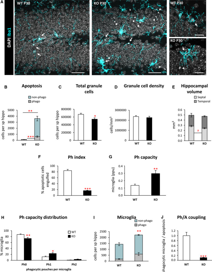
FIGURE 1.

Microglial phagocytosis is impaired in the dentate gyrus (DG) of postnatal day 30 (P30) Cstb knockout (KO) mice. A, Representative confocal images of the DG in wild‐type (WT) and Cstb KO P30 mice. Healthy or apoptotic (pyknotic/karyorrhectic) nuclear morphology was visualized with 4,6‐diamidino‐2‐phenylindole (DAPI; white), and microglia were stained for Iba1 (cyan). High‐magnification examples of phagocytic microglia following the typical “ball‐and‐chain” form (upper right panel), with the tip of their processes or phagocytosing with their soma (lower right panel). Arrows point to apoptotic cells engulfed by microglia (Iba1+), and arrowheads point to nonphagocytosed apoptotic cells. B, Number of apoptotic cells (pyknotic/karyorrhectic) in the septal hippocampus (sp hippo; n = 6 animals per condition) showing both phagocytosed (phago) and nonphagocytosed (non‐phago) apoptotic cells. C, Total number of granule cells per septal hippocampus. D, Density of granule cells (per mm3) in the septal hippocampus of both WT and Cstb KO P30 mice. E, Proportion (in mm3) between the volume of the septal and the temporal portions of the hippocampus in WT and Cstb KO P30 mice. F, Phagocytic index (Ph index; in % of apoptotic cells being engulfed by microglia) in the septal hippocampus. G, Weighted phagocytic capacity (Ph capacity) of DG microglia (in parts per unit [ppu]). H, Histogram showing the Ph capacity distribution of DG microglia (in % of microglial cells with 0‐2 phagocytic pouches [Ph]). I, Number of phagocytic (Iba1 + with DAPI inclusions) and nonphagocytic microglial cells (Iba1 + with no DAPI inclusions) per septal hippocampus. J, Phagocytosis/apoptosis (Ph/A; in fold change) in the septal hippocampus. Bars represent the mean ± standard error of the mean. *P < .05, **P < .01, ***P < .001, a P = .052 by one‐tailed Student t test. Scale bars = 40 µm (A, low magnification), 20 µm (A, high magnification); z‐thickness = 7 µm (A, low magnification), 3.5 µm (A, high magnification)
