Description
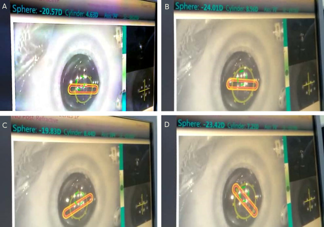
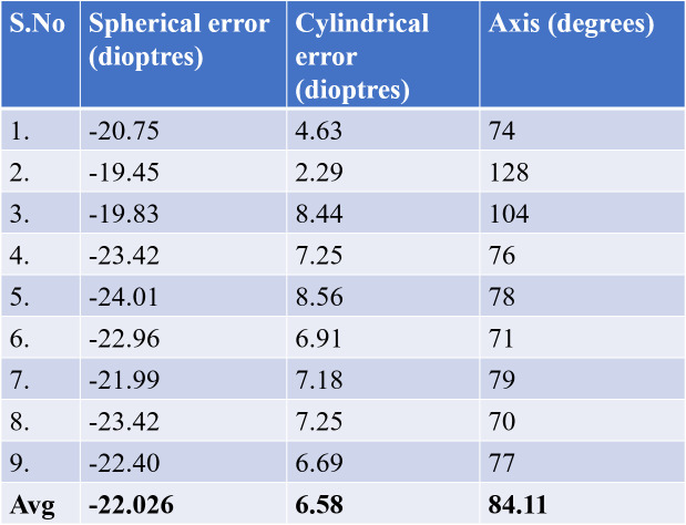
A 30-year-old man already operated for vitreoretinal surgery in the right eye and silicone oil in situ 5 months ago (for rhegmatogenous retinal detachment) developed cataract and was posted for lens aspiration and intraocular lens (IOL) insertion with silicone oil removal in the same sitting. Keratometry of the eye was 44.81/46.46 D at 99/9°, anterior chamber depth was 2.78 mm (IOLMaster, Zeiss). Optical biometry (IOLMaster, Zeiss) showed axial length of 22.69 mm and IOL power of 22.5 D (0.17 myope) in silicone oil-filled mode; and axial length of 23.36 mm and IOL power of 20 D (0.11 myope) on post vitrectomy mode. Three standard pars plana ports were made. Lens aspiration was performed through a superotemporal clear corneal 2.2 incision with co-axial irrigation–aspiration probe. Silicone oil removal was then performed through the posterior route. In the fluid-filled eye, optiwave refractive analysis (ORA) was performed to calculate the best IOL power intraoperatively, which the machine showed to be 22.5 D. The same power hydrophobic single piece acrylic lens was inserted through the superotemporal 2.2 incision and the incision sutured with 10-0 monofilament nylon suture. After fluid–air exchange was performed and the posterior segment completely filled with air, ORA was captured intraoperatively to measure the on-table refractive error. Nine readings with least amount of cylindrical error and best horizontal and vertical alignments were taken (figure 1A–D show four such readings, figure 2 compiles all). The average spherical and cylindrical errors were −22.026 D and 6.58 D, respectively, amounting to a spherical equivalent of −18.736 D.
Figure 1.
(A–D) Four sample readings of refractive error through intraoperative aberrometry.
Figure 2.
Nine values of refractive errors and their average calculation.
There has been no study or report of immediate refraction in a gas-filled posterior segment post vitrectomy to the best of our knowledge. Since the refractive index of air is different from normal vitreous, refractive status of the eye is bound to be different in case the posterior segment is filled with air versus vitreous.1 Pars plana vitrectomy is one of the most commonly performed posterior segment surgeries and air is commonly used for intraocular tamponade.2 Air takes around 1 week postoperatively to get absorbed.3 Retinoscopy in air-filled eye is challenging due to poor reflex and unpredictability of refractive error with no literature available on it. To circumvent the problem, we thought of using ORA for intraoperative on-table refraction to get an estimate of the refractive status of the eye.4 ORA projects light onto the retina and the reflected images pass through the optical system of the eye, distorting its wavefront, which is subsequently analysed according to optical and mathematical principles proprietary to the device. ORA uses a superluminescent light-emitting diode and Talbot-Moire’ interferometer to take 40 measurements, which are analysed. The Talbot-Moire’ fringe patterns are produced by the reflected wavefront after it passes through two gratings placed at a specific distance and angle to each other. The resulting fringe patterns provide information about the spherical, cylindrical and axis components of the refractive error.4 ORA has never been tried in air-filled eye in the literature. In our case, we found the eye highly myopic, as shown above. Refraction after 3 weeks (complete resolution of air) was −2.95 D sphere and 2.46 D cylinder at 69° (spherical equivalent −1.72 D) on ORA. So, the refractive error attributed to air inside the posterior segment was −17.016 D. Though this is theoretical and seldom used, still, seeing how the eye behaves when filled with air should be documented in the literature. The drawbacks include more patients being needed to validate the result. Accurate measurement of the posterior segment length can help us calculate the refractive error/mm induced by air in the posterior segment, which was not done in this case.
To conclude, we report for the first time the refractive status of an eye with air in situ in the posterior segment.
Learning points.
Change in posterior segment contents changes refractive status of the eye.
Air in eye makes it significantly myopic to the order of −17 D.
Footnotes
Contributors: SK: Concept and planning. AAB: Surgery and preparation of manuscript. AD: Video editing. LK: Video retrieval.
Funding: The authors have not declared a specific grant for this research from any funding agency in the public, commercial or not-for-profit sectors.
Competing interests: None declared.
Patient consent for publication: Obtained.
Provenance and peer review: Not commissioned; externally peer-reviewed.
References
- 1.Gao Q, Chen X, Ge J, et al. Refractive shifts in four selected artificial vitreous substitutes based on Gullstrand-Emsley and Liou-Brennan schematic eyes. Invest Ophthalmol Vis Sci 2009;50:3529–34. 10.1167/iovs.08-2802 [DOI] [PubMed] [Google Scholar]
- 2.Ramulu PY, Do DV, Corcoran KJ, et al. Use of retinal procedures in Medicare beneficiaries from 1997 to 2007. Arch Ophthalmol 2010;128:1335–40. 10.1001/archophthalmol.2010.224 [DOI] [PubMed] [Google Scholar]
- 3.Kumar M, Varshney A. Case report: biometry in air-filled eye following retinal surgery. Kerala J Ophthalmol 2019;31:232 10.4103/kjo.kjo_82_19 [DOI] [Google Scholar]
- 4.Hemmati HD, Gologorsky D, Pineda R. Intraoperative wavefront aberrometry in cataract surgery. Semin Ophthalmol 2012;27:100–6. 10.3109/08820538.2012.708809 [DOI] [PubMed] [Google Scholar]


