Abstract
Objective:
The aim of this study was to evaluate the effect of a calcium hydroxide (CH) dressing on the tubular penetration of two endodontic sealers, AH Plus (Dentsply Maillefer, Ballaigues, Switzerland) and MTA Fillapex (Angelus, Londrina, Brazil).
Methods:
Seventy-two mandibular premolars with a single root canal were prepared with ProFile.04 rotary instruments (Dentsply Maillefer) and divided into four groups. In two groups, an intracanal CH dressing was placed for 15 days. The obturations were performed with lateral condensation of gutta-percha in combination with one of the tested sealers. The roots were transversely sectioned at the apical and middle levels. The percentage of sealer penetration in the root canal walls and the percentage of impregnated dentin area in the transverse sections were obtained using confocal laser scanning microscopy. Statistical analysis was performed using one-way analysis of variance (ANOVA) and Games-Howell test.
Results:
The CH dressing reduced the mean value of tubular penetration in the middle third of teeth obturated with AH Plus (P<0.01), whereas no difference was observed at the apical sections for both sealers.
Conclusion:
The CH dressing did not interfere with the apical penetration of both tested sealers, however, decreased the tubular penetration in the middle third of the AH Plus root canal fillings. Overall, MTA Fillapex presented higher tubular penetration than AH Plus obturations.
Keywords: Calcium hydroxide, confocal laser scanning microscopy, epoxy resin-based root canal sealer, mineral trioxide aggregate-based sealer
HIGHLIGHTS.
MTA Fillapex (Angelus, Londrina, Brazil) presented higher tubular penetration than AH Plus (Dentsply Maillefer, Ballaigues, Switzerland).
CH dressing did not affect the apical tubular penetration of each sealer.
CH dressing reduced the tubular penetration of AH Plus in the middle third.
INTRODUCTION
A root canal preparation aims to shape the endodontic space, properly remove pulp tissue and microorganisms, and establish conditions for obturation (1). The root canal filling should ideally prevent fluid penetration, proliferation of bacteria remaining in the canal, or a reinfection after root canal obturation has been performed (2).
Microorganisms that remain in the root canal system after root preparation may perpetuate infections upon coming in contact with periapical tissues. In an attempt to improve root canal disinfection, the calcium hydroxide (CH) dressing is used before the canal obturation (3). However, it has been demonstrated that the CH dressing is very difficult to remove, especially in the apical third of the root canal (4). None of the currently available methods efficiently removes the CH dressing completely from the canal walls (5). The presence of CH dressing inside the canal may obstruct the dentinal tubules and thereby prevent sealer penetration and affect the apical sealing of the obturation (5, 6).
Achieving sealer penetration into dentinal tubules is desirable because it improves the adaptation and retention of the root filling and acts like a physical barrier entombing remaining bacteria inside dentinal tubules (7, 8). Adequate flowability allows the sealer to fill irregularities, tubules and lateral canals in a better way (9). AH Plus sealer (Dentsply Maillefer, Ballaigues, Switzerland) is an endodontic sealer that presents excellent physicochemical properties that have been widely studied and referenced as the standard for evaluating new cements (10). MTA Fillapex sealer (Angelus, Londrina, Brazil). Composed of mineral trioxide aggregate (MTA), silica, bismuth oxide, salicylate resin and natural resin and has adequate physical properties to be used in endodontic therapy (9, 11). According to Silva et al. (12), both sealers show acceptable flow values.
The effect of CH dressing on the push-out bond strength of AH Plus and MTA Fillapex was already evaluated (13-15). But, until the present moment, there are no studies evaluating the influence of using CH dressing in the tubular penetration of these two sealers. Thus, the aim of this study was to evaluate the influence of the use of CH dressing before root canal filling on the sealer penetration of AH Plus and MTA Fillapex and compare their tubular penetration ability, using confocal laser scanning microscopy (CLSM). Null hypotheses were that the tubular penetration of the two sealers would not be affected by using CH as the intracanal medication before the final root canal filling and that there was no difference between the tubular penetrations of both sealers.
MATERIALS AND METHODS
After approval from Pontifical Catholic University of Parana Ethics Committee (0347.0.084.000-11), 72 human extracted mandibular premolars, obtained by the Human Teeth Bank from Pontifical Catholic University of Parana, with single root canal were selected. The crowns were removed by using a diamond disc to obtain roots with a standardised length (14 mm). A size 15 stainless steel K-file was inserted into the root canal, until the tip of the instrument was first visible at the foramen, and 1 mm was deducted from this measure to obtain the working length (WL). Apical patency was confirmed with a 25 K-File (Dentsply Maillefer). The root canals were shaped in the crown-down technique by using rotary ProFile.04 instruments (Dentsply Maillefer) up to a size 40.04 at the WL.
During the shaping procedure, the root canals were irrigated with 2.5% sodium hypochlorite (NaOCl). After instrumentation, a final flush was applied using 3 mL of 17% ethylenediaminetetraacetic acid (EDTA) for 3 minutes and 5 mL of 2.5% NaOCl for 1 minute. The root canals were dried with paper points (Dentsply Industry and Commercial Ltda., Petropolis, RJ, Brazil).
The prepared roots were randomly divided into four groups of 18 roots each, according to the endodontic sealer utilized and the placement of an intracanal CH dressing: (1) CH/AH Plus group (i.e. CH dressing before obturation with gutta-percha and AH Plus); (2) AH Plus group (i.e. obturation with gutta-percha and AH Plus); (3) CH/MTA Fillapex group (i.e. CH dressing before obturation with gutta-percha and MTA Fillapex) and (4) MTA Fillapex group (i.e. obturation with gutta-percha and MTA Fillapex).
The dressing was prepared by mixing CH powder and propylene glycol 400 (1.4 g/mL). The paste was inserted into the canal using a lentulo-spiral carrier size 25 (Dentsply Maillefer) inserted until 3 mm from the WL. Mesio-distal and bucco-lingual radiographs verified the adequate placement of the CH dressing. Access cavities were filled with glass ionomer (Vidrion R; SS White, Rio de Janeiro, RJ, Brazil). Samples were stored in 100% relative humidity at 37°C for 15 days. Afterwards, the CH was manually removed using a master apical file (40.04) in rotary motion up to the WL and 5 mL of 2.5% NaOCl irrigation solution. The final irrigation was performed with 3 mL of 17% EDTA for 3 minutes and 5 mL of distilled water. Finally, they were dried with paper points.
To enable visualization under the confocal laser scanning microscope, the sealers were mixed with 0.1% fluorescent Rhodamine B dye and then prepared according to the manufacturer’s instructions. The root canals were filled by the lateral condensation technique. The Rhodamine-sealer mixture was delivered into the canal with a lentulo-spiral carrier size 30 (Dentsply Maillefer). A master gutta-percha cone 40.04 was entirely coated with a labelled sealer and then inserted into the root canal. A size B finger spreader (Dentsply Maillefer) was used to introduce accessory cones (which were also coated with a sealer) until the complete obturation of the root canal. The teeth were radiographed to validate the quality of the canal obturations. After the final obturation, they were stored for 15 days (37°C, 100% relative humidity) to permit the complete setting of the sealers.
The specimens were horizontally sectioned under continuous water cooling. The 2 mm thick slices were obtained at 3 mm (apical) and 6 mm (middle) from the apical foramen, and their surfaces were polished by the Labopol 21 polisher (Struers AS, Ballerup, Denmark) with a diamond paste (Arotec, Cotia, SP, Brazil) to eliminate dentin debris. The coronal surface of each slice was examined under CLSM (Olympus Fluoview 1000; Olympus Corporation, Tokyo, Japan).
The absorption wavelength for Rhodamine B was 540 nm and the emission wavelength was 590 nm. The samples were analysed using a 10´ lens, approximately 14 μm below the surface, and recorded in a format of 512´512 pixels using the FV10-ASW 3.1 Viewer software (Olympus Corporation).
The percentage of sealer penetration around the root canal was calculated using Image Tool 3.0 software (University of Texas Health Science Center at San Antonio-UTHSCSA, San Antonio, Texas, USA). First, the circumference of the canal was determined using the rule tool. Second, only the segments along the canal circumference in which there was sealer penetration into the dentinal tubules were measured, regardless of the depth of penetration. These data were converted to the percentage of the root canal wall with sealer penetration. The percentage of the impregnated dentin area was also obtained by using the Adobe Photoshop CS6 software (Adobe Systems, San Jose, CA), as follows: the total area of the image was acquired in pixels; the root canal area was subsequently measured with the ‘lasso’ tool and subtracted from the total area of the image, which defined the total dentin area. Next, the area of impregnated dentin, which showed sealer penetration into dentinal tubules, was calculated. With these data, the percentage of impregnated dentin was determined.
Statistical analysis
The data were analysed using Kolmogorov-Smirnov test for distribution, Levene test for equality of variances, one-way ANOVA for variance and Games-Howell post hoc test. The significance level was set at 5%. The Pearson correlation test was used to determinate the level of correlation between the evaluation methods. The software used was Statistical Package for Social Sciences version 19 (IBM Corp.; Armonk, NY, USA).
RESULTS
The results showed normal distribution (P>0.05) and heterogeneity between groups (P<0.05). The means and standard deviations for the percentage of root canal wall having sealer penetration and the percentage of impregnated dentin are presented in Table 1 (1, 2).
TABLE 1.
Tubular sealer penetration per third, mean and standard deviation values (percentage)
| Group | n | a-Sealer penetration segments (%) | b-Dentin impregnated area (%) | ||
|---|---|---|---|---|---|
| Apical third | Middle third | Apical third | Middle third | ||
| CH/AH Plus | 18 | 30.25±16.17AB | 47.90±20.65A | 7.54±5.82A | 15.69±11.53A |
| AH Plus sealer | 18 | 19.38±10.35A | 76.86±21.86B | 5.10±3.36A | 34.02±12.64BD |
| CH/MTA Fillapex | 18 | 50.66±29.04BC | 75.62±18.66B | 33.97±23.60B | 47.76±22.28CD |
| MTA Fillapex sealer | 18 | 55.76±22.01C | 81.61±15.66B | 32.48±25.39B | 67.02±24.24C |
Different uppercase letters in the same column indicate a statistically significant difference, CH/AH Plus: Calcium Hydroxide dressing used before obturation with AH Plus sealer, CH/MTA-Fillapex: Calcium Hydroxide dressing used before obturation with MTA-Fillapex sealer
For the apical third, both analyses showed that the CH did not affect the tubular penetration of the sealers (P>0.05). With CH dressing, there was no difference between MTA Fillapex and AH Plus (P>0.05). However, without CH, MTA Fillapex showed significantly higher tubular penetration than AH Plus (P<0.05).
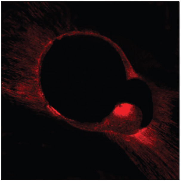
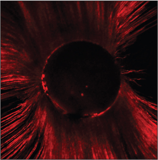
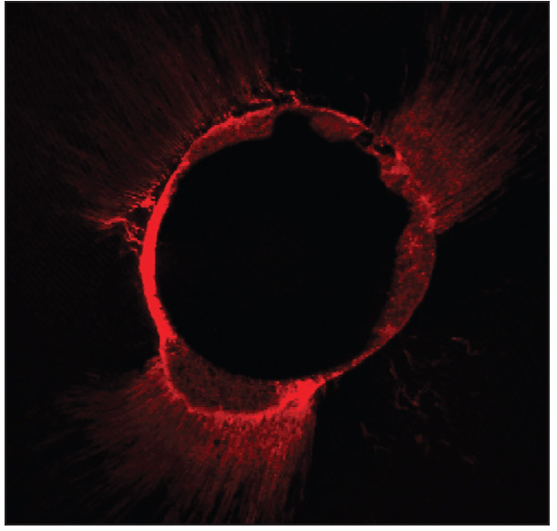
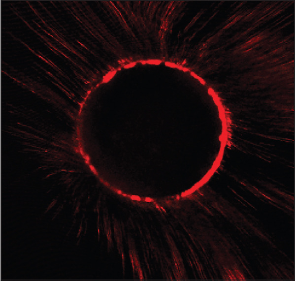
In the middle third, CH dressing significantly decreased the tubular penetration of AH Plus (P<0.05) and did not interfere in MTA Fillapex penetration (P>0.05). Figure 1-4 show representative samples of this result. The AH Plus obturations had a lower percentage of impregnated dentin when compared to the MTA Fillapex obturations regardless of the presence or absence of CH (P<0.05). There was no significant difference between MTA Fillapex and AH Plus without CH in the sealer penetration segments evaluation (P>0.05).
Figure 1.

Representative CLSM image (10×), showing the sealer penetration in dentinal tubules at middle third of CH dressing/AH Plus group
Figure 4.

Representative CLSM image (10×), showing the sealer penetration in dentinal tubules at middle third of MTA Fillapex group
The Pearson correlation coefficient showed a strong relationship (0.783) between both the analyses: the percentage of impregnated dentin and the percentage of sealer penetration segments.
Figure 2.

Representative CLSM image (10×), showing the sealer penetration in dentinal tubules at middle third of AH Plus group
Figure 3.

Representative CLSM image (10×), showing the sealer penetration in dentinal tubules at middle third of CH dressing/MTA Fillapex group
DISCUSSION
Sealer penetration into the dentinal tubules is required to inhibit the growth of remaining bacteria and to prevent reinfection after the root canal treatment (16). If some leakage occurs in the obturation core, a high degree of sealer penetration could prevent bacterial repopulation by entombing the bacteria in the tubules (17). It was also demonstrated that the physical barrier created by sealer penetration into dentinal tubules persists even after endodontic retreatment procedures (6).
Confocal laser scanning microscopy provides reliable information concerning sealer distribution along the canal wall circumference in greater detail, compared with scanning electronic microscopy (18). The small amount of 0.1% Rhodamine B added to the sealer to produce fluorescence has no effect on the sealer’s chemical or mechanical properties, and it allows the observation of the sealer inside the dentinal tubules using CLSM (19).
According to Gharib et al. (19), sealer penetration can be evaluated by measuring the area of the canal circumference in which the sealer is present and the depth of the tubular penetration. However, the clinical relevance of the maximum depth of penetration is questioned (20). In addition, the magnification of CLSM used in this research did not allow the analysis of the maximum penetration up to the cement-enamel junction (6). Therefore, to evaluate the influence of CH on sealer penetration in the present study, we measured the percentage of walls where the sealer was present and the percentage of impregnated dentin. The results of both analyses showed a strong correlation, which indicated that the interference of CH in the tubular penetration of the tested sealers could be observed in both analyses.
During endodontic obturation procedures, residual CH may act as a barrier and lower the volume of fluid that can penetrate the obturation voids; it also prevents the sealer from penetrating inside tubules (5). Despite various proposed techniques, no technique thoroughly removes the CH dressing (4, 21). The conventional syringe irrigation was used to remove the CH dressing before obturation procedures because this method does not require special devices, since the syringe is already used in root canal treatment. It has been demonstrated that this method is less efficient than passive ultrasonic irrigation and the Self-Adjusting File (SAF) system (ReDent-Nova, Ra’anana, Israel), although no method removes the CH dressing completely from the root canal. Since the goal of this study was to evaluate the effect of CH on the sealers penetration into dentinal tubules, a method less efficient, but also commonly used, seemed to be a right choice.
The first null hypothesis was rejected, as the CH dressing reduced the sealer penetration into the canal wall and the percentage of impregnated dentin in the middle third of the root canals obturated with AH Plus. The sealer penetration into dentinal tubules is related to its physicochemical characteristics as composition, film thickness and flow (16, 22). The effect of CH on sealer tubular penetration could be related to the properties of the sealer because it depends on the interaction between the sealer and the residual CH (5, 23, 24). When CH was used, MTA Fillapex showed better tubular penetration than AH Plus in the middle third. This can be explained by the affinity between the MTA compounds and CH. Supposedly, a merge could have happened between CH residues and the sealer affecting the readings.
The fluidity of some sealers, such as Canals (ShowaYakuhin), Ketac-Endo (Espe, Seefeld, Germany) and Sealapex (Kerr, Romulus, MI, USA), is reduced when they are in contact with CH, although the overall values remain in accordance with the ISO-6876 (4). Addition of 10% CH to AH Plus sealer reduced its fluidity, whereas 5% CH had no such effect (25).
Tubular penetration was higher in the middle third than in the apical third, regardless of which sealer was used or whether CH dressing was used or not. This is in agreement with previous studies (19). Anatomical characteristics influence sealer penetration. The apical third has a smaller diameter and lesser density of dentinal tubules, compared to the middle third and cervical third (26, 27). The obturation technique also contribute with lower penetration in apical third, since the pressure of cold lateral condensation could be lower apically (28). It can be suggested that the influence of CH in the penetration of AH Plus could be more apparent in middle third, since the apical third has less dentinal tubules The lower sealer penetration in the apical third is also attributed to the ineffective delivery of irrigants and the reduced effectiveness of smear layer removal techniques in the apical region. CH removal is more difficult in the apical third; despite this, it interestingly did not have a role in the tubular penetration of both sealers in the 3-mm level segments (4, 21).
In this study, MTA Fillapex obturations showed significantly higher tubular penetration than AH Plus in both thirds, according to the dentin impregnated area evaluation and in the apical third showed by the sealers penetration segments data. These results contradict the second hypothesis. In contrast, Akcay et al. (29) demonstrated that these two sealers had similar penetrability. Different results may be achieved because of different methodology as the obturation technique used. The results can be explained by the different physical properties of the sealers as the complex viscosity that evolves both. According to Rai et al. (30), the complex viscosity of MTA Fillapex is lower than AH Plus even with increase of temperature from 25°C to 37°C. This represents a higher flowability of MTA Fillapex allowing it to achieve a better tubular penetration. MTA Fillapex has physical properties that make it suitable for use as an endodontic sealer, although further investigation is necessary to identify whether any other physicochemical characteristics such as higher pH and the presence of MTA components may have facilitated the dentinal penetration of this sealer.
CONCLUSION
MTA Fillapex obturations presented a higher tubular penetration than AH Plus obturations. The use of the intracanal CH dressing did not interfere with the penetration of either sealer in the apical third of the canal. However, it decreased the tubular penetration in the middle third when AH Plus sealer is used.
Footnotes
Ethical Approval: Ethics committee approval was received for this study from the Ethics Committee of Pontifical Catholic University of Parana (Decision Date: 19.10.2011/Decision No: 347.0.084.000-11).
Informed Consent: N/A
Peer-review: Externally peer-reviewed.
Authorship Contributions: Concept - U.X.S.N.; Design - A.T.G.C., U.X.S.N.; Supervision - V.P.D.W., U.X.S.N.; Resource - A.T.G.C., F.S.G., L.P., C.W., V.P.D.W., E.C., L.F.F., U.X.S.N.; Materials - E.C., L.F.F.; Data Collection and/or Processing - A.T.G.C., C.W.; Analysis and/or Interpretation - E.C., L.F.F.; Literature Review - A.T.G.C.; Writer - A.T.G.C., L.P.; Critical Review - L.P., U.X.S.N.
Conflict of Interest: No conflict of interest was declared by the authors.
Financial Disclosure: The authors declared that this study has received no financial support.
REFERENCES
- 1.Nawal RR, Parande M, Sehgal R, Naik A, Rao NR. A comparative evaluation of antimicrobial efficacy and flow properties for Epiphany, Guttaflow and AH-Plus sealer. Int Endod J. 2011;44(4):307–13. doi: 10.1111/j.1365-2591.2010.01829.x. [DOI] [PubMed] [Google Scholar]
- 2.Haapasalo M, Udnæs T, Endal U. Persistent, recurrent, and acquired infection of the root canal system post-treatment. Endod Top. 2003;6(1):29–56. [Google Scholar]
- 3.Vera J, Siqueira JF Jr, Ricucci D, Loghin S, Fernandez N, Flores B, et al. Oneversus two-visit endodontic treatment of teeth with apical periodontitis:A histobacteriologic study. J Endod. 2012;38(8):1040–52. doi: 10.1016/j.joen.2012.04.010. [DOI] [PubMed] [Google Scholar]
- 4.Hosoya N, Kurayama H, Iino F, Arai T. Effects of calcium hydroxide on physical and sealing properties of canal sealers. Int Endod J. 2004;37(3):178–84. doi: 10.1111/j.0143-2885.2004.00781.x. [DOI] [PubMed] [Google Scholar]
- 5.Böttcher DE, Hirai VH, Silva Neto UX, Grecca FS. Effect of calcium hydroxide dressing on the long-term sealing ability of two different endodontic sealers:an in vitro study. Oral Surg Oral Med Oral Pathol Oral Radiol Endod. 2010;110(3):386–9. doi: 10.1016/j.tripleo.2010.05.007. [DOI] [PubMed] [Google Scholar]
- 6.Kok D, Rosa RA, Barreto MS, Busanello FH, Santini MF, Pereira JR, et al. Penetrability of AH plus and MTA fillapex after endodontic treatment and retreatment:a confocal laser scanning microscopy study. Microsc Res Tech. 2014;77(6):467–71. doi: 10.1002/jemt.22371. [DOI] [PubMed] [Google Scholar]
- 7.Silva RV, Silveira FF, Horta MC, Duarte MA, Cavenago BC, Morais IG, et al. Filling effectiveness and dentinal penetration of endodontic sealers:a stereo and confocal laser scanning microscopy study. Braz Dent J. 2015;26(5):541–6. doi: 10.1590/0103-6440201300138. [DOI] [PubMed] [Google Scholar]
- 8.Kokkas AB, Boutsioukis ACh, Vassiliadis LP, Stavrianos CK. The influence of the smear layer on dentinal tubule penetration depth by three different root canal sealers:an in vitro study. J Endod. 2004;30(2):100–2. doi: 10.1097/00004770-200402000-00009. [DOI] [PubMed] [Google Scholar]
- 9.Zhou HM, Shen Y, Zheng W, Li L, Zheng YF, Haapasalo M. Physical properties of 5 root canal sealers. J Endod. 2013;39(10):1281–6. doi: 10.1016/j.joen.2013.06.012. [DOI] [PubMed] [Google Scholar]
- 10.Marin-Bauza GA, Rached-Junior FJ, Souza-Gabriel AE, Sousa-Neto MD, Miranda CE, Silva-Sousa YT. Physicochemical properties of methacrylate resin-based root canal sealers. J Endod. 2010;36(9):1531–6. doi: 10.1016/j.joen.2010.05.002. [DOI] [PubMed] [Google Scholar]
- 11.Silva EJ, Rosa TP, Herrera DR, Jacinto RC, Gomes BP, Zaia AA. Evaluation of cytotoxicity and physicochemical properties of calcium silicate-based endodontic sealer MTA Fillapex. J Endod. 2013;39(2):274–7. doi: 10.1016/j.joen.2012.06.030. [DOI] [PubMed] [Google Scholar]
- 12.Silva RV, Silveira FF, Horta MCR, Duarte MAH, Cavenago BC, et al. Evaluation of cytotoxicity and physicochemical properties of calcium silicate-based endodontic sealer MTA Fillapex. J Endod. 2013;39(2):274–7. doi: 10.1016/j.joen.2012.06.030. [DOI] [PubMed] [Google Scholar]
- 13.Guiotti FA, Kuga MC, Duarte MA, Sant'Anna AJ, Faria G. Effect of calcium hydroxide dressing on push-out bond strength of endodontic sealers to root canal dentin. Braz Oral Res. 2014;28(1):1–6. doi: 10.1590/S1806-83242014.50000002. [DOI] [PubMed] [Google Scholar]
- 14.Akcay M, Arslan H, Topcuoglu HS, Tuncay O. Effect of calcium hydroxide and double and triple antibiotic pastes on the bond strength of epoxy resin-based sealer to root canal dentin. J Endod. 2014;40(10):1663–7. doi: 10.1016/j.joen.2014.05.006. [DOI] [PubMed] [Google Scholar]
- 15.Gokturk H, Bayram E, Bayram HM, Aslan T, Ustun Y. Effect of double antibiotic and calcium hydroxide pastes on dislodgement resistance of an epoxy resin-based and two calcium silicate-based root canal sealers. Clin Oral Investig. 2017;21(4):1277–82. doi: 10.1007/s00784-016-1877-1. [DOI] [PubMed] [Google Scholar]
- 16.Ordinola-Zapata R, Bramante CM, Graeff MS, del Carpio Perochena A, Vivan RR, Camargo EJ, et al. Depth and percentage of penetration of endodontic sealers into dentinal tubules after root canal obturation using a lateral compaction technique:a confocal laser scanning microscopy study. Oral Surg Oral Med Oral Pathol Oral Radiol Endod. 2009;108(3):450–7. doi: 10.1016/j.tripleo.2009.04.024. [DOI] [PubMed] [Google Scholar]
- 17.Marciano MA, Ordinola-Zapata R, Cunha TV, Duarte MA, Cavenago BC, Garcia RB, et al. Analysis of four gutta-percha techniques used to fill mesial root canals of mandibular molars. Int Endod J. 2011;44(4):321–9. doi: 10.1111/j.1365-2591.2010.01832.x. [DOI] [PubMed] [Google Scholar]
- 18.Marciano MA, Guimarães BM, Ordinola-Zapata R, Bramante CM, Cavenago BC, Garcia RB, et al. Physical properties and interfacial adaptation of three epoxy resin-based sealers. J Endod. 2011;37(10):1417–21. doi: 10.1016/j.joen.2011.06.023. [DOI] [PubMed] [Google Scholar]
- 19.Gharib SR, Tordik PA, Imamura GM, Baginski TA, Goodell GG. A confocal laser scanning microscope investigation of the epiphany obturation system. J Endod. 2007;33(8):957–61. doi: 10.1016/j.joen.2007.03.011. [DOI] [PubMed] [Google Scholar]
- 20.Guimarães BM, Amoroso-Silva PA, Alcalde MP, Marciano MA, de Andrade FB, Duarte MA. Influence of ultrasonic activation of 4 root canal sealers on the filling quality. J Endod. 2014;40(7):964–8. doi: 10.1016/j.joen.2013.11.016. [DOI] [PubMed] [Google Scholar]
- 21.Capar ID, Ozcan E, Arslan H, Ertas H, Aydinbelge HA. Effect of different final irrigation methods on the removal of calcium hydroxide from an artificial standardized groove in the apical third of root canals. J Endod. 2014;40(3):451–4. doi: 10.1016/j.joen.2013.10.019. [DOI] [PubMed] [Google Scholar]
- 22.Mamootil K, Messer HH. Penetration of dentinal tubules by endodontic sealer cements in extracted teeth and in vivo. Int Endod J. 2007;40(11):873–81. doi: 10.1111/j.1365-2591.2007.01307.x. [DOI] [PubMed] [Google Scholar]
- 23.Bidar M, Disfani R, Gharagozloo S, Khoynezhad S, Rouhani A. Medication with calcium hydroxide improved marginal adaptation of mineral trioxide aggregate apical barrier. J Endod. 2010;36(10):1679–82. doi: 10.1016/j.joen.2010.06.010. [DOI] [PubMed] [Google Scholar]
- 24.Porkaew P, Retief DH, Barfield RD, Lacefield WR, Soong SJ. Effects of calcium hydroxide paste as an intracanal medicament on apical seal. J Endod. 1990;16(8):369–74. doi: 10.1016/S0099-2399(06)81908-4. [DOI] [PubMed] [Google Scholar]
- 25.Duarte MA, Ordinola-Zapata R, Bernardes RA, Bramante CM, Bernardineli N, Garcia RB, et al. Influence of calcium hydroxide association on the physical properties of AH Plus. J Endod. 2010;36(6):1048–51. doi: 10.1016/j.joen.2010.02.007. [DOI] [PubMed] [Google Scholar]
- 26.Moon Y, Shon WJ, Baek SH, Bae KS, Kum KY, Lee W. Effect of final irrigation regimen on sealer penetration in curved root canals. J Endod. 2010;36(4):732–6. doi: 10.1016/j.joen.2009.12.006. [DOI] [PubMed] [Google Scholar]
- 27.Jardine AP, Rosa RA, Santini MF, Wagner M, So MV, Kuga MC, et al. The effect of final irrigation on the penetrability of an epoxy resin-based sealer into dentinal tubules:a confocal microscopy study. Clin Oral Investig. 2016;20(1):117–23. doi: 10.1007/s00784-015-1474-8. [DOI] [PubMed] [Google Scholar]
- 28.Kuçi A, Alaçam T, Yavaş Ö, Ergul-Ulger Z, Kayaoglu G. Sealer penetration into dentinal tubules in the presence or absence of smear layer:a confocal laser scanning microscopic study. J Endod. 2014;40(10):1627–31. doi: 10.1016/j.joen.2014.03.019. [DOI] [PubMed] [Google Scholar]
- 29.Akcay M, Arslan H, Durmus N, Mese M, Capar ID. Dentinal tubule penetration of AH Plus, iRoot SP, MTA fillapex, and guttaflow bioseal root canal sealers after different final irrigation procedures:A confocal microscopic study. Lasers Surg Med. 2016;48(1):70–6. doi: 10.1002/lsm.22446. [DOI] [PubMed] [Google Scholar]
- 30.Rai RU, Singbal KP, Parekh V. The effect of temperature on rheological properties of endodontic sealers. J Conserv Dent. 2016;19(2):116–9. doi: 10.4103/0972-0707.178683. [DOI] [PMC free article] [PubMed] [Google Scholar]


