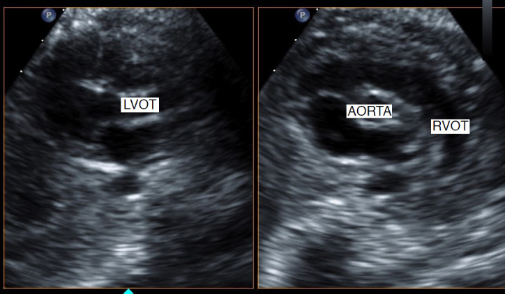
Fig. 5. Use of matrix probe.

Image of the left ventricular outflow (LVOT) on the left and the right ventricular outflow (RVOT) or short axis on the right are obtained simultaneously. Cross section of the LVOT/aorta is seen on the right since this is a perpendicular view to the RVOT view.
