Figure 2.
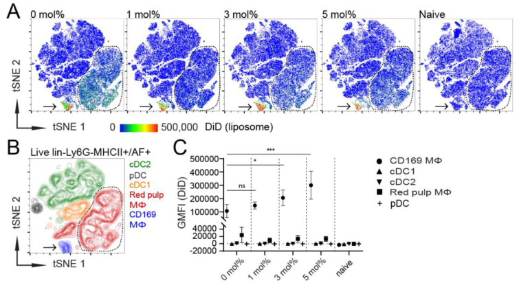
CD169+ MΦ are the main antigen-presenting cells (APCs) that take up liposomes, and the inclusion of 3–5 mol% GM3 augments targeting efficiency. (A) DiD-containing liposomes (22.5 nmol of phospholipid), supplemented with adjuvant, were intravenously (IV) injected into mice. After 2 h, splenocytes were stained and major splenic APC populations were clustered using high-dimensional data reduction analysis (pre-gated on live, lineage−, Ly6G− and MHCII+/AF+ cells). Liposomal DiD fluorescence in APC populations is depicted for 0, 1, 3 or 5 mol% GM3-containing liposomes and non-injected naive mice. (B) CD169+ MΦ, red pulp MΦ, cDC1, cDC2 and pDC were manually gated and overlaid to identify the clusters that were obtained by high-dimensional data reduction. Arrow points to CD169+ MΦ, and red pulp MΦ are bordered by a line. (C) Quantification of DiD fluorescence within various splenic APC populations. Indicated is the average GMFI ± SD (n = 6 for CD169+ MΦ, red pulp MΦ, cDC1 and cDC2 and n = 3 for pDC). * p < 0.05, *** p < 0.001, ns: no significance.

