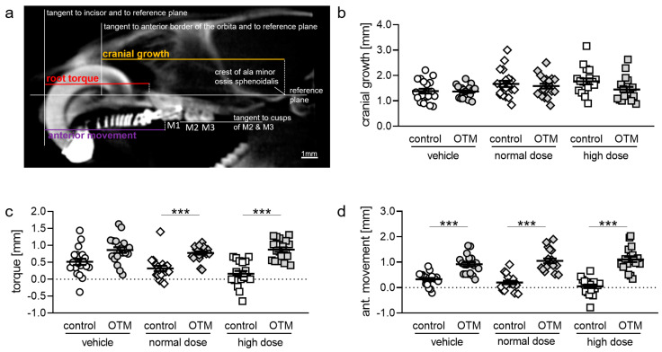
Figure 3.
(a) Radiological cephalometry of cranial growth, root torque, and anterior movement of the first upper molar (M1) in reproducible two-dimensional sagittal cone-beam computed tomography (CBCT) planes of the rat skull based on the method described by Kirschneck et al. [32]. (b) Cranial growth, (c) root torque, and (d) anterior movement of M1 determined with CBCT. n ≥ 19. Statistics: (b,c) Welch-corrected ANOVA followed by Games–Howell multiple comparison tests. (d) Ordinary ANOVA followed by Bonferroni multiple comparison tests *** p < 0.001.

