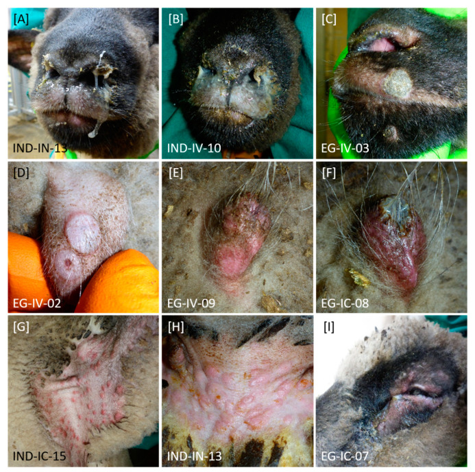
Figure 2.
Clinical signs observed after experimental infection of sheep with SPPV-“India/2013/Surankote” and SPPV-“Egypt/2018”, respectively. Differences in type of clinical signs between both groups could not be detected. (A–C) In diseased animals, nasal discharge with different severity could be observed. (C–I) In addition, sheep showed pox lesions characteristic for SPPV infections. Here, (D–F) especially prepuce and other regions without wool like (G) axilla and (H) groin were affected. Furthermore, these lesions could be observed (C) around the nose and mouth as well as (I) around the eyes. (I) Few animals additionally displayed ocular discharge.

