Figure 2.
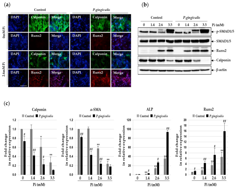
Effect of P. gingivalis on Pi-induced osteogenic transition in VSMCs. P. gingivalis-infected A7r5 cells were cultured for five days in calcification medium containing 2.6 mM Pi. (a) Higher magnification images showed immunoreactivity of Runx2 (red) and calponin (green) in P. gingivalis-infected cells under calcification conditions. (original magnification, ×400). Scale bar: 50 μm. (b) Protein levels of Runx2, calponin, p-SMAD1/5, and SMAD1/5 were examined by Western blotting using their corresponding antibodies. β-actin served as the loading control. (c) Total RNA was isolated and analyzed by real-time PCR using primers specific for rat Runx2, ALP, calponin, and α-SMA. The expression levels of the control (untreated) were set to 1, and the values were normalized to β-actin mRNA levels. * p < 0.05; ** p < 0.01 vs. 0 mM Pi, # p < 0.05; ## p < 0.01 vs. control, one-way ANOVA followed by a Student’s t-test. Data shown are the mean ± SD, obtained for at least three independent experiments.

