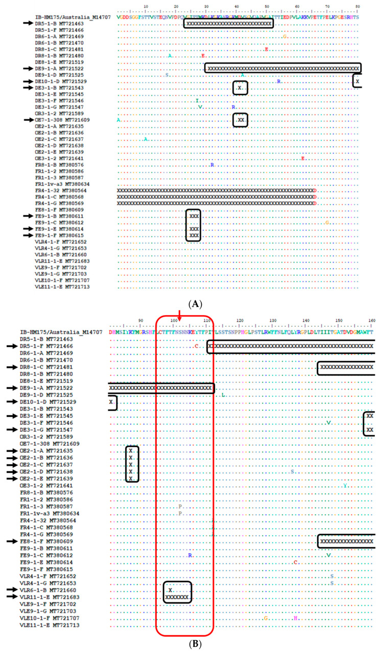
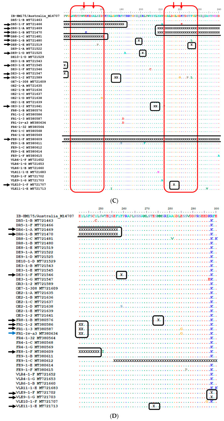
Figure 6.
Alignment of the deduced amino acid sequences of the VP1 region of the HM175 strain and hepatitis A virus (HAV) strains carrying in-frame deletions (A–D). Conserved sites, substitutions and deletions are represented by dots, single-letter abbreviation and the letter “X”, respectively. The red arrows and blocks highlight amino acid change at epitopes 102 (B), 171, 176, 217 and 221 (C), and their surroundings. The closed black blocks highlight in-frame deletions within a subfigure. The open black blocks highlight in-frame deletions that span between two or more subfigures. The black arrows point to HAV strains with in-frame deletion(s). The blue arrow points to a HAV strain with in-frame deletion, which was characterised after viability treatment. The sequence alignment used to construct Figure 6 is provided as Supplementary file 5 (Fasta file).


