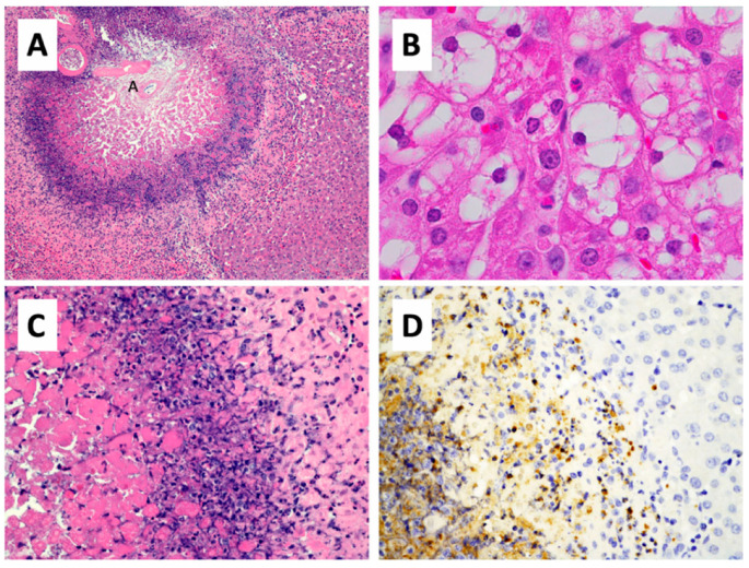
Figure 5.
Histopathology of thermoembolization. (A) In this section of pig liver that was located downstream from a much larger lesion, thermoembolization results in a targetoid lesion with locally extensive coagulative necrosis that is centered on the hepatic arteriole (annotated as small “A”) and radiates peripherally to encompass portions of several hepatic lobules. Hepatic lobular architecture is preserved centrally, even though constituent cells are necrotic. Zones of lytic necrosis and inflammation are visible as the basophilic zone. In less affected regions of liver (B) located more distal to another large thermoembolization lesion, many centrilobular hepatocytes are vacuolated and scattered apoptotic cells are present. (C) Non-discrete zones of coagulative necrosis, lysed necrotic hepatocytes, neutrophilic infiltrate, apoptotic cells, and injured but potentially viable hepatic parenchyma are present from left to right in a higher magnification image of a portion of panel A. (D) The infiltration of neutrophils is documented by myeloperoxidase immunostaining of a parallel field. Hematoxylin and eosin staining (A–C) and immunohistochemical staining for myeloperoxidase. Magnification: 100× (A), 1000× (B), 400× (C,D).

