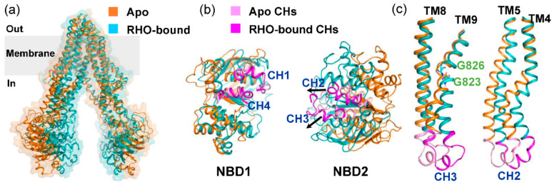
Figure 6.
Conformational changes upon rhodamine-123 binding. (a) Global superposition of the apo (PDB code: 4Q9H) and RHO-bound structures. The RHO-bound snapshot was the representative frame taken from the second populated cluster, as shown in Figure 4b. (b) Extracellular view of the NBDs after the structural superposition of TMDs. Arrows indicate movements of CHs. (c) Close-up view of the TM helix pairs TM4-TM5 and TM8-TM9. Two glycine residues located on TM9 were displayed as sticks.

